Blood Res2016;51:133-47. bloodresearch.or.kr
144 Letters to the Editor
MM with bortezomib, a proteasome inhibitor known for its remarkable efficacy in treating extramedullary MM.
Although the pathogenesis of MPE is unknown, it is theor- ized that it may be a direct extension of thoracic myeloma- tous involvement. A review of 57 cases [9] demonstrated that half of the patients with MPE had concomitant thoracic skeletal, lung parenchymal, or chest wall plasmacytomas, which would provide a source for MPEs. Similarly, both of our patients had a pulmonary nodule, which likely repre- sented metastatic disease.
Genetic analysis showed that the patient in our first case had a trisomy at chromosome 3 and monosomy at chromo- some 13. In addition to the t(4;14) translocation, this com- plex karyotype is associated with unfavorable prognosis [5].
Given that the median survival time for high-risk patients without malignant pleural effusions is 3 years, it is likely that the progression of the myeloma and development of the pleural effusions contributed significantly to the even- tual death of the first patient. In our second case, the patient had no trisomy, but she did have monosomy of chromosome 13 in addition to the t(4;14) translocation. This chromosome 13 abnormality was also seen in 77.8% of patients in the Cho et al. [10] case series.
Although rare, more cases of MPE are being described in the literature, with evidence indicating its poor prognosis and lack of efficacious treatment [5, 12]. Because of the severity of MPE, we recommend that patients with pleural effusions and suspicion of myeloma undergo protein electro- phoresis, flow cytometry, cytologic examination of the pleu- ral fluid, or pleural biopsy examination to identify MPE and begin treatment promptly [12, 13].
Akshay Amaraneni1, Usman Saeed2, Devin Malik1, Megan Brown3, Sreenivasa R. Chandana4
1Department of Internal Medicine, 2Department of Internal Medicine-Pediatrics, Western Michigan University, Homer Stryker M.D. School of Medicine, Kalamazoo, 3Michigan State University, College of Human Medicine, East Lansing,
4Division of Hematology and Oncology, West Michigan Cancer Center, Kalamazoo, MI, USA Correspondence to: Sreenivasa R. Chandana
Division of Hematology and Oncology, West Michigan Cancer Center, 200 N Park Street, Kalamazoo, MI 49007, USA
E-mail: [email protected]
Received on May 9, 2015; Revised on May 27, 2015; Accepted on Jun. 15, 2015 http://dx.doi.org/10.5045/br.2016.51.2.142
AuthorsÊ Disclosures of Potential Conflicts of Interest No potential conflicts of interest relevant to this article were reported.
REFERENCES
1. Siegel R, Naishadham D, Jemal A. Cancer statistics, 2013. CA
Cancer J Clin 2013;63:11-30.
2. Kumar SK, Rajkumar SV, Dispenzieri A, et al. Improved survival in multiple myeloma and the impact of novel therapies. Blood 2008;111:2516-20.
3. Bladé J, Rosiñol L. Complications of multiple myeloma. Hematol Oncol Clin North Am 2007;21:1231-46.
4. Yosunkaya S, Maden E, Toy H, Yazici R, Ozer F, Reisli I. A multi- ple myeloma case presenting with bilateral pleural involvement.
Tuberk Toraks 2007;55:285-9.
5. Mikhael JR, Dingli D, Roy V, et al. Management of newly diag- nosed symptomatic multiple myeloma: updated Mayo strat- ification of myeloma and risk-adapted therapy (mSMART) con- sensus guidelines 2013. Mayo Clin Proc 2013;88:360-76.
6. Kintzer JS Jr, Rosenow EC 3rd, Kyle RA. Thoracic and pulmonary abnormalities in multiple myeloma. A review of 958 cases. Arch Intern Med 1978;138:727-30.
7. Meoli A, Willsie S, Fiorella R. Myelomatous pleural effusion.
South Med J 1997;90:65-8.
8. Kamble R, Wilson CS, Fassas A, et al. Malignant pleural effusion of multiple myeloma: prognostic factors and outcome. Leuk Lymphoma 2005;46:1137-42.
9. Kim YJ, Kim SJ, Min K, et al. Multiple myeloma with myeloma- tous pleural effusion: a case report and review of the literature.
Acta Haematol 2008;120:108-11.
10. Cho YU, Chi HS, Park CJ, Jang S, Seo EJ, Suh C. Myelomatous pleural effusion: a case series in a single institution and literature review. Korean J Lab Med 2011;31:225-30.
11. Mangiacavalli S, Varettoni M, Zappasodi P, Pica G, Lazzarino M, Corso A. A striking response to bortezomib in a patient with pleu- ral localization of multiple myeloma. Leuk Res 2009;33:577-8.
12. Keklik M, Sivgin S, Pala C, et al. Flow cytometry method as a diag- nostic tool for pleural fluid involvement in a patient with multiple myeloma. Mediterr J Hematol Infect Dis 2012;4:e2012063.
13. Oudart JB, Maquart FX, Semouma O, Lauer M, Arthuis- Demoulin P, Ramont L. Pleural effusion in a patient with multiple myeloma. Clin Chem 2012;58:672-4.
A rare case of diffuse large B cell lymphoma-associated
hemophagocytic syndrome initially present in the bone marrow with a favorable clinical course
TO THE EDITOR: Lymphoma-associated hemophagocytic syndrome (LAHS) is a hematological disorder associated with malignant lymphoma. It is characterized by clinical features and laboratory findings associated with hemophago- cytic lymphohistiocytosis (HLH), such as fever, cytopenia, hyperferritinemia, hypofibrinogenemia, and hemophagocy- tosis in the bone marrow (BM) [1]. The development of LAHS can be accounted for by various types of lymphoma,
bloodresearch.or.kr Blood Res 2016;51:133-47.
Letters to the Editor 145
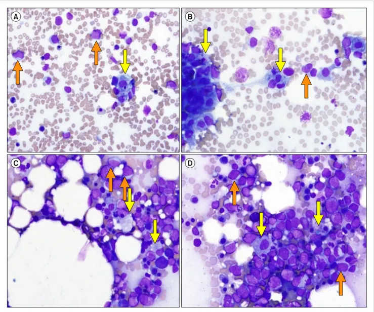
Fig. 1. Bone marrow aspirates (A, B, Wright stain, ×400) and touch print (C, D, Wright stain, ×400). The bone marrow aspirates and touch print show normocellular marrow with infiltration of neoplastic lymphoid cells (20.0% of total nucleated cells, orange arrows). In addition, hemophagocytic histiocytes (11.0% of total nucleated cells, yellow arrows) were identified.
but most of them are associated with T cell or natural killer (NK) cell lymphomas such as aggressive NK/T cell lympho- ma, peripheral T cell lymphoma, anaplastic large cell lym- phoma, or extranodal NK/T cell lymphoma [2-7]. Develop- ment of LAHS from B cell lymphoma is rarely reported, and occurs in old age with a low frequency of BM involve- ment at diagnosis [6, 7]. To date, seven cases of LAHS asso- ciated with large B cell lymphoma initially manifesting in the BM have been reported and the majority of these cases were of the non-germinal center (GC) type, with a complex karyotype and very poor prognosis [8]. We report here a rare case of diffuse large B cell lymphoma (DLBCL)-associated hemophagocytic syndrome initially manifesting in the BM and exhibiting a favorable clinical course.
In February 2015, a 73-year-old female was admitted with fever (body temperature, 38.1oC), poor oral intake,
and general weakness, which had developed 2 weeks prior to admission. She was diagnosed as diabetes mellitus (DM) 8 years ago and had been on treatment with a hypoglycemic agent, but had no medical history related to hematological malignancy, including hepatosplenomegaly. The patient’s hemogram results at admission showed pancytopenia (white blood cell [WBC] count 3.57×10⁹/L [absolute neutrophil count 2.82×10⁹/L], hemoglobin 7.6 g/dL, platelet count 32.0×10⁹/L). Her peripheral blood smear showed the pres- ence of atypical lymphocytes (3% of total nucleated cells).
She underwent BM aspiration and biopsy due to pan- cytopenia with presence of atypical lymphocytes.
The patient’s BM aspirates (Fig. 1A, B) and touch print (Fig. 1C, D) showed normocellular marrow with increased infiltration of large-sized neoplastic lymphoid cells with a frequency of 20.0% (indicated by the orange arrows).
Blood Res2016;51:133-47. bloodresearch.or.kr
146 Letters to the Editor
Fig. 2. Bone marrow biopsy (A, hematoxylin and eosin stain, ×400) and immunohistiochemical staining results for CD3, CD20 and CD68 (B–D,
×400, respectively). The bone marrow biopsy shows normocellular marrow with diffuse infiltration of CD20-positive neoplastic lymphoid cells (orange arrows) accompanied by an increase of CD68-positive histiocytes with occasional hemophagocytosis (yellow arrows), indicating the presence of B-cell lymphoma associated with hemophagocytic histiocytosis.
In addition, histiocytes with active hemophagocytosis were occasionally identified with a frequency of 11.0% (indicated by the yellow arrows). Flow cytometric analysis results of BM aspirates demonstrated the presence of a clonal B lym- phocyte population with kappa light chain restriction: 19.0%
positivity for CD19, 22.6% positivity for CD20, 35.0% pos- itivity for kappa light chain, and 5.2% positivity for lambda light chain. However, T-cell markers showed no evidence of clonality, with variable positivity for CD3, CD2, CD5, CD7, CD4 and CD8, (range, 10.9–48.1%).
The BM biopsy (Fig. 2A) showed that 65% of the marrow was cellular. Diffuse infiltration of CD20-positive neoplastic lymphoid cells (indicated by the orange arrows) and in- creased infiltration of CD68-positive hemophagocytic his- tiocytes (indicated by the yellow arrows) was demonstrated by immunohistochemical stains for CD3, CD20 and CD68
(DAKO, Glostrup, Denmark) (Fig. 2B–D, respectively).
Serum ferritin, fibrinogen, triglyceride, and soluble inter- leukin-2 (IL-2) receptor levels were 1,198.0 ng/mL (reference range, 33–298 ng/mL), 300.5 mg/dL (reference range, 170–
380 mg/dL), 149 mg/dL (reference range, 58–250 mg/dL) and 6,530.0 U/mL (reference range, 124–466 U/mL), respec- tively. The patient’s karyotype analysis results showed 46–
48,XX,add(1)(q25),add(1)(q42),add(2)(q33),del(4)(q21),del (5)(p13p15.3),add(6)(q21),del(8)(q22),+16,add(17)(q25),add (19)(p13.3),+20[cp11]/46,XX[9], indicating the presence of a complex karyotype in 55% of all analyzed cells. Based on these results, and because her clinical and laboratory findings fulfilled the 2008 diagnostic criteria of HLH (fever, cytopenia, hyperferritinemia, hemophagocytosis in BM, high soluble IL-2 receptor levels) [9], the patient was initially diagnosed with B cell LAHS which was finally revised as
bloodresearch.or.kr Blood Res 2016;51:133-47.
Letters to the Editor 147
DLBCL (non-GC type)-associated hemophagocytic syndrome.
She was treated with 5 cycles of R-CHOP (cyclophospha- mide, doxorubicin, vincristine, prednisone plus rituximab) chemotherapy. At follow-up after 3 months of chemo- therapy, the patient’s hemogram results had improved sig- nificantly (WBC 4.45×10⁹/L, hemoglobin 10.7 g/dL, platelet count 148×109/L). In the BM study, residual neoplastic lym- phoid cells were not identified and active hemophagocytosis was not observed, despite persistent histiocytic hyperplasia.
The patient has been discharged in an improved condition and is waiting to undergo additional chemotherapy.
According to the literature, B cell LAHS cases are rather uncommon compared with T- or NK-cell lymphoma cases and the most common type is DLBCL, which occurs in older patients [6, 7]. To date, there have been seven reports of large B cell lymphoma initially manifesting in the BM, with consistent clinical features such as occurrence in old age (range, 54–80 yr), non-GC type (six cases), a complex karyotype, aggressive clinical course (four cases died before treatment), and no association with Epstein-Barr virus (EB) infection (all seven cases) [8]. The clinical features of our patient were generally similar to those of the previous cases:
a complex karyotype, non-GC type of DLBCL, old age at diagnosis. However, our patient also demonstrated an un- common clinical feature of B cell LAHS initially manifesting in the BM: a favorable clinical outcome after chemotherapy.
Since our patient did not undergo serologic or molecular EBV studies, we could not confirm that EBV infection was not associated with B cell LAHS initially manifesting in the BM, which was suggested by the previous study [8].
Further studies will be required to confirm the clinical fea- tures of B cell LAHS initially manifesting in the BM.
In conclusion, we report a rare case of DLBCL-associated hemophagocytic syndrome initially manifesting in the BM and exhibiting a favorable clinical outcome. More extensive studies will be needed to investigate the clinical features of these patients.
*This work was supported by the year 2015 clinical re- search grant from Pusan National University Hospital.
Sang Hyuk Park1, Eun Yup Lee1, Joo Seop Chung2
1Department of Laboratory Medicine and Biomedical Research Institute, 2Division of Hematology-Oncology, Department of Internal Medicine, Pusan National University Hospital, Pusan National University School of Medicine, Busan, Korea
Correspondence to: Sang Hyuk Park Department of Laboratory Medicine and Biomedical Research Institute, Pusan National University Hospital, 179 Gudeok-ro, Seo-gu, Busan 49241, Korea
E-mail: [email protected]
Received on May 26, 2015; Revised on Jun. 3, 2015; Accepted on Jun. 18, 2015 http://dx.doi.org/10.5045/br.2016.51.2.144
AuthorsÊ Disclosures of Potential Conflicts of Interest No potential conflicts of interest relevant to this article were reported.
REFERENCES
1. Usmani GN, Woda BA, Newburger PE. Advances in under- standing the pathogenesis of HLH. Br J Haematol 2013;161:609- 22.
2. Cheung MM, Chan JK, Lau WH, et al. Primary non-Hodgkin's lymphoma of the nose and nasopharynx: clinical features, tumor immunophenotype, and treatment outcome in 113 patients. J Clin Oncol 1998;16:70-7.
3. Falini B, Pileri S, De Solas I, et al. Peripheral T-cell lymphoma associated with hemophagocytic syndrome. Blood 1990;75:434- 44.
4. Florena AM, Iannitto E, Quintini G, Franco V. Bone marrow bi- opsy in hemophagocytic syndrome. Virchows Arch 2002;441:
335-44.
5. Shimazaki C, Inaba T, Shimura K, et al. B-cell lymphoma asso- ciated with haemophagocytic syndrome: a clinical, immuno- logical and cytogenetic study. Br J Haematol 1999;104:672-9.
6. Han AR, Lee HR, Park BB, et al. Lymphoma-associated hemo- phagocytic syndrome: clinical features and treatment outcome.
Ann Hematol 2007;86:493-8.
7. Sano H, Kobayashi R, Tanaka J, et al. Risk factor analysis of non-Hodgkin lymphoma-associated haemophagocytic syn- dromes: a multicentre study. Br J Haematol 2014;165:786-92.
8. Yeh YM, Chang KC, Chen YP, et al. Large B cell lymphoma pre- senting initially in bone marrow, liver and spleen: an aggressive entity associated frequently with haemophagocytic syndrome.
Histopathology 2010;57:785-95.
9. Jordan MB, Filipovich AH. Hematopoietic cell transplantation for hemophagocytic lymphohistiocytosis: a journey of a thou- sand miles begins with a single (big) step. Bone Marrow Transplant 2008;42:433-7.