Veterinary Science
DOI: 10.4142/jvs.2010.11.4.299
Received: 06 Jan. 2010, Accepted: 29 Mar. 2010
Original Article
*Corresponding author
Tel: +82-63-270-4030; Fax: +82-63-270-4028
E-mail: [email protected]
Expression of KA1 kainate receptor subunit in the substantia gelatinosa of the trigeminal subnucleus caudalis in mice
Seon Ah Park
1, Soo Joung Park
1, Seong Kyu Han
1,2,*
1
Department of Oral Physiology and BK21 program, and
2Laboratory for Oral Disease-Related Compounds, School of Dentistry and Institute of Oral Bioscience, Chonbuk National University, Jeonju 561-756, Korea
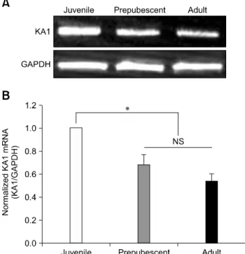
The KA1 kainate receptor (KAR) subunit in the substantia gelatinosa (SG) of the trigeminal subnucleus caudalis (Vc) has been implicated in the processing of nociceptive information from the orofacial region. This study compared the expression of the KA1 KAR subunit in the SG of the Vc in juvenile, prepubescent and adult mice. RT-PCR, Western blot and immunohistochemistry analyses were used to examine the expression level in SG area. The expression levels of the KA1 KAR subunit mRNA and protein were higher in juvenile mice than in prepubescent or adult mice.
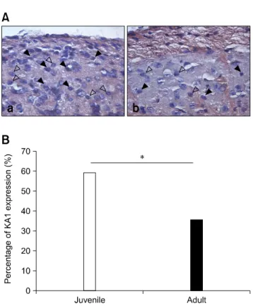
Quantitative data revealed that the KA1 KAR subunit mRNA and protein were expressed at levels approximately two and three times higher, respectively, in juvenile mice than in adult mice. A similar expression pattern of the KA1 KAR subunit was observed in an immunohistochemical study that showed higher expression in the juvenile (59%) than those of adult (35%) mice. These results show that the KA1 KAR subunits are expressed in the SG of the Vc in mice and that the expression level of the KA1 KAR subunit decreases gradually with postnatal development. These findings suggest that age-dependent KA1 KAR subunit expression can be a potential mechanism of age-dependent pain perception.
Keywords: immunohistochemistry, KA1, RT-PCR, substantia gelatinosa, trigeminal subnucleus caudalis, Western blot
Introduction
The substantia gelatinosa (SG) laminar II of the trigeminal subnucleus caudalis (Vc) is a critical site for orofacial nociceptive processing because it receives the synaptic inputs from primary myelinated Aδ and unmyelinated C fibers [38]. The SG neurons function as excitatory and inhibitory interneurons and regulate the output of projection
neurons in lamina I and IV, which transmit noxious information to a higher brain center [8,18,23,24,30].
Kainate receptors (KARs) belong to the ionotropic glutamate receptor families, which also include α -amino- 3-hydroxy-5-methyl-4-isoxazolepropionic acid and N- methyl-D-aspartate subunits. Native KARs are formed by the heteromeric combination of five subunits, GluR5-7 (GluK1-3) and KA1-2 (GluK4-5). The KARs are expressed in nociceptive pathways including the dorsal root ganglion, spinal cord, thalamus and cortex [45], particularly at the spinal dorsal horn and Vc, which are involved in pain processing [3,7,15]. Among the KAR subunits, GluR5- or GluR6-containing KARs are involved in nociceptive transmission [39,46,47]. Because the KA1 subunit contributes to functional KARs with GluR5/6 subunits, the level of KA1 expression in the SG area can provide key information regarding pain processing. However, there is little information available regarding KA1 KAR subunit expression in the SG area of the Vc in mice. This study examined the KA1 KAR subunit mRNA and protein level in the SG area of the Vc using RT-PCR, Western blotting and immunohistochemistry and compared the expression levels according to the postnatal stage.
Materials and Methods Brain slice preparation
All experiments were approved by the Experimental
Animal Care and Ethics Committee of Chonbuk National
University. The mice (Damul Science, Korea) were housed
under 12 h light : 12 h dark cycles (lights on at 07:00 h) with
access to food and water ad libitum. Juvenile (postnatal
days 7-14), prepubescent (postnatal days 25-35) and adult
(postnatal days 42 over) mice were decapitated between
10:00 and 12:00 h, after which their brains were removed
rapidly and placed in an ice-cold bicarbonate-buffered
artificial cerebrospinal fluid (ACSF) with the following
composition (in mM): 126 NaCl, 2.5 KCl, 2.4 CaCl
2, 1.2
MgCl
2, 11
D-glucose, 1.4 NaH
2PO
4 and 25 NaHCO
3 (pH