Blood Res2020;55:112-127. bloodresearch.or.kr
120 Letters to the Editor
AuthorsÊ Disclosures of Potential Conflicts of Interest No potential conflicts of interest relevant to this article were reported.
REFERENCES
1. Rison RA, Beydoun SR. Paraproteinemic neuropathy: a practical review. BMC Neurol 2016;16:13.
2. Kelly JJ Jr, Kyle RA, O'Brien PC, Dyck PJ. Prevalence of mono- clonal protein in peripheral neuropathy. Neurology 1981;31:
1480-3.
3. Jain A, Haynes R, Kothari J, Khera A, Soares M, Ramasamy K.
Pathophysiology and management of monoclonal gammopathy of renal significance. Blood Adv 2019;3:2409-23.
4. Fermand JP, Bridoux F, Dispenzieri A, et al. Monoclonal gamm- opathy of clinical significance: a novel concept with therapeutic implications. Blood 2018;132:1478-85.
5. Nobile-Orazio E, Bianco M, Nozza A. Advances in the treatment of paraproteinemic neuropathy. Curr Treat Options Neurol 2017;19:43.
6. Dimachkie MM, Barohn RJ, Katz J. Multifocal motor neuro- pathy, multifocal acquired demyelinating sensory and motor neuropathy, and other chronic acquired demyelinating poly- neuropathy variants. Neurol Clin 2013;31:533-55.
7. Sandes AF, de Lourdes Chauffaille M, Oliveira CR, et al. CD200 has an important role in the differential diagnosis of mature B-cell neoplasms by multiparameter flow cytometry. Cytometry B Clin Cytom 2014;86:98-105.
8. Swerdlow SH, Campo E, Harris NL, et al, eds. WHO classification of tumours of haematopoietic and lymphoid tissues. Revised 4th ed. Lyon, France: IARC Press, 2017.
9. Wang HY, Zu Y. Diagnostic algorithm of common mature B-cell lymphomas by immunohistochemistry. Arch Pathol Lab Med 2017;141:1236-46.
10. Singh G. Serum free light chain assay and / ratio performance in patients without monoclonal gammopathies: high false-pos- itive rate. Am J Clin Pathol 2016;146:207-14.
11. Drayson M, Tang LX, Drew R, Mead GP, Carr-Smith H, Bradwell AR. Serum free light-chain measurements for identifying and monitoring patients with nonsecretory multiple myeloma. Blood 2001;97:2900-2.
12. Stübgen JP. Autoantibody-mediated sensory polyneuropathy as- sociated with indolent B-cell non-Hodgkin’s lymphoma: a report of two cases. J Clin Neurol 2015;11:283-6.
13. Chen LY, Keddie S, Lunn MP, et al. IgM paraprotein-associated peripheral neuropathy: small CD20-positive B-cell clones may predict a monoclonal gammopathy of neurological significance and rituximab responsiveness. Br J Haematol 2020;188:511-5.
14. Briani C, Visentin A, Salvalaggio A, et al. Peripheral neuropathies in chronic lymphocytic leukemia: a single center experience on 816 patients. Haematologica 2017;102:e140-3.
Large granular lymphocytes (LGL) in primary Sjögren syndrome (pSS):
immunophenotype and review on the pathological role of T cells in pSS
TO THE EDITOR: Primary Sjogren syndrome (pSS) is a chronic autoimmune systemic disease that mainly affects the exocrine glands, causing severe inflammation with ac- companying destruction of the gland. It is also characterized by systemic symptoms and laboratory findings of polyclonal B-lymphocyte activation, hypergammaglobulinemia, and positive autoantibodies. Its pathophysiology is not yet fully understood; genetic factors seem to play a relatively minor role, while environmental ones, most likely infections, con- tribute to disease onset and progression by activation of the innate and adaptive immune systems. Although the role of B cells in the disease is better characterized, little is known about the involvement of T cells in pSS [1-3].
We report a case of a pSS patient who was followed up at our hematology unit for monoclonal CD8+ T lymphocytosis. We have discussed the immunophenotype of CD8+ T lymphocytes and reviewed the involvement of pathological CD8+ T lymphocytes in pSS.
Case report
In September 2012, a 39-year-old woman was referred to our outpatient service because of unexplained lymphocy- tosis, mild anemia, and thrombocytopenia. Together with the lymphocytosis, the patient developed xerophthalmia and xerostomia with anti-nuclear, extractable nuclear antigen, and Ro-SSA antibody positivity. A diagnosis of pSS was made following salivary gland biopsy. Clinical evaluation showed slight dryness of the mouth and eyes with no alter- ations to the spleen, liver, and lymph nodes. The tests per- formed on September 22, 2012 were significant for 9.61×10⁹/L leukocytes, 8.19×10⁹/L lymphocytes, 106×109/L platelets, and 11.8 g/dL hemoglobin; normal liver and kidney function values were seen with a slight polyclonal rise in the immunoglobulin dosage. In addition, hepatitis markers (A, B, and C serology) and parasitological stool assays were negative.
Therefore, to investigate a possible lymphoproliferative disorder, bone marrow and imaging studies were carried out. Bone marrow biopsy showed an interstitial and often intra-sinusoidal infiltration by small-medium sized CD8+
T lymphocytes, which had partial CD5 expression. However, no other sites appeared to be involved since a total body CT examination showed no adenopathies or liver or spleen enlargement.
Flow cytometric analyses were performed in the periph- eral blood and bone marrow samples using a FacsCanto II cytometer (BD Biosciences, Franklin Lakes, NJ, USA) equipped with three lasers (405, 488, 633 nm). A total of
bloodresearch.or.kr Blood Res 2020;55:112-127.
Letters to the Editor 121
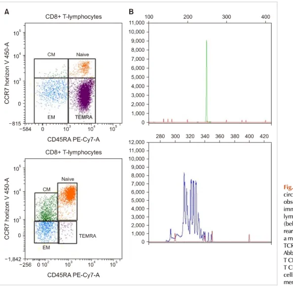
Fig. 1. (A) Immunophenotyping of circulating lymphocytes at the last observation (above) compared with immunophenotyping of circulating lymphocytes in a healthy donor (below). (B) TCR and receptor rearrangements. TCR (above) shows a monoclonal rearrangement, while TCR (below) is polyclonal.
Abbreviations: CM, central memory T CD8+ cells; EM, effector memory T CD8+ cells; naïve, naïve T CD8+ cells; TEMRA, terminal effector memory T CD8+ cells.
100,000 events/tube were acquired, and fluorochrome-con- jugated antibodies were used to investigate different lym- phoid antigens (CD3, CD4, CD5, CD8, CD7, TCR , TCR
, CD45RA, CD45RO, CD57, CD2, CD16-56, CD19, CD20, CD22, CD10, CCR7, CD27, CD28, and and light chains).
The analysis of the peripheral blood confirmed lymphocy- tosis (7.15×10⁹/L lymphocytes) determined by an increase in CD8+ T lymphocytes (6.15×10⁹/L) which had normal expression of CD3, CD2, and CD7 markers, but weak CD5 expression and partial (50%) expression of CD57. Further, CD8+ lymphocytes were positive for CD45RA, but they did not express CCR7 (Fig. 1A), which are features found in terminal effector memory T lymphocytes (TEMRA) [4].
The bone marrow analysis revealed the presence of a very similar population, which accounted for 88% of all lymphocytes. Furthermore, they all appeared to present the
T-cell receptor (TCR-), and polymerase-chain re- action analysis of the TCR genes confirmed a clonal re- arrangement of TCR , while the TCR gene showed a polyclonal rearrangement (Fig. 1B).
This clinical and immunophenotypic condition is well identified as CD8+ T cell large granular lymphocytic (LGL) leukemia [5].
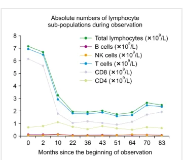
In November 2012, the patient started SS therapy with Hydroxychloroquine (Plaquenil) 200 mg once daily and pre- dnisone 4 mg once daily, and in the following months, the number of lymphocytes returned closer to normal (6.65×10⁹/L in February 2013) and the mild thrombocytope- nia initially noted remained stable. Therefore, close mon- itoring, done with periodic testing and annual flow cyto- metric analysis of the peripheral blood, was done. The num- ber of lymphocytes normalized within one year and has since remained constant (about 2×10⁹/L lymphocytes), but CD8+ cells have continued to remain higher than normal, representing 80 to 89% of the total T lymphocyte population, and have also continued to represent an important pro- portion of the total number of lymphocytes (54% of all lymphocytes in 2015, 42% in 2016 and 2017, 52% and then 57% in 2018, and 61% in 2019; see Fig. 2 for a graphical representation). Moreover, throughout the years, CD57 con- tinued to be expressed by about 50% of these cells.
During the last visit in November 2019, a more in-depth analysis was performed, including CD27 and CD28 de- tection: naïve and central memory cells and over 70% of effector memory cells were found to express CD27 but not CD28, while 93.4% of TEMRA cells did not express either
Blood Res2020;55:112-127. bloodresearch.or.kr
122 Letters to the Editor
Fig. 2. Graph detailing variations in lymphocyte numbers during observation.
CD27 or CD28 (data not shown).
Discussion
LGL leukemia is a rare condition accounting for 2-3%
of all mature lymphoid leukemias [6]. The finding of LGL in the context of autoimmune disorders is common, but it often occurs in rheumatoid arthritis or in the presence of less specific autoimmune features (such as positivity to autoantibodies, e.g., antinuclear antibodies); in pSS, less than 15 cases have been described [7-9].
LGL disorders are characterized by proliferation of LGL cytotoxic lymphocytes of either T-cell (mainly CD3+
TCR+CD8+CD57+CD56+/- CD16+/-, rarely CD4+CD8+/-) or, less frequently, NK-cell (CD3-CD2+CD16+CD D56+
CD57+/-) origin [10].
In our case, the LGL immunophenotype was characterized as CD3+, CD8+, TCR+, CD57+, CD45RA+, CD62L-, CD5dim, CD27, and CD28, ascribable to a sub-population of TEMRA lymphocytes [4]. This physiological population arises from central memory cells in a context of homeostatic proliferation in the absence of an antigen, appearing for example after the acute phase of a viral infection [11]; sim- ilarly, LGL cells are thought to originate from chronic in- flammation, in which prolonged antigen stimulation act as the triggering event [10]. However, while physiological TEMRA cells are characterized by a low proliferative capacity and a high rate of cell death [11, 12], LGL cells have pro- longed survival and activity mainly through pathological activation of the STAT pathway [10].
The relationship between pSS and LGL is not at all clear.
One analysis of circulating T cell subpopulations in pSS had identified no difference in numbers of circulating CD57+
cells between pSS patients and healthy controls and had only found a decrease in CD8bright, CD27+ and CD57+
cells (which could correspond to a TEMRA sub-population)
in patients with anti-SSA/SSB antibodies compared to those without antibodies [2]. On the other hand, a recent multi- disciplinary study has associated TEMRA cells with pSS-specif- ic patterns of gene transcription and protein dysregulation–
meaning that this cell population presents modifications in gene transcription and protein processing which are spe- cific to disease (although the relationship between these cells and transcriptome and proteomic changes are likely more subtle and in need of further study) [13].
The fact that the LGL in this case, as well as in all others reported [7-9], was determined by CD8+ T cells could repre- sent a further argument that CD8+ cells undergo important modifications in pSS, which should be taken in consider- ation. Indeed, in our case, the diagnosis of LGL was made together with that of pSS, suggesting that the two diseases share at least part of the pathogenesis, if not a common etiology, as has been suggested in a previous report [7].
With regards to the former, one hypothesis which has al- ready been proposed [10] is that the continuous immune stimulation enabled a mutated clone to develop and lose its typical high turnover rate. In line with this hypothesis, therapy aimed at immune system control would lead to lymphocyte count normalization [7, 8]; indeed, our patient was managed with immunosuppressive therapy and is still in good health, with a good lymphocytosis control even years later.
However, further research is needed to answer the re- maining open questions about the origin of this population, its nature, and its actions.
Rita Tavarozzi, Giovanni Carulli, Enrica Manzato, Paola Sammuri, Elena Ciabatti, Mario Petrini Department of Clinical and Experimental Medicine, Section
of Haematology, University of Pisa, Pisa, Italy
Correspondence to: Rita Tavarozzi Department of Clinical and Experimental Medicine, Section of Haematology, University of Pisa, via Roma 67, Pisa 56126, Italy E-mail: [email protected]
Received on Mar. 19, 2020; Revised on Apr. 7, 2020; Accepted on Apr. 16, 2020 https://doi.org/10.5045/br.2020.2020052
ACKNOWLEDGMENTS
We would like to thank Dr. Linda Carli for her help in ensuring a correct follow-up of the patient.
AuthorsÊ Disclosures of Potential Conflicts of Interest No potential conflicts of interest relevant to this article were reported.
REFERENCES
1. Singh N, Cohen PL. The T cell in Sjogren’s syndrome: force ma- jeure, not spectateur. J Autoimmun 2012;39:229-33.
bloodresearch.or.kr Blood Res 2020;55:112-127.
Letters to the Editor 123
Fig. 1. Spectrum of circulating lymphoid cells in the peripheral blood in this case. Note the absence of characteristic circumferential hair-like projections in most cells (Leishman Giemsa stain, ×400).
2. Sudzius G, Mieliauskaite D, Siaurys A, et al. Distribution of pe- ripheral lymphocyte populations in primary Sjögren's syndrome patients. J Immunol Res 2015;2015:854706.
3. Björk A, Mofors J, Wahren-Herlenius M. Environmental factors in the pathogenesis of primary Sjögren's syndrome. J Intern Med 2020;287:475-92.
4. Mahnke YD, Brodie TM, Sallusto F, Roederer M, Lugli E. The who’s who of T-cell differentiation: human memory T-cell subsets. Eur J Immunol 2013;43:2797-809.
5. Semenzato G, Zambello R, Starkebaum G, Oshimi K, Loughran TP Jr. The lymphoproliferative disease of granular lymphocytes:
updated criteria for diagnosis. Blood 1997;89:256-60.
6. Swerdlow SH, Campo E, Harris NL, et al, eds. WHO classification of tumours of haematopoietic and lymphoid tissues. 4th ed. Lyon, France: IARC Press, 2008.
7. Molad Y, Okon E, Stark P, Prokocimer M. Sjögren's syndrome associated T cell large granular lymphocyte leukemia: a possible common etiopathogenesis. J Rheumatol 2001;28:2551-2.
8. Franco G, Palazzolo R, Liardo E, Tripodo C, Mancuso S. T cell large granular lymphocytic leukemia in association with Sjögren’s syndrome. Acta Haematol 2010;124:5-8.
9. Baber A, Nocturne G, Krzysiek R, et al. Large granular lympho- cyte expansions in primary Sjögren's syndrome: characteristics and outcomes. RMD Open 2019;5:e001044.
10. Lamy T, Moignet A, Loughran TP Jr. LGL leukemia: from patho- genesis to treatment. Blood 2017;129:1082-94.
11. Geginat J, Lanzavecchia A, Sallusto F. Proliferation and differ-
entiation potential of human CD8+ memory T-cell subsets in re- sponse to antigen or homeostatic cytokines. Blood 2003;101:
4260-6.
12. Brenchley JM, Karandikar NJ, Betts MR, et al. Expression of CD57 defines replicative senescence and antigen-induced apoptotic death of CD8+ T cells. Blood 2003;101:2711-20.
13. Tasaki S, Suzuki K, Nishikawa A, et al. Multiomic disease sig- natures converge to cytotoxic CD8 T cells in primary Sjögren's syndrome. Ann Rheum Dis 2017;76:1458-66.
Atypical presentation of hairy cell leukemia: a report and
comprehensive review
TO THE EDITOR: Pulmonary medicine consultation was sought for a 65-year-old man referred from an outside hospi- tal with complaints of breathlessness, mediastinal lympha- denopathy, and intermittent low-grade fever accompanied by worsening anemia and low total leukocyte count. The patient had a history of one month of first-line anti-tuber- culous therapy (ATT) based on a provisional diagnosis of pulmonary tuberculosis. His chest radiography and thoracic contrast-enhanced computerized tomography (CECT) scan