REVIEW ARTICLE
외래에서 우연히 발견된 췌장 낭성 종양에 대한 임상적 접근
우용식, 이규택
1한림대학교 의과대학 내과학교실, 성균관대학교 의과대학 내과학교실1
Clinical Approach to Incidental Pancreatic Cystic Neoplasm in Outpatient Clinics
Young Sik Woo and Kyu Taek Lee1
Department of Internal Medicine, Hallym University College of Medicine, Chuncheon, Department of Internal Medicine, Sungkyunkwan University School of Medicine, Seoul1, Korea
Cystic lesions of the pancreas are increasingly observed due to increased use of abdominal images. The malignant rate of pancreas cystic lesion varies widely between various types. Identification of malignant or high-risk lesions is important when determining the appropriate course of management. Using these image findings, including cyst size, presence of solid components, and pancreatic duct involvement, the 2012 International Association of Pancreatology (IAP) and the 2015 American Gastroenterological Association (AGA) guidelines provide a rationale in identifying higher risk patients requiring further workups using an endoscopic ultrasound (EUS).
EUS with fine needle aspiration and cytology allows confirmation of the cyst type and determines the risk of malignancy. Small cysts with no suspicious features may undergo the regular imaging study for regular surveillance due to low risk for malignancy. In this re- view, the differences between the 2012 IAP and 2015 AGA guidelines are presented, In addition to possible recommendations for management and surveillance. (Korean J Gastroenterol 2017;70:13-20)
Key Words: Pancreatic cyst; Pancreatic neoplasms; Endosonography; Endoscopic ultrasound-guided fine needle aspiration
Received June 8, 2017. Revised June 26, 2017. Accepted June 27, 2017.
CC This is an open access article distributed under the terms of the Creative Commons Attribution Non-Commercial License (http://creativecommons.org/licenses/
by-nc/4.0) which permits unrestricted non-commercial use, distribution, and reproduction in any medium, provided the original work is properly cited.
Copyright © 2017. Korean Society of Gastroenterology.
교신저자: 이규택, 06351, 서울시 강남구 일원로 81, 성균관대학교 의과대학 삼성서울병원 소화기내과
Correspondence to: Kyu Taek Lee, Division of Gastroenterology, Department of Internal Medicine, Samsung Medical Center, Sungkyunkwan University School of Medicine, 81 Irwon-ro, Gangnam-gu, Seoul 06351, Korea. Tel: +82-2-3410-3409, Fax: +82-2-3410-6983, E-mail: [email protected]
Financial support: None. Conflict of interest: None.
서 론
최근 건강검진의 보편화와 복부 초음파(ultrasonography), 복부 전산화단층촬영(computed tomography, CT), 복부 자 기공명영상(magnetic resonance imaging, MRI) 등 점점 더 많은 영상 검사가 임상에서 사용됨에 따라 우연히 발견되는 췌장 낭성 병변이 증가 추세에 있다.1영상 검사에서 발견된 췌장 낭종의 유병률은 2-16%이며, 고령에서 증가하는 것으로 보고된다.2-4 과거에는 췌장 낭성 병변 중 대부분이 가성낭종 (pseudocyst)으로 보고되었으나,5 최근에는 이들 췌장 낭성 병변의 상당수는 낭성 종양으로 보고되고 있다. 또한 수술 받 은 환자를 대상으로 한 연구에서 전암성 및 악성 병변의 빈도
는 30-47%로 보고되었다.6,7 일반적으로 췌장 낭성 종양은 CT 혹은 MRI에서 확인되는 영상학적 특징으로 최초 진단된다.
내시경 초음파(endoscopic ultrasonography, EUS)는 높은 해상도로 추가적인 영상학적 정보를 제공할 수 있고, EUS 유 도하 미세바늘 흡인술(endoscopic ultrasound guided fine needle aspiration, EUS-FNA)로 얻은 낭액의 분석은 췌장 낭종의 감별 진단에 도움을 줄 수 있다. 이러한 진단을 바탕으 로 수술적 절제술이나 추적 관찰을 결정하게 된다. 하지만 실 제 임상에서는 병변의 악성화 위험도와 췌장 수술의 위험도, 환자의 연령, 병변의 위치 등 여러 가지 변수를 함께 고려하게 된다. 최근 췌장 낭성 종양의 자연경과나 예후인자 등에 대한 지식이 늘어나면서 치료 방안은 지속적으로 발전하고 있다.
Table 1. Chracteristics of Pancreatic Cystic Lesions
Type of cyst Key features Malignancy rate, %
Intraductal papillary mucinous neoplasm
Main duct Mucinous. Segmental or diffuse dilatation of MPD.
MPD diameter >10 mm is highly suggestive of malignancy and 5-9 mm is moderately suggestive of malignancy.
38-689,14-16
Branch duct Mucinous. Communicates with MPD and no MPD dilatation. 12-4716-20
Mixed Satisfy criteria for both main duct and branch duct IPMN. 38-659,14-16
Mucinous cystic neoplasm Mucinous. No communication with duct.
Occurs exclusively in middle age female (mean age, 48-55 y).
Body and tail of pancreas most common locations.
10-1723,24
Solid pseudopapillary neoplasm Rare. Occurs more often in young female (mean age, 30 y).
Large, mixed solid and cystic lesion.
8-2026-28
Serous cystic adenoma Serous. Honeycomb appearance and central scar.
Macrocystic variant appears similar to MCN.
0
MPD, main pancreatic duct; IPMN, intraductal papillary mucinous neoplasms; MCN, mucinous cystic neoplasm.
2006년 췌장 낭성 병변의 치료 방침으로 International Sendai Consensus Guideline이 발표되었고,8그 이후 여러 개정 사 항들이 제안되고 있다.9,10 본고에서는 최근 발표된 진료지침 들을 중심으로 우연하게 발견된 췌장 낭종의 진단적 접근과 치료 방법 즉, 경과 관찰, 수술적 치료에 대하여 고찰해 보고 자 한다.
본 론
1. 췌장 낭성 종양의 분류
췌장 낭종은 종류에 따라서 다양한 악성 위험도가 있는 것으 로 알려져 있다(Table 1). 점액성 낭성 종양(mucinous cystic neoplasm, MCN)과 췌관 내 유두상 점액성 종양(intraductal papillary mucinous neoplasm, IPMN) 같은 점액성 종양은 악성화 가능성이 있는 전암성 병변으로 생각한다. 고형 가유 두상 종양(solid pseudopapillary neoplasm, SPN)의 경우 위 험도는 낮으나 전암성 병변으로 간주하고 있다. 이에 반해 장액 성 낭성 종양(serous cystic neoplasm, SCN)은 악성화 가능성 이 거의 없어 양성 종양으로 판단한다. 그 외에도 내분비 종양 (endocrine tumor)이나 관상선암(ductal adenocarcinoma)에 서 내부괴사로 인한 낭성 변화를 동반한 고형 종양도 영상학 적인 넓은 의미의 낭종이라 볼 수 있다. 실제 발생률은 정확하 게 알려져 있지 않으나 수술로 확진된 환자를 대상으로 한 국내 연구의 결과 IPMN이 40%로 가장 높았고, MCN 25%, SCN 18%, SPN이 15%였다.11수술로 확진된 환자를 대상으로 한 외 국 연구에서 추정 상대 발생률은 IPMN이 51%, MCN이 11-18%, SCN이 13-23%, SPN이 2-5% 순이었다.12
IPMN은 유두상으로 성장하는 점액성 췌관세포에 의해 점 액이 다량 생산되어 췌관이 확장하게 되는 질환이며, 전암성 병변으로 인식된다. 환자는 무증상일 수도 있고, 15%의 IPMN
환자에서 확장된 췌관 소견이 보이며, 점액 분비로 인하여 췌 관이 막혀 폐쇄성 췌장염이 발생할 수 있다.13 IPMN은 췌관 에서 발생하는 종양으로 발생 부위에 따라서 주췌관형(main duct type, 다른 폐색의 원인을 배제할 수 있으면서 주췌관 직경이 5 mm 이상 분절형 혹은 미만형으로 확장된 경우), 분지췌관형(branch duct type, 주췌관의 확장 없이 췌관과 교 통이 되는 분지췌관에서 발생하는 경우), 혼합형(mixed type, 주췌관 및 분지췌관형 모두를 만족하는 경우)으로 분류된다.9 주췌관형 IPMN (main duct-intraductal papillary mucinous neoplasm, MD-IPMN)은 대부분 악성으로 진행된다고 알려 져 있고, 분지췌관형 IPMN (branch duct-intraductal papil- lary mucinous neoplasm, BD-IPMN)은 그중 일부만이 악성 으로 이행된다고 알려져 있다. MD-IPMN의 악성화 가능성은 38-68%로 보고되고 있고,9,14-16혼합형 IPMN (mixed type- in- traductal papillary mucinous neoplasm, mixed-IPMN)에 서도 비슷한 수준으로 알려져 있다. BD-IPMN의 경우 12-47%로 악성화 가능성이 보고되어 있으나,16-20 대부분의 연구가 수술 받은 환자를 대상으로 이루어져 실제 악성화 빈 도는 낮을 것으로 생각된다. MCN은 주췌관과의 교통은 없으 며 낭 내부는 점도가 높은 액체로 채워져 있는 것이 특징이다.
주로 중년의 여성에서 호발하고, 췌장의 체부 및 미부에서 발 생한다.21 MCN의 가장 큰 조직학적 특징은 난소 간질(ovarian stroma)이라고 불리는 상피 바로 아래에 세포밀도가 높은 간 질층(stromal layer)이 있다는 것이며, 이것이 IPMN과의 차 이점이다.22 MCN의 악성화 빈도는 10-17%로 보고되고 있
고,23,24 선종에서부터 침습성 선암종까지 다양한 악성도를 보
이나 대다수의 환자에서 악성 종양으로 진행할 수 있어 수술 적 절제가 원칙이다. SPN은 처음에는 고형 종양으로 시작되 나 병변이 커지면서 병변 내부가 괴사하고, 낭성 변화된 부위 가 혼재되는 특징이 있다. 발생 빈도는 매우 낮으며, 주로
Table 2. Cystic Fluid Analysis for Incidental Pancreatic Cystic Lesion
Diagnostic performance Positive result
String sign (>1 cm, >1 sec) 95% specificity44 Mucinous
CEA >192 ng/mL 73% sensitivity, 84% specificity45 Mucinous
CEA <5 ng/mL 100% sensitivity, 86% specificity46,47 Serous
Amylase <250 U/L 44% sensitivity, 98% specificity48 Exclude pseudocyst
Cytology Poor sensitivity, high specificity Malignant
CEA, carcinoembryonic antigen.
20-30대 젊은 여성의 영상학적 검사에서 고형 또는 낭성으로 나타날 수 있고, 경계가 명확하며 중심부에 석회화가 동반되 는 특징을 보인다.25 8-20%에서 주변 침범이나 전이가 가능하
여26-28 수술적 절제가 권고된다. SCN은 악성화의 가능성이
매우 낮아 증상이 있는 경우를 제외하고 수술적 절제술은 권 고되지 않는다. Microcystic SCN의 경우 영상 검사에서 벌 집 모양의 소낭으로 나타나고, 중심 반흔 및 특징적인 모양의 석회화 소견이 관찰된다.29,30 2 cm 이상 크기의 macrocystic SCN의 경우 때때로 다른 점액성 낭종과 감별이 어려울 수 있다.31
2. 우연히 발견된 췌장 낭성 종양에 대한 임상적 접근과 치료 방침: 권고사항 중심으로
통증을 동반한 췌장 낭종의 경우 전암성 혹은 악성화 병변 의 가능성이 높다.6 임상적 특징과 악성화 유무의 연관성을 분석한 연구에서도 증상의 유무가 독립적인 위험인자로 보고 되고 있다. MD-IPMN과 mixed-IPMN에서 BD-IPMN보다 증 상을 보이는 경우가 더 많고, 악성화 비율도 높다. IPMN 종류 와 관련 없이 증상 발생 기간이 길어질수록 악성화 발생 가능 성이 증가하는 것으로 알려져 있다.32,33 따라서 증상을 가진 췌장 낭종 환자에서 수술적 치료를 적극적으로 고려해야 한다.
복부 CT 혹은 MRI 영상 검사 소견 중 악성화와 관련된 특징이 알려져 있다. 주췌관 직경 10 mm 이상과 조영 증강되 는 고형 병변과 같은 영상학적 특징은 췌장 낭종의 악성화 고위험인자로 알려져 있다. 다른 위험인자로는 낭종의 크기가 3 cm 이상, 낭종 벽이 두꺼워지거나 조영 증강되는 경우, 조영 증강되지 않는 벽결절(mural nodule), 주췌관 직경이 5-9 mm, 주췌관 직경의 갑작스러운 변화 및 원위부 췌장 위축, 림프절 종대가 있다. 10 mm 이상 주췌관 확장 소견은 고위험 MD- IPMN, mixed-IPMN에서 특징적인 소견으로, 많은 연구에서 악성화 예측의 독립적인 인자로 확인되었다.15,17 5-9 mm 주 췌관 확장 소견은 적은 비율이지만 악성화와 관련 있는 것으 로 보고되었다.16,34,35 낭종 내 고형종괴(solid component) 혹 은 벽결절의 존재는 낭종의 크기나 종류와 관련 없이 MCN, IPMN 모두에서 악성화와 높은 관련성을 보인다.18,34,36 또한 낭종의 크기가 크거나 추적 관찰 중 크기가 증가하는 경우
벽결절이 동반될 가능성이 있다.37,38낭종 크기를 수술적 치료 의 단독 지표로 사용하는 데 논란이 있는데, BD-IPMN에서 직경 30 mm 이상의 낭종은 악성과의 연관성이 그리 높지 않은 것으로 보고되고 있고,39 직경 30 mm 이하의 부췌관형 IPMN에서도 침윤성 암이 발견되었다는 보고가 있으므로 낭종의 크기가 절대적인 악성화의 예측인자는 아니다.40 직경이 3 cm 이상인 낭종에 대하여 수술적 치료를 권고한 2006 International Association of Pancreatology (IAP)와 다르게 2012 IAP에 서는 걱정스러운 특징(worrisome features, WFs)이 없는 3 cm 이상의 병변은 내시경 초음파를 시행하여 벽결절이나 낭 종벽의 두께 증가가 없으면 특히 고령에서는 수술적 치료보다 는 관찰을 권고하였다.
최초 영상 검사 결과를 바탕으로 다음과 같은 경우에는 추 가 검사 없이 추적 관찰이 가능하다. Microcystic SCN을 시 사하는 벌집 모양과 중심 반흔의 소견이 관찰되는 낭종의 경 우 악성화 가능성이 거의 없기 때문에 증상이 없는 경우 추적 관찰이 가능하다. Macrocystic SCN의 경우 영상 검사 자체 만으로 MCN과 구분이 불가능하기 때문에 EUS를 시행하여 낭종의 모양을 관찰하거나 낭종 천자액의 carcinoembryonic antigen (CEA)을 확인하는 것이 필요하다. 위험인자가 없는 무 증상 낭종을 가진 1,735명을 대상으로 23.5개월 동안 추적 관찰 을 시행한 연구에서 1 cm 미만 0.3%, 1-2 cm 0.6%, 2-3 cm 1.3%에서 낮은 악성화 빈도를 보고하였다.41
CT와 MRI의 발전에도 불구하고 췌장 낭성 병변, 특히 크 기가 작은 경우 감별 진단에는 제한점이 있으며, EUS의 역할 이 중요하다. EUS는 내부 구조를 관찰하는 것이 유리하여 악 성 낭종을 진단하거나 고위험 낭종을 확인하는 데 도움을 줄 수 있다. EUS는 단독으로 낭종 크기를 확인하는 데 CT와 대 등하고, 벽결절을 확인하는 데 뛰어나다.42,43 수술을 시행한 154명의 췌장 낭성 병변에 대한 EUS의 민감도는 76%로, CT 와 MRI보다 민감도에서 각각 48%, 34% 더 우수하다고 보고 하였다. 하지만 시술자 간 차이점이 있고, 수술을 시행한 환자 만 포함되어 표본의 편중(selection bias)이 있다는 문제점이 있다.
EUS-FNA 시행이 CT와 MRI에 비해서 각각 36%, 54% 진 단 정확도의 상승을 보이고, 감별이 어려운 낭종의 경우는 객
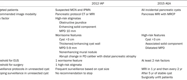
Table 3. Difference between the 2012 IAP and the 2015 AGA Guidelines for the Management of Pancreatic Cysts9,10
2012 IAP 2015 AGA
Targeted patients Suspected MCN and IPMN All incidental pancreatic cysts
Recommended image modality Pancreatic protocol CT or MRI Pancreas MRI with MRCP
Risk factor High-risk stigmatas
Obstructive jaundice Enhancing solid component MPD 10 mm
Worrisome features High-risk features
Cyst >3 cm Cyst >3 cm
Thickened/enhancing cyst wall Associated solid component
MPD 5-9 mm Dilatated MPD
Nonenhancing mural nodule
Abrupt change in PD caliber with distal pancreatic atrophy
Threshold for EUS 1 worrisome feature At least 2 risk factors
Threshold for surgery 1 high-risk stigmata
Surveillance protocols in unresected cyst Frequent surveillance based on cyst size MRI in 1 yr and then every 2 yr Stopping surveillance in unresected cyst No recommendation to stop After 5 yr of stable cyst
Surgically unfit patients
IAP, International Association of Pancreatology; AGA, American Gastroenterological Association; EUS, endoscopic ultrasonography; MCN, mucinous cystic neoplasm; IPMN, intraductal papillary mucinous neoplasm; CT, computed tomography; MRI, magnetic resonance imaging; MPD, main pancreatic duct; PD, pancreatic duct; MRCP, magnetic resonance cholangiopancreatography.
관성과 정확도를 높이기 위해서 EUS-FNA를 시행하여 낭종 액과 분석함으로써 진단에 도움이 된다. 낭액 천자로 낭액의 점도 검사, 생화학적 분석, 종양표지자 검사, 세포 검사 등으 로 감별을 시도할 수 있다(Table 2). 낭액의 점도를 판단할 수 있는 string sign (천자침 끝에서 1 cm, 1초 이상 점도 유지) 양성인 경우 MCN이나 IPMN으로 진단하는데, 95% 특 이도를 보인다.44췌장 낭성 병변에서의 낭액의 종양표지자 중 CEA가 가장 민감도가 높은 표지자로 알려져 있고, 점액성 낭종과 비점액성 낭종을 감별하는 데 도움을 준다. CEA는 192 ng/mL 이상일 때 점액성 종양 진단의 민감도는 73%, 특이도 84%로 보고되었다.45 CEA가 5 ng/mL 이하일 때 SCN, 가성낭종 진단의 민감도는 100%, 특이도 86%로 보고하였
다.46,47 낭액의 아밀라제 수치가 많이 높은 경우는 가성낭종을
강하게 의심할 수 있으나, 250 U/L 이하인 경우 가성낭종은 배제할 수 있다.48 췌장 낭종의 천자액 세포 검사의 가장 큰 단점은 췌장 낭종액에는 세포수가 적어서 진단이 어렵고 민감 도가 낮다는 점이지만, 악성 변화가 있을 때에 민감도가 올라 가고 특이도는 매우 높다. 우연하게 발견된 췌장 낭종 병변을 임상적으로 접근할 때 (1) 낭종의 종류, 점액성 혹은 비점액성 낭종, (2) 현재 악성화 소견의 유무, (3) 악성화 진행 가능성의 유무를 고려하는 것이 중요하다. 현재까지 췌장의 낭성 종양 의 전향적 연구가 부족한 상태로, 췌장 낭성 종양의 자연경과 와 예후에 대한 정보는 제한적이다. 또한 수술적 절제 후에 종양성 병변의 감별과 악성화 유무는 정확한 진단이 가능하기 때문에, 수술을 하지 않은 상태에서 영상학적 정보를 바탕으
로 악성화에 대한 위험도 분류 및 적절한 추적 관찰은 실제 임상에서 매우 어려운 과제이다. 2006년에 IPMN과 MCN에 대한 국제적 합의 진료지침(international consensus guide- lines, IAP)이 발표된 이후, 최근 여러 진료지침들이 발표되었 다. 2012년에 국제췌장학회 개정판(2012 IAP)이 발표되어 현재 까지 많이 이용되고 있다.8,9또한, 최근 2015년에는 미국 소화기 학회(American Gastroenterology Association, 2015 AGA) 에서 무증상 췌장 낭성 종양에 국한하여 새로운 진료지침을 발표하였다.10 2012 IAP에서는 IPMN과 MCN에 대한 진료지 침으로 복부 CT 또는 MRI의 영상 소견을 바탕으로 고위험인 자(high-risk stigmatas)와 WFs로 분류하였다. 2015 AGA 가 이드라인은 우연하게 발견된 무증상 췌장 낭종에 대한 진료지 침으로 세 가지 고위험인자(high-risk features, HRF)를 제시 하였다(Table 3).
2012 IAP에서 폐쇄성 황달, 주췌관 직경 10 mm 이상(주췌 관형이나 혼합형)과 조영 증강되는 고형 병변을 고위험인자 (high-risk stigmatas)로 정의하였고, 수술적 절제를 권고하 였다. WFs (췌장염, 낭종 직경 ≥3 cm, 조영 증강되는 낭종 벽이 두꺼워진 경우, 조영 증강이 되지 않는 벽결절, 주췌관 직경 5-9 mm, 췌장 실질의 위축을 동반한 주췌관 직경의 갑 작스런 변화) 중 하나 이상을 가진 환자에서 EUS를 실시하여 EUS에서 벽결절이 관찰되거나 주췌관 침범 소견, 세포 검사 에서 양성인 경우 수술적 절제를 권고하였다. 낭종의 크기가 3 cm 이상인 젊은 환자의 경우, 위험인자를 보이지 않는 경우 에도 수술을 적극적으로 고려하도록 권고하였다. 2015 AGA
Table 5. Recommended Indications for Resection of Pancreatic Cysts in Accordance to the 2012 IAP and the 2015 AGA Guidelines9,10
Diagnosis 2012 IAP 2015 AGA
MCN Resection Resection
MD-IPMN Resection Resectiona
Mixed-IPMN Resection Resectiona
BD-IPMN Pancreatitis (for relief of symptoms) Obstructive jaundice
Solid component MPD >10 mm
Cytologic features of adenocarcinoma Definite mural nodule on EUS
MPD features suspicious for involvementc
>3 cm cyst in young surgically fit patient
Solid component and MPD >5 mm (both on EUS and MRI) and/or concerning features on EUS and EUS-FNAb
IAP, International Association of Pancreatology; AGA, American Gastroenterological Association; MCN, mucinous cystic neoplasm; MD-IPMN, main duct–intraductal papillary mucinous neoplasm; Mixed-IPMN, mixed type-intraductal papillary mucinous neoplasm; BD-IPMN, branch duct–
intraductal papillary mucinous neoplasm; MPD, main pancreatic duct; EUS, endoscopic ultrasonography; MRI, magnetic resonance imaging;
EUS-FNA, endoscopic ultrasound guided fine needle aspiration.
aPresence of a nodule or malignant cytologic features; bDefinite mural nodule, positive cytology for malignancy; cPresence of thickened walls, intraductal mucin, or mural nodules is suggestive of MPD involvement; in their absence, MPD involvement is inconclusive.
Table 4. Recommended Indication for Endoscopic Ultrasound Guided Fine Needle Aspiration of Pancreatic Cysts
Cyst size ≥3 cm
Diameter of main pancreatic duct >5 mm Solid component
Thickened or enhanced cyst wall Change of cyst size during surveillance
가이드라인에서는 우연히 발견된 췌장 낭종의 HRF (낭종 직 경 ≥3 cm, 고형 성분, 주췌관 확장)를 제시하였다. 2개 이상 의 HRF가 발견되었거나 추적 관찰 중 의미 있는 변화가 있는 경우 EUS-FNA를 실시하도록 권고하고 있다. 각각의 진료지 침에서 EUS-FNA 권고 기준의 차이를 보이지만 단일 위험 요인을 가지는 경우에도 악성화 가능성이 있으므로 적극적으 로 EUS-FNA를 고려하는 것이 필요하다(Table 4). EUS에서 고형 성분과 주췌관 확장 소견이 둘 다 있는 경우 혹은 악성화 소견을 시사하는 세포가 확인되는 경우에 수술을 권고하고 있 다(Table 5).
내시경 초음파 유도하 췌장 낭종 절제술(endoscopic ultra- sound guided pancreatic cyst ablation, EUS-PCA)은 기저 질환이나 다른 이유로 수술 대상이 되지 못하는 경우 차선책 의 치료 방법으로 소개되었다.49 일반적으로 EUS 유도하에 22 G의 세침으로 병변을 천자 후 낭액을 흡인하여 성상을 확 인하고, 흡인한 낭액의 양만큼의 80-100% 에탄올의 주입과 흡인을 반복하거나 주입하여 3분 경과한 뒤 제거한다. 세침을 제거하기 전 paclitaxel 또는 gemcitabine과 같은 항암제를 주입할 수 있다.50,51 EUS-PCA 시술 후 CT를 통하여 추적 관 찰하여 반응 여부를 판단하게 되고, 현재까지 보고에 따르면 33-79%까지 완전 소실(complete resolution)을 보였다. 시술
과 관련된 합병증은 약 8-9%에서 발생하였고, 발열, 췌장염, 낭 종 내 출혈, 낭종 주변으로 유출, 비정맥 폐색, 간문맥 혈전증 등이 보고되어 비교적 안전한 시술로 판단된다.49,52,53 EUS-PCA 의 적응증은 아직 정립되어 있지 않으나 일반적으로 다음과 같은 경우 고려할 수 있다. 첫째로 2-4 cm 크기의 unilocular 또는 oligolocular 형태로 췌관 교통이 없고 악성화 증거를 보 이지 않는 낭종, 둘째로 추적 기간 동안 크기가 증가한 낭종, 셋째로 수술을 거부하거나 수술 고위험 환자들의 낭종이 대상
이다.54,55 낭성 종양이 서서히 자라고 악성으로 진행하는 데
5-10년 이상의 시간이 걸리고, EUS-PCA를 시행한 후 치료 효과에 대한 병리학적 확인이 어렵기 때문에 시술에 대한 적 응이 되는 대상을 선정하는 것과 치료 효과에 대한 논란이 있다. 최근 연구에서 완전 관해를 보인 환자 중 6년 추적 관찰 에서 98.3%에서 관해 유지가 보고되어53 향후 췌장 낭종의 치료 방법으로 임상적 유용성이 기대된다.
영상 검사에서 악성을 시사하는 위험인자가 없는 저위험도 췌장 낭종의 경우 추적 관찰이 가능하다. 낭종의 크기가 3 cm 이하, 주췌관 확장이 없는 경우, 고형 성분이 관찰되지 않는 경우는 악성 위험도가 낮으므로 수술 치료하지 않고 추적 관 찰이 필요하다. 추적 관찰을 위한 영상 검사에는 EUS, CT, MRI가 이용되고 있다. MRI의 경우 CT보다 낭종의 췌관과의 교통, 결절의 해상도가 우수하고, 방사선 피폭을 피할 수 있는 장점이 있다. Gadolium을 사용하지 않은 non-contrast MRI 에서도 악성화를 감별하는 데 비슷한 효과를 보인다는 보고가 있다.56 EUS도 방사선 피폭이 없고, 좋은 해상도를 가졌다는 장점이 있지만 시술자 간 소견의 차이를 보일 수 있다는 단점 이 있다.
2012 IAP 가이드라인에서는 크기에 따라 관찰 간격을 제시
하고 있다. 2-3 cm인 경우 MRI와 EUS를 번갈아 가면서 3-6 개월마다 추적 검사할 것을 권고하고 있다. 또한 낭종의 크기 가 1-2 cm이면 CT나 MRI로 해마다 추적 검사를 하고 변화 가 없으면 검사 간격을 늘리고, 1 cm 미만이면 2-3년마다 추 적 검사할 것을 권고하고 있다.9 AGA 가이드라인은 단순하 게 1년 후 MRI를 시행하고, 이후 2년마다 MRI 시행을 권고하 고 있다. 5년 동안 추적 관찰을 하였지만 의미 있는 변화가 관찰되지 않는 경우, 추적 관찰을 종료할 것을 권고하고 있다.
또한 수술적 치료의 위험성이 더 높아서 수술적 치료 대상이 되지 않는 경우에는 추적 관찰을 권고하지 않았다.10 하지만 이를 뒷받침할 수 있는 췌장 낭성 종양의 자연경과에 대한 연구는 부족한 실정이다. 최근 후향적 연구로 310명의 환자를 대상으로 5년 이상 악성화 유무를 확인하였는데, 이 중 1%인 3명의 환자에서만 침윤성 암종이 발견되었고, 고위험인자가 없는 환자에서는 침윤성 암종이 발생하지 않았다는 보고가 있 다.57 의료수가가 비싸고 비용 대비 효율성을 중시하는 미국의 경우 이 1%의 의미를 5년 이후에는 추적 검사가 불필요하다 는 쪽으로 해석하지만, 췌장암의 치명성과 위험성을 강조하는 나라에서는 이 때문에 5년 이후에도 지속적으로 추적 검사가 필요하다고 해석할 수 있다. 추적 검사의 중단 시기를 결정하 는 것에 있어서 또 하나의 고려사항은 기대여명이다. 20년 이 상의 기대여명이 예상되는 젊은 나이의 환자에서는 중단 시기 에 관한 진료지침을 그대로 적용하는 것보다는 지속적으로 추 적 관찰을 하는 것이 이득이 될 수도 있을 것이다. 실제 임상 에서는 각각의 환자의 특성과 상황에 맞는 추적 관찰 방법과 중단 시기를 선택해야 하겠다.
이러한 진료지침의 정확도는 명확하게 알려져 있지 않으나 수술 전 영상을 바탕으로 낭종을 분석하고 수술 후 병리학적 결과를 비교한 평가한 연구들이 있다. 수술을 받은 317명의 낭종 환자의 결과를 2012 IAP 진료지침에 따라서 분류하였 고, 양성 예측도와 음성 예측도는 각각 88%, 92.5%였다. 비록 고이형성증이나 침습암 소견을 가지지 않는 일부 낭종 환자가 불필요한 수술을 시행 받았지만, 2006 IAP 진료지침과 비교 하여 상대적으로 더 많은 환자가 불필요한 수술을 피할 수 있었다.35 두 진료지침은 IPMN의 수술 기준에서 차이를 보이 는데, 2012 IAP와 다르게 AGA 진료지침에서는 악성 세포 검사 소견이나 벽결절 없이 주췌관 확장만 있는 경우 수술을 권고하지 않는 좀 더 보수적인 입장을 보였다. AGA 유효성을 분석한 연구에서 고이형성증 낭종이나 악성 낭종을 가진 환자 가 누락되었다는 연구 결과들이 있었다.58,59 최근 115명의 수 술적 치료를 받은 환자에서 두 가지 진료지침을 비교하였고, AGA 진료지침에서는 전암성 낭종, 악성 낭종을 가진 환자 중 11%가 누락되었다. 이에 반해 IAP 진료지침 기준에서는 누락된 전암성 낭종, 악성 낭종을 가진 환자가 없었다.60
결 론
최근 우연히 발견된 췌장 낭성 병변은 지속적으로 증가하 고 있고, 비종양성 병변에서 악성 종양까지 병리적으로 다양 한 질환을 구성하고 있다. 악성화 위험이 있는 점액성 종양 (MCN, MD-IPMN)과 증상이 있는 낭성 종양은 수술이 원칙 이다. 최근 진료지침에서 악성화 위험도 예측 가능한 영상의 학 소견을 제시하였다. 낭종 내 벽결절, 10 mm 이상의 주췌 관의 확장, 세포 검사에서 비전형적 세포는 수술적 절제가 필 요한 고위험인자이다. 3 cm 이상의 낭종 크기, 5 mm 이상의 주췌관의 확장, 낭종 벽이 두꺼워지거나 조영 증강되는 경우 는 내시경 초음파 시행이 필요한 위험인자이다. 췌장 낭종에 대한 진료지침들이 존재하지만 각각 장단점을 보이고 있다.
또한 수술에 대한 위험성이 상존하므로 개개의 환자에서 치료 와 추적 관찰에 대한 결정은 병변 악성화 위험도, 환자의 연 령, 낭종의 위치, 수술 위험도 등을 함께 고려하여야 한다.
REFERENCES
1. Edirimanne S, Connor SJ. Incidental pancreatic cystic lesions.
World J Surg 2008;32:2028-2037.
2. de Jong K, Nio CY, Hermans JJ, et al. High prevalence of pancre- atic cysts detected by screening magnetic resonance imaging examinations. Clin Gastroenterol Hepatol 2010;8:806-811.
3. de Jong K, Bruno MJ, Fockens P. Epidemiology, diagnosis, and management of cystic lesions of the pancreas. Gastroenterol Res Pract 2012;2012:147465.
4. Chang YR, Park JK, Jang JY, Kwon W, Yoon JH, Kim SW. Incidental pancreatic cystic neoplasms in an asymptomatic healthy pop- ulation of 21,745 individuals: large-scale, single-center cohort study. Medicine (Baltimore) 2016;95:e5535.
5. Balthazar EJ, Chako AC. Computed tomography of pancreatic masses. Am J Gastroenterol 1990;85:343-349.
6. Fernández-del Castillo C, Targarona J, Thayer SP, Rattner DW, Brugge WR, Warshaw AL. Incidental pancreatic cysts: clin- icopathologic characteristics and comparison with symptomatic patients. Arch Surg 2003;138:427-423; discussion 433-424.
7. Goh BK, Tan YM, Cheow PC, et al. Cystic lesions of the pancreas:
an appraisal of an aggressive resectional policy adopted at a sin- gle institution during 15 years. Am J Surg 2006;192:148-154.
8. Tanaka M, Chari S, Adsay V, et al. International consensus guide- lines for management of intraductal papillary mucinous neo- plasms and mucinous cystic neoplasms of the pancreas.
Pancreatology 2006;6:17-32.
9. Tanaka M, Fernández-del Castillo C, Adsay V, et al. International consensus guidelines 2012 for the management of IPMN and MCN of the pancreas. Pancreatology 2012;12:183-197.
10. Vege SS, Ziring B, Jain R, Moayyedi P; Clinical Guidelines Committee; American Gastroenterology Association. American gastroenterological association institute guideline on the diag- nosis and management of asymptomatic neoplastic pancreatic
cysts. Gastroenterology 2015;148:819-822; quize12-13.
11. Yoon WJ, Yoon YB, LEE KH, et al. The cystic neoplasms of the pan- creas in Korea. Korean J Med 2006;70:261-267.
12. Gaujoux S, Brennan MF, Gonen M, et al. Cystic lesions of the pan- creas: changes in the presentation and management of 1,424 patients at a single institution over a 15-year time period. J Am Coll Surg 2011;212:590-600; discussion 600-603.
13. Madura JA, Wiebke EA, Howard TJ, et al. Mucin-hypersecreting in- traductal neoplasms of the pancreas: a precursor to cystic pan- creatic malignancies. Surgery 1997;122:786-792; discussion 792-793.
14. Ferrone CR, Correa-Gallego C, Warshaw AL, et al. Current trends in pancreatic cystic neoplasms. Arch Surg 2009;144:448-454.
15. Shimizu Y, Yamaue H, Maguchi H, et al. Predictors of malignancy in intraductal papillary mucinous neoplasm of the pancreas:
analysis of 310 pancreatic resection patients at multiple high-volume centers. Pancreas 2013;42:883-888.
16. Hackert T, Fritz S, Klauss M, et al. Main-duct intraductal papillary mucinous neoplasm: high cancer risk in duct diameter of 5 to 9 mm. Ann Surg 2015;262:875-880; discussion 880-881.
17. Schmidt CM, White PB, Waters JA, et al. Intraductal papillary mu- cinous neoplasms: predictors of malignant and invasive pathology.
Ann Surg 2007;246:644-651; discussion 651-654.
18. Correa-Gallego C, Do R, Lafemina J, et al. Predicting dysplasia and invasive carcinoma in intraductal papillary mucinous neo- plasms of the pancreas: development of a preoperative nomogram.
Ann Surg Oncol 2013;20:4348-4355.
19. Pelaez-Luna M, Chari ST, Smyrk TC, et al. Do consensus in- dications for resection in branch duct intraductal papillary muci- nous neoplasm predict malignancy? A study of 147 patients. Am J Gastroenterol 2007;102:1759-1764.
20. Fritz S, Schirren M, Klauss M, et al. Clinicopathologic character- istics of patients with resected multifocal intraductal papillary mucinous neoplasm of the pancreas. Surgery 2012;152(3 Suppl 1):S74-S80.
21. Reddy RP, Smyrk TC, Zapiach M, et al. Pancreatic mucinous cystic neoplasm defined by ovarian stroma: demographics, clinical fea- tures, and prevalence of cancer. Clin Gastroenterol Hepatol 2004;2:1026-1031.
22. Murakami Y, Uemura K, Ohge H, Hayashidani Y, Sudo T, Sueda T. Intraductal papillary-mucinous neoplasms and mucinous cyst- ic neoplasms of the pancreas differentiated by ovarian-type stroma. Surgery 2006;140:448-453.
23. Yamao K, Yanagisawa A, Takahashi K, et al. Clinicopathological features and prognosis of mucinous cystic neoplasm with ovar- ian-type stroma: a multi-institutional study of the Japan pancreas society. Pancreas 2011;40:67-71.
24. Park JW, Jang JY, Kang MJ, Kwon W, Chang YR, Kim SW. Mucinous cystic neoplasm of the pancreas: is surgical resection recom- mended for all surgically fit patients? Pancreatology 2014;14:
131-136.
25. Chang H, Gong Y, Xu J, Su Z, Qin C, Zhang Z. Clinical strategy for the management of solid pseudopapillary tumor of the pan- creas: aggressive or less? Int J Med Sci 2010;7:309-313.
26. Goh BK, Tan YM, Cheow PC, et al. Solid pseudopapillary neo- plasms of the pancreas: an updated experience. J Surg Oncol
2007;95:640-644.
27. Yang F, Jin C, Long J, et al. Solid pseudopapillary tumor of the pan- creas: a case series of 26 consecutive patients. Am J Surg 2009;198:210-215.
28. Sunkara S, Williams TR, Myers DT, Kryvenko ON. Solid pseudopa- pillary tumours of the pancreas: spectrum of imaging findings with histopathological correlation. Br J Radiol 2012;85:e1140- e1144.
29. Goh BK, Tan YM, Yap WM, et al. Pancreatic serous oligocystic ad- enomas: clinicopathologic features and a comparison with se- rous microcystic adenomas and mucinous cystic neoplasms.
World J Surg 2006;30:1553-1559.
30. Lee SE, Kwon Y, Jang JY, et al. The morphological classification of a serous cystic tumor (SCT) of the pancreas and evaluation of the preoperative diagnostic accuracy of computed tomography.
Ann Surg Oncol 2008;15:2089-2095.
31. Khurana B, Mortelé KJ, Glickman J, Silverman SG, Ros PR.
Macrocystic serous adenoma of the pancreas: radiologic-pathologic correlation. AJR Am J Roentgenol 2003;181:119-123.
32. Salvia R, Fernández-del Castillo C, Bassi C, et al. Main-duct intra- ductal papillary mucinous neoplasms of the pancreas: clinical predictors of malignancy and long-term survival following resection. Ann Surg 2004;239:678-685; discussion 685-677.
33. Moriya T, Hashimoto Y, Traverso LW. The duration of symptoms predicts the presence of malignancy in 210 resected cases of pancreatic intraductal papillary mucinous neoplasms. J Gastrointest Surg 2011;15:762-770; discussion 770-771.
34. Chung JW, Chung MJ, Park JY, et al. Clinicopathologic features and outcomes of pancreatic cysts during a 12-year period.
Pancreas 2013;42:230-238.
35. Goh BK, Thng CH, Tan DM, et al. Evaluation of the Sendai and 2012 international consensus guidelines based on cross-sec- tional imaging findings performed for the initial triage of muci- nous cystic lesions of the pancreas: a single institution experi- ence with 114 surgically treated patients. Am J Surg 2014;208:
202-209.
36. Sahora K, Mino-Kenudson M, Brugge W, et al. Branch duct intra- ductal papillary mucinous neoplasms: does cyst size change the tip of the scale? A critical analysis of the revised international consensus guidelines in a large single-institutional series. Ann Surg 2013;258:466-475.
37. Uehara H, Ishikawa O, Katayama K, et al. Size of mural nodule as an indicator of surgery for branch duct intraductal papillary mu- cinous neoplasm of the pancreas during follow-up. J Gastroenterol 2011;46:657-663.
38. Das A, Wells CD, Nguyen CC. Incidental cystic neoplasms of pan- creas: what is the optimal interval of imaging surveillance? Am J Gastroenterol 2008;103:1657-1662.
39. Walsh RM, Vogt DP, Henderson JM, et al. Management of sus- pected pancreatic cystic neoplasms based on cyst size. Surgery 2008;144:677-684; discussion 684-685.
40. Serikawa M, Sasaki T, Fujimoto Y, Kuwahara K, Chayama K.
Management of intraductal papillary-mucinous neoplasm of the pancreas: treatment strategy based on morphologic classification.
J Clin Gastroenterol 2006;40:856-862.
41. Wu BU, Sampath K, Berberian CE, et al. Prediction of malignancy
in cystic neoplasms of the pancreas: a population-based cohort study. Am J Gastroenterol 2014;109:121-129; quiz 130.
42. Leeds JS, Nayar MN, Dawwas M, et al. Comparison of endoscopic ultrasound and computed tomography in the assessment of pan- creatic cyst size using pathology as the gold standard. Pancreatology 2013;13:263-266.
43. Zhong N, Zhang L, Takahashi N, et al. Histologic and imaging fea- tures of mural nodules in mucinous pancreatic cysts. Clin Gastroenterol Hepatol 2012;10:192-198, 198.e1-2.
44. Bick BL, Enders FT, Levy MJ, et al. The string sign for diagnosis of mucinous pancreatic cysts. Endoscopy 2015;47:626-631.
45. Brugge WR, Lewandrowski K, Lee-Lewandrowski E, et al. Diagnosis of pancreatic cystic neoplasms: a report of the cooperative pan- creatic cyst study. Gastroenterology 2004;126:1330-1336.
46. Hammel P, Levy P, Voitot H, et al. Preoperative cyst fluid analysis is useful for the differential diagnosis of cystic lesions of the pancreas. Gastroenterology 1995;108:1230-1235.
47. Attasaranya S, Pais S, LeBlanc J, McHenry L, Sherman S, DeWitt JM. Endoscopic ultrasound-guided fine needle aspiration and cyst fluid analysis for pancreatic cysts. JOP 2007;8:553-563.
48. van der Waaij LA, van Dullemen HM, Porte RJ. Cyst fluid analysis in the differential diagnosis of pancreatic cystic lesions: a pooled analysis. Gastrointest Endosc 2005;62:383-389.
49. Gan SI, Thompson CC, Lauwers GY, Bounds BC, Brugge WR.
Ethanol lavage of pancreatic cystic lesions: initial pilot study.
Gastrointest Endosc 2005;61:746-752.
50. Oh HC, Seo DW, Kim SH, Min B, Kim J. Systemic effect of endo- scopic ultrasonography-guided pancreatic cyst ablation with ethanol and paclitaxel. Dig Dis Sci 2014;59:1573-1577.
51. Moyer MT, Dye CE, Sharzehi S, et al. Is alcohol required for effec- tive pancreatic cyst ablation? The prospective randomized CHARM trial pilot study. Endosc Int Open 2016;4:E603-E607.
52. DeWitt J, McGreevy K, Schmidt CM, Brugge WR. EUS-guided ethanol versus saline solution lavage for pancreatic cysts: a randomized, double-blind study. Gastrointest Endosc 2009;70:
710-723.
53. Choi JH, Seo DW, Song TJ, et al. Long-term outcomes after endo- scopic ultrasound-guided ablation of pancreatic cysts. Endoscopy 2017 May 16. [Epub ahead of print]
54. DeWitt J. Endoscopic ultrasound-guided pancreatic cyst ablation.
Gastrointest Endosc Clin N Am 2012;22:291-302, ix-x.
55. Oh HC, Brugge WR. EUS-guided pancreatic cyst ablation: a crit- ical review (with video). Gastrointest Endosc 2013;77:526-533.
56. Nougaret S, Reinhold C, Chong J, et al. Incidental pancreatic cysts: natural history and diagnostic accuracy of a limited serial pancreatic cyst MRI protocol. Eur Radiol 2014;24:1020-1029.
57. Kwong WT, Hunt GC, Fehmi SM, et al. Low rates of malignancy and mortality in asymptomatic patients with suspected neo- plastic pancreatic cysts beyond 5 years of surveillance. Clin Gastroenterol Hepatol 2016;14:865-871.
58. Singhi AD, Zeh HJ, Brand RE, et al. American Gastroenterological Association guidelines are inaccurate in detecting pancreatic cysts with advanced neoplasia: a clinicopathologic study of 225 patients with supporting molecular data. Gastrointest Endosc 2016;83:1107-1117.e2.
59. Ridtitid W, DeWitt JM, Schmidt CM, et al. Management of branch-duct intraductal papillary mucinous neoplasms: a large single-center study to assess predictors of malignancy and long-term outcomes. Gastrointest Endosc 2016;84:436-445.
60. Lekkerkerker SJ, Besselink MG, Busch OR, et al. Comparing 3 guidelines on the management of surgically removed pancreatic cysts with regard to pathological outcome. Gastrointest Endosc 2017;85:1025-1031.