I. 서론
치아우식증, 치주질환 혹은 외상 등에 의해서 상실 된 한 개의 치아는 기능 및 심미적인 이유로 대개의 경우 보철적 수복을 필요로 한다.1 가장 일반적인 단 일치아 수복치료방법은 인접 자연치를 삭재한 후, 이 를 지대치로 사용하는 고정성 계속가공의치(fixed partial denture)이다. 하지만 지대치 삭재시, 치수손 상(pulp injury)에 의한 치근단 병소, 부적절한 지대 치 유지형태(retention form) 및 접착시멘트의 용해 (cement washout)에 의한 보철물 탈락, 지대치 파절, 금관 변연부에 발생하는 이차 치아우식증 등은 고정 성 계속가공의치의 장애 요인이다.2 레진접착에 의 하여 유지를 얻는 레진접착성 보철물(resin bonded denture)은 건전한 인접치의 삭재량은 줄일 수 있으 나, 잦은 보철물 탈락3과 치아의 두께가 얇은 경우 투 과현상에 의해 금속이 비치는 심미적 문제가 장애요 인이 된다. 교정치료에 의해 단일치아 상실로 인한 치간 공간을 없애는 방법도 시도되지만, 새로운 위치 로 이동한 치아의 심미적 치아형태로 만들기 위해, 레진(resin build-up)이나 금관(crown)등 부가적인 치료가 필요하며, 장기간의 치료시간이 단점으로 지
적된다4. 가철성 국소의치(removable partial den- ture) 역시 환자의 이물감호소나 발음장애 등이 문제 점으로 지적된다.
이러한 전통적인 단일 상실치아의 보철치료 방법 의 한계를 극복할 수 있는 대안으로 제시된 치료방 법은 단일 임플란트 지지에 의한 수복(single-tooth implant replacement)이며, 건전한 인접자연치의 삭 재가 필요없고, 가공의치(pontic)와 달리 임플란트에 의해 수복된 금관이 자연치아처럼 치조골에서 자연 스럽게 연조직을 뚫고 나오므로 심미적으로 유리 할 수 있다.5원래 무치악 환자의 치료에 사용되었던 임 플란트에 의한 보철치료는 장기적 추적연구들6,7에 서 높은 성공률을 보였으며, 이후 부분 무치악 환자 의 치료8로 확대되어 역시 좋은 결과를 보였다. 그리 고 마침내 단일 상실치아의 수복까지 그 사용이 확 대되었다.9 이러한 도입 과정의 결과, 무치악이나 부 분 무치악 환자의 치료에 비해 단일치아상실환자의 치료에 사용된 임플란트에 관한 연구논문은 숫자가 적고, 추적기간도 상대적으로 짧다.
일반적으로 임플란트 시술에서 3개 이하의 임플란 트 사용은 기계 역학적으로 불리한 것으로 알려져 있으며, 단일 임플란트는 굴곡모멘트(bending
단일 임플란트 지지에 의한 보철물의 생존율에 관한 문헌 연구
장문택
전북대학교 치과대학 치주과학교실, 구강생체과학연구소
대한치주과학회지 : Vol. 32, No. 1, 2002
*본 연구는 전북대학교병원 특수목적연구비의 지원에 의하여 이루어진 것임.
교신 저자: 장문택, 전라북도 전주시 덕진구 금암동 634-18 전북대학교 치과대학 치주과학교실, 우편번호: 561-712
moment)에 아주 취약하므로, 특히 교합력이 강하게 작용하는 구치부에서 이의 사용은 더욱 금기로 여겨 졌다.10실재로 초기의 단일 임플란트 연구에서 교합 이 가해진 단일 임플란트지지 금관의 나사 풀림 현 상(screw loosening)은 자주 관찰되었으며, 특히 구 치부에서 그 빈도는 급속히 증가되었다.11,12,13,14더불 어 심미적 이유로 연조직 깊이 매식된 임플란트 주 위의 연조직 문제, 치관의 심미적 문제 등이 초기의 연구들에서 자주 관찰되었다.12하지만 임플란트 부 품15과 술식의 발달16등으로 상당부분 위에서 언급된 문제들이 해결되어가고 있음이 최근의 연구들17,18에 서 보고되고 있다.
임플란트의 장기적 성공에 필수적인 성공적인 골 유착(osseointegration)은 임플란트의 재료(implant material), 임플란트의 모양(implant design), 임플란 트표면의 성질(surface quality), 임플란트가 식립될 부위의 골상태(status of the bone), 수술방법(surgical technique), 임플란트에 교합을 가하는 방법(implant loading conditions) 등의 6가지 요소에 크게 영향을 받는 것으로 알려져 있다.19이러한 요소들이 임플란 트의 생존에 어떻게 영향을 주는가에 대하여 단일 임플란트에 관한 연구는, 부분 무치악이나 완전 무치 악 환자의 치료에 관한 연구들만큼 많지는 않지만, 최근 그 숫자가 점차 증가하고 있다.
이 종설 논문의 목적은 문헌에 보고된 단일 치아 수복에 사용된 임플란트의 성공률을 골유착에 영향 을 주는 여러 요인들에 따라 분석하는 것이다.
II. 연구재료 및 방법
의학 정보검색프로그램 PubMed (http://www.
ncbi.nih.gov/entrez/query.fcgi)에서“single”,
“dental”, “implant”의 검색어(keyword)를 사용하여 단일 임플란트에 관한 논문을 검색하였으며, 이와 더 불어 검색된 논문들의 참고문헌목록에서 추가로 단 일임플란트에 관한 논문들을 수작업(hand search- ing)으로 찾았다. 이렇게 검색된 모든 논문들을 대상 으로 다음과 같은 선정기준(inclusion criteria)을 사 용하여 연구대상 논문을 최종적으로 선택하였다;
1. 단일 임플란트 생존율/성공률이 구체적으로 조 사, 기재된 논문
2. 영어로 발간된 논문
3. 평가위원(referees)에 의해 검토되는 학술지 (peer reviewed journal)에 실린 논문
4. 환자를 대상으로 한 임상연구(human clinical trials)
5. 추적연구(follow-up study)
선택된 논문들의 결과는 표를 사용하여 다음 항목 별로 분류하여 내용을 정리하였다.
1. 발행년도
2. 연구방법(prospective/retrospective) 3. 연구대상 임플란트
①. 종류(재료, 모양, 표면)
②. 개수
4. 연구대상 환자 수 5. 임플란트 식립부위
6. 실패한 임플란트 수와 그 이유 7. 추가 술식(예, bone graft, sinus lift etc.)
8. 치료 후 발생한 문제점(complication); 나사풀림 (screw loosening)
결과 분석은 통계프로그램 SPSS 10.0을 사용하였다.
III. 결과 및 고안
총 65 개의 논문들11-14, 20-80이 선정기준을 만족하였 으며, 이 가운데 같은 환자를 대상으로 한 7개 논문
12,28,30,31,42,43,67을 제외한 58개의 논문을 최종적 연구
대상으로 하였다. 1990년 Jemt 등11의 논문이 발표된 이후 해마다 논문의 숫자는 계속 증가하여 2001년에 는 12편의 논문이 단일 임플란트의 성공률을 보고하 였다(Figure 1). 논문의 86%(n=50)는 단일 임프란트 만을 대상으로 한 연구였으며, 나머지는 단일 임플란 트를 포함한 여러 가지 유형의 임플란트 보철치료를 연구한 논문이었다. 22개(37%)는 전향적(prospec- tive)연구였으며, 나머지 37개(63%)는 후향적(retro- spective)연구였다.69* (69:두 가지 유형의 연구가 행 해짐). 추적기간은 6개월에서42,80최고 13년68까지이 며, 한 개의 논문에서 조사된 단일 임플란트는 12개78
에서 314개23(평균 79개), 연구대상 환자의 숫자는 10 명57에서 314명23까지 다양하게 분포하였다. 총 5025 개의 단일 임플란트가 식립되었으며, 197개의 실패 가 보고되었다.
1. 임플란트 종류(Implant type)
연 구 에 사 용 된 임 플 란 트 는 Branemark implant11,13,14,24,27,32-36,38-40,48-50,53,58,59,65,68-71, Astra tech implant41,44,47,66,74, ITI implant41,54,56,61,64, Frialit implant20,25,45,46,76, 3i implant26,62,72, Ankylos implant60, Sulzer Calcitek implant51,57, Spectra system(Paragon)63, Screw-vent78, Genetics implant system75, Steri-Oss implant73,80, AlphaBio implant73, Mac System77, Endopore79,미상(not identified)23,52등이었다.
2. 임플란트 재료(Implant material)
대부분의 단일 임플란트(Branemark, Astra, ITI, 3i, Frialit-2, Ankylos, Mac system)는 순수 타이타니움 (commercially pure titanium)으로 만들어졌지만, Screw-vent, Genetics implant system은 타이타니움
합금(titanium alloy)을, Frialit implant의 초기모델인 Frialit-1(Tubingen) implant는 Al2O3ceramic으로 만 들어졌다. 이상 모든 재료는 생체조직에 적합(bio- inert)하며, 골과 유착(osseointegration)하는 것으로 알려져 있다. 하지만 세라믹은 파절 위험 때문에 주 로 전치부 단일 치아의 수복에 사용되었으며, 높은 실패율(13%)25과 금속재질의 임플란트에 비하여 월 등히 높은 실패율23때문에 지금은 많이 사용되지 않 는다. 타이타니움 합금은 순수 타이타니움에 비하여 기계적 강도는 우수하나10,81골과 유착하는 면적이나 임플란트 제거 토크(removal torque)가 상대적으로 떨어지는 것이 단점으로 지적되었다82.
3. 임플란트 모양(Implant design)
단일 임플란트 연구에 사용된 임플란트의 대부분 은 나사가 있는 스크루(threaded screw)모양이지만, 일부 임플란트(IMZ, Sulzer Calcitek, Steri-Oss, Endopore)는 원통(cylinder)모양, 그리고 ITI 초기모 델처럼 원통형의 가운데가 비어있는 (hollow cylin- der)모양도 사용되고 있다. 전치부 치아를 발치하고 즉시 식립 할 때 많이 사용되는 Frialit 임플란트는 초 Figure 1. 단일 임플란트 생존율을 보고한 논문의 년간 발행 숫자
14
12
10
8
6
4
2
0
Study No.(n=58) 1990 1993 1994 1995 1996 1997 1998 1999 2000 2001
기모델(Tubingen, Frialit-1)에서는 세라믹으로 층이 진 원통(stepped cylinder)모양으로 만들어졌다가, 최 근(Frialit-2)에는 타이타니움을 소재로 나사가 있는 층이 진 스크루(threaded, stepped screw)형태로 제작 된다. 그리고 구치부에 사용되어 좋은 결과를 얻은60 Ankylos implant는 나사가 있으며(threaded), 끝 부분 이 점점 가늘어지는(progressive)형태로 제작되었다.
임플란트와 지대주(abutment) 결합부분의 형태도 평평한 면끼리 만나는 butt joint(Branemark, 3i 등)와 경사진 임플란트 내면에 지대주가 결합하는 morse taper(Astra, ITI, Ankylos, Mac)가 있다. Morse taper 형태의 기계적 우수성이 in vitro study83,84에서 제기 되었지만, 동물실험85에서 다른 임플란트-지대주 접 합방법을 가진 Branemark, Astra, ITI 임프란트들 간 에 임플란트 주위 연조직 및 골조직의 제원(dimen- sion)의 차이가 없음이 관찰되었다. 그리고 최근의 Branemark implant와 Astra implant를 임상적, 방사 선학적, 미생물학적으로 비교한 임상실험86에서 Branemark implant가 Astra tech implant 보다 임플 란트 주위의 탐침 깊이가 더 깊고(0.6mm), 임플란트 와 골접촉 부분의 최상부가 임플란트-지대주 연결부 위에 더 가까웠다(1.6mm). 저자들은 이러한 차이를 임플란트의 임플란트-지대주 연결부위의 모양에 따 른 생물학적 고경(biologic width)확립이라고 주장하 였다. 하지만 이러한 차이가 통계적 유의성은 있지 만 임상적 유의성은 없다고 하였다.
4. 임플란트 직경(Implant diameter)
Rangert등의 파절된 39개의 Branemark implant를
분석한 연구10에서, 35개(90%)의 임플란트 파절이 구 치부에서 일어났으며, 30개(77%)는 1 개나 2 개의 임 플란트에 의해 지지받고 있었다. 174개의 ITI single- tooth implant를 대상으로 한 연구54에서는 보철물 장 착 6개월까지 1개, 그 이후에 3 개의 파절이 관찰되 었다. 파절 임플란트는 모두 가운데가 빈(hollow) 직 경 3.5 mm 임플란트이고, 위치는 하악 구치부에 식 립된 임플란트 이었다. 반대로 4.1 mm 직경의 속이 찬 나사형(solid screw) 임플란트는 평균 40.3개월의 추적기간중 단 한 개의 파절도 없었다. 위와 같은 연 구 결과를 고려할때, 구치부 단일 임플란트는 위치적 으로, 숫적으로 파절 가능성은 더욱 증가할 것이다.
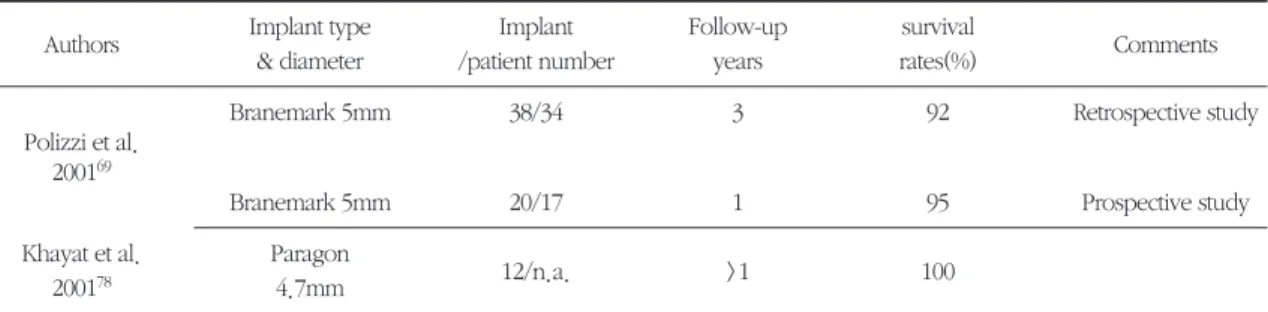
구치부의 과도한 교합력에 저항하고69, 골질이 떨어 지거나87, 짧은 길이의 임플란트를 사용해야 할 때88, 굵은 직경(wide implant)의 임플란트를 사용하거나
69, 두 개의 임플란트를 연결하여 사용하기를 권한다
36. 동물실험에서 굵은 직경을 가진 임플란트는 골 접촉면적이 넓어지므로, 직경이 작은 임플란트보다 제거할 때 드는 힘(removal torque)이 통계적으로 유 의성있게 컸다89. 단일 임플란트의 임상연구에서 굵 은 직경의 임플란트는 좋은 결과를 보였지만(Table 1), 부분 무치악환자에 사용된 임플란트의 임상연구
90에서는 상악 29 %, 하악 19 %의 높은 실패율을 보 였고, 다른 임상연구에서도91굵은 직경(5mm)의 임 플란트에서 일반 임플란트(3.75mm) 보다 오히려 더 많은 실패가 관찰되었다. 저자들은 그 이유를 굵은 직경의 임플란트가 주로 실패한 임플란트를 제거한 후 대체용(rescue) 임플란트 혹은 골질이 좋지 않은 (poor bone quality)경우에 사용되고, 임플란트 형태 의 변화(changed implant design), 초기의 시행착오
Table 1. 굵은 직경의 단일 임플란트를 사용한 논문
Authors Implant type Implant Follow-up survival
Comments
& diameter /patient number years rates(%)
Branemark 5mm 38/34 3 92 Retrospective study
Polizzi et al.
200169
Branemark 5mm 20/17 1 95 Prospective study
Khayat et al. Paragon
12/n.a. >1 100
200178 4.7mm
(learning curve)때문 등으로 해석하였다.
굵은 직경의 임플란트의 경우와 대조적으로 좁은 치간골(reduced interradicular bone), 얇은 치조골 두께(thin alveolar crest), 하악 전치같이 치아의 치 경부 직경이 작은 경우(small cervical diameter)등에 는 작은 직경의 임플란트가 사용되어 좋은 결과를 얻었다(Table 2). 하지만 이들 연구들은 모두 전치부 에 국한되어 사용되었음을 상기하고, 사용 전 항상 기계역학적인 면을 고려하여 파절의 위험성에 대비 하야 한다92.
5. 임플란트 표면(Surface quality)
임플란트 표면은 기계로 매끈하게 깍은 표면
(machined smooth surface; Branemark system, 초기 Astra tech implant)과 여러 가지 방법으로 거칠게 (rough) 처리한 표면으로 분류할 수 있다. 임플란트 의 표면이 거칠면 골과 접촉하는 면적이 증가되므로 초기고정에 유리하다고 믿어졌다93v ITI implant의 TPS(Titanium Plasma Spray)와 SLA(Sand-blasted Large-grit Acid-etched), Ankylos implant의 sand blast- ing, 3i implant에 사용된 sand blasting후 acid etching, Astra tech implant의 TiO2blasting, hydroxyapatitie coating(Calcitek implant, Frialit-2 implant, SteriOss implant, IMZ implant, Spectra system; Paragon implant), Endopore implant처럼 타이타이늄 합금을 구슬 모양으로 표면에 용융(sintered spheroidal parti- cles of titanium alloy)하는 등 다양한 방법으로 표면 Table 2. 가는 직경의 단일 임플란트를 사용한 논문
Authors Implant type Implant/patient Follow-up
survival rates(%)
& diameter number periods(years)
Polizzi et al. Branemark
30/21 3-7 93.3
199959 3.0mm
Vigolo & Givani 3i
52/44 5 94.2
200062 2.9mm
Andersen et al. 3i
32/28 3 93.8
200172 3.25mm
Table 3. 임플란트 종류로 분류한 단일 임프란트에 관한 논문 및 생존률 Included
Fixture type Reference no. implant Failed implant
Survival rate % (SD)
numbers numbers
Branemark 13,14,24,27,29,32-36,38-40, 2021 62 96.3(3.7)
48-50,53,58,59,65,68-71
Astra 41*,44,47,66,74 209 2 99.2(1.7)
ITI 41*,54,56,61,64 471 12 96.3(3.1)
Frialit 20,25,45,46,76 581 33 91.8(6.4)
3i 26,62,72 179 8 95.6(2.9)
Mixed
22,37,51,55,73 260 9 96.0(6.9)
types**
Other types*** 21,23,52,57,60,63,75,77-80 1304 71 96.6(3.7)
Total 5025 197 96.2(4.2)
* 41은 Astra 와 ITI의 생존률 결과가 분리되어 보고됨
** 한 연구에 두 가지 이상의 임플란트가 사용되고, 생존율 결과는 분리 보고되지 않음
*** 사용된 임플란트 종류가 2개 이하인 연구
을 처리하여 거칠게 만들었다. Mazor등57은 상악 구 치부의 결손부에 sinus augmentation후 10개의 Hydroxyapatite coated surface를 가진 임플란트를 식 립하여 100% 성공하였으며, Schwartz-Arad등52도 구 치부에서 HA coated된 implant를 사용했을때 machined surface를 가진 임플란트보다 더 높은 성공 률을 관찰하였다. 하지만 임플란트 주위염증(peri- implantitis)등으로 인한 골흡수 때문에 연조직이 퇴축 되면, 노출된 임플란트의 거친 표면에 침착된 치태제 거가 어려운 점이 문제점으로 지적되고 있다94.
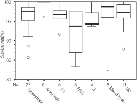
이상의 다양한 임플란트 재료, 임플란트 디자인, 임플란트 표면을 가진 여러 가지 단일 임플란트들을 대상으로 한 연구들의 생존율을 비교해봤을 때, 대부 분 95 % 이상의 높은 생존율을 보였다(Table 3). 낮 은 성공률을 보이는 몇 개의 연구는 1980년대 단일 임플란트가 처음 식립될 때20,23,25, 혹은 발치 후 즉시 식립하고 즉시 보철물을 제작 장착하여 부하를 가하 는 특이한 경우를 실험한 경우 등65,73에서 아주 한정 적으로 보고되었다(Figure 2).
6. 수술 수기(Surgical technique)
골조직은 아주 열에 민감하므로, 임플란트 식립부 위를 드릴링할 때 냉각된 식염수로 충분히 관수(irri- gation)하여 적절한 온도를 유지하도록 하여야 한 다.95굵고 긴 임플란트가 사용되거나,96골질이 아주 단단한 경우에는 더욱 열에 의한 손상의 위험성이 높으므로 드릴링할 때 회전속도를 줄여야한다.97 드 릴이 무딘 경우에도 새 드릴을 사용할 때 보다 열이 더 발생하므로, 일정 횟수 이상 사용된 드릴은 새 드 릴로 교체되어야한다98.
지대주 연결방식에 따른 1 단계형(one stage) 혹은 2 단계형(two stages) 임플란트 술식의 장단점과 장 기적 결과에 대하여 많은 논쟁이 있었다.99 하지만 현 재 두 가지 방법 모두 장기적으로 높은 성공률을 보 임100에 따라 이에 대한 논쟁은 일단락되었으며, 여러 가지 상황에 따라 두 가지 방법 가운데 적절한 술식 을 선택하여 사용하고 있다.
7. 부족한 골양을 해결하기 위한 특별한 수술방 법들(Special surgical techniques in compromised situation)
Figure 2. 임플란트 종류에 따른 단일 임플란트 생존률(Box plots) 100
95
90
85
80
N= 27 5 5 5 4 6 11
Branemark Astra tech ITI Frialit 3i Mixed types etc.
Survival rate(%)
해부학적 한계, 즉 협설로 좁은 치조골, 그리고 상 악동, 하악치조관에 근접하여 임플란트 식립에 필요 한 골 양이 부족한 경우, 이 같은 한계를 극복하기 위 한 다양한 술식이 개발되어 사용되고 있다.101 최근 에는 골 신장술(Distraction osteogenesis)102도 임플 란트 식립에 부족한 골 양을 수직적으로 증가시키기 위해 시도되고 있다.
단일 임프란트의 경우, 임플란트 식립에 부족한 골 양을 증가시키기 위해, mandibular symphysis, max- illary tuberosity, mandibular retromolar 부위를 공여 부로 하는 자가골 이식(autogenous bone graft)37,51, membrane을 이 용 한 GBR(Guided Bone Regeneration),26,35,51,58 상악 전치부의 얇은 잔존골을 ostetome과 dilator를 사용하여 협설 두께를 증가시 키는 방법등68이 보고되었다. 하지만 단일 임플란트 를 식립하기 위해 하악골을 상악 전치부에 이식했을 때, 약 4개월 후 이미 60% 정도의 골흡수를 관찰하였 다.103 상악 구치부의 잔존골이 상악동의 확장 (pneumatization)으로 아주 얇아져서 임플란트의 식 립이 어려운 경우, 상악동을 거상시키고, 여러 가지 종류의 골이식재를 넣는 방법으로 수직고경을 증가 시켜 단일 임플란트를 식립하여 3년의 추적기간동안 좋은 결과를 얻었다.57(Table 4).
하지만 길이가 긴 임플란트를 식립하기 위한 위와 같은 부가적 술식없이 기존의 골 양에 맞는 짧은 임 플란트(<7mm)를 사용하여서도 좋은 결과를 얻은 연 구도 있다104. 그리고 Palmer등43은 15개의 단일 Astra implant를 식립할 때, 관찰된 3개의 fenestration, 8개 의 dehiscence에 아무런 추가 술식 없이도 5 년간의
추적연구66에서 특별한 문제를 발견할 수 없었다.
Lekholm등105도 임플란트의 노출과 생존율 사이에 아무런 상관관계도 발견할 수 없었다. 이 결과 저자 들은 단지 임플란트 식립시 노출된 임플란트 표면을 골로 덮기 위해, 추가 술식(bone graft, GBR)을 시도 하는 것에 대해 의문을 표시했다.
8. 골 상태(Status of the bone)
골질이 불량한 부위에 임플란트를 식립하면 초기 고정을 얻기가 어려우므로, 실패율이 증가하는 것으 로 알려져 있다. 단일 임플란트에 관한 연구에서도 임플란트의 실패원인을 식립부위의 골질이 불량했 기 때문으로 보고한 연구가 있다12. 골질이 좋지 않을 때, 길거나(>13mm), 굵은 직경의 임플란트를 사용하 거나, 거친 표면을 가진 임플란트를 사용하여 좋은 결과를 얻었다106. 단일 임플란트 연구의 경우 Deporter 등79이 상악 구치부에 시술한 경우와 Mazor등57이 상악동 거상술 후 상악 구치부에 식립 한 경우를 제외하고는, 대부분의 임플란트가 전치부 혹은 하악 구치부등 비교적 골질이 좋은 부분에 식 립되었다.
통상적으로 임플란트는 치아를 발치하고 그 자리 가 완전히 치유된 다음 식립하지만, 치료기간이 길어 지고, 치유기간의 발치와 흡수가 단점으로 지적된 다. 또 발치와 즉시 식립은 발치수술과 임플란트 수 술을 한꺼번에 할 수 있다는 것이 다른 장점이 될 것 이다107. 물론 초기 고정을 얻을 수 있는 발치와 상 (하)방의 골이 어느 정도 존재해야 하고, 발치창과 임
Table 4. 추가 술식을 사용한 단일 임플란트 논문
Implant Augmentatio Implant
Follow-up Survival Comments Authors
type n type /patient
periods rates(%) number
Raghoebar et Branemark Autogenous
31/27 24-68 months 100 Maxillary
al. 199637 & ITI bone graft anterior ridge
de Wijs et
Frialit-2 Bone
68/54 >4 years 93.7 Anterior area
al. 199740 splitting
Mazor et al. Sulzer Sinus
10/10 3 years 100 No bone loss
199957 calcitek augmentation
플란트의 틈이 너무 크지 않아야 임플란트 상부의 골유착을 기대할 수 있을 것이다. 단일 임플란트 영 역에서는 끝으로 갈수록 굵기가 좁아지는 층이진 형 태의 단일 Frialit 임플란트를 전치부 발치창에 즉시 식립하여 추적한 연구가 자주 발견된다.20, 25, 45, 76구 치부에서도 Branemark implant를 발치와에 즉시 식 립하여 높은 성공률을 얻었다14.
9. 임플란트 교합 부하방법(Implant loading conditions)
임플란트가 교합에 조기노출(premature loading) 되면, 임플란트에 미세한 움직임(micromovement) 을 일으켜 임플란트와 주위 골 사이에 골유착 대신 연조직이 게재된 섬유성 골결합(fibro-osteal inte- gration)을 초래하여 실패한다고 믿어졌다108. 그러 므로 Albrektsson등19은 임플란트 식립 후, 최소한 3-4개월의 치유기간이 교합전에 필요하다고 하였 다. 이러한 이유에서 Branemark implant는 임플란 트 수술 후 연조직으로 임플란트를 덮고, 치유가 일 어난 다음에 지대주를 연결하는 2차 수술법(2 stage procedure)을 따랐다. 하지만 최근에는 하악에 3개 의 임플란트를 식립하고, 수술 당일 보철물을 제작 하여 장착시키는 Branemark Novum이라고 불리는 즉시 교합시키는 방법109이 시도되어, 3년 추적연구
에서 98%의 높은 성공률을 보였다. 그리고 환자 만 족도, 골 소실양 등의 임상검사에서 통상의 치유기 간을 허용한 기존 술법과 다름없는 좋은 결과를 보 였다.
단일 임플란트의 즉시 교합부하에 관한 연구 (Table 5)로는 Ericsson등이65 Branemark 단일 임플 란트를 1-stage 방법으로 시술하고, 24 시간 내에 임 시수복금관으로 즉시 교합시킨 실험군을 기존의 2 stage 술식으로 시술된 대조군에 비교 연구하였다.
18 개월간 추적결과, 골소실의 양은 차이가 없었지 만, 실험군에서는 2개의 임플란트가 상실되었고, 반 면 대조군에서는 모두 성공하였다. 저자들은 이러한 즉시 부하법이 일반적으로 시술되기 전에 잘 조절된 실험방법으로 여러 기관에서 실행한 연구(controlled multi-center study)가 더 필요하다고 하였다. Cooper 등74도 1 stage 술식으로 상악 전치부에 Astra 임플란 트를 시술하고, 3주 후 지대주를 연결할때 임시 수복 물로 교합시키고, 7 내지 9주 후 영구 보철물을 장착 하였다. 전향적 다기관 연구(12 months prospective multicenter study)방법으로 이들 58개 임플란트 보 철물을 12개월 후에 임상 및 방사선 검사를 하여 생 존율 96.2%, 평균 골소실 양은 0.4 mm로 보고하였 다. 저자들은 시간에 따른 임플란트주위 연조직의 증가를 관찰하여, 이 즉시 부하 법이 심미적으로 좋 은 결과를 얻을 수 있다고 하였다.
Table 5. 즉시 교합이 가해진 단일 임플란트 논문 Implant
Authors Implant type /patient Follow-up Survival rates
Loading condition
number periods(months) (%)
Ericsson
Branemark 14/14 18 12/14 within 24 hours
et al. 200065 after implantation
immediately after
17/17 up to 24 82.4 implantation on
Chaushu Steri-Oss extraction socket
et al. 200173 & Alpha Bio immediately after
9/9 up to 24 100 implantation on
healing socket Cooper
Astra 53/47 12 96.2 3weeks after
et al. 200174 implantation
이상의 연구는 발치창이 완전히 치유된 치조골에 임플란트를 수술하고, 즉시 혹은 조기에 부하시킨 경 우이지만 Chaushu 등73은 발치후 단일 임플란트를 즉시 식립하고, 즉시 부하시킨 임플란트 시술 증례를 보고하였다. 대조군의 완전히 치유된 발치창에 식립 하고, 즉시부하시킨 경우에 비하여 낮은 생존율을 관 찰하여(82.4% vs 100%), 발치창에 즉시 식립하고 즉 시 부하시키는 방법의 위험성을 보고하였다.
임플란트에 교합을 가하는 방법과 더불어, 과도한 교합력에 노출되는 것도 임플란트의 파절 혹은 임플 란트 주위 골소실로 생존율에 영향을 줄 것으로 여 겨진다. 임플란트 직경에서 언급되었듯이, 강한 교 합력이 가해지는 구치부의 단일 임플란트는 위와 같 은 위험성에 크게 노출된다. 하지만 최근 여러 연구 에서 굵은 직경69혹은 표면이 거칠거나52,57, 두 개의 임프란트를36 사용하여 좋은 결과를 얻었다(Table 6). 하지만 잦은 나사의 풀림이 다른 문제점으로 지 적된다14.
10. 기타 문제점(Complications)
단일 임플란트 지지에 의한 금관의 문제점 가운데 하나는 잦은 나사 풀림 현상이다. 90년대 초반의 연 구에서 43%27에서 65%11에 이르는 높은 빈도로 보고 되었던 이러한 문제점은 Jorneus 등15의 연구결과를 따라, 임플란트와 같은 재질의 타이타니움 나사를 금 으로 된 나사로 교체하고, 나사를 잠글 때의 힘을 기 계를 사용하여 일정하게 조절하여(35N) 크게 줄일 수 있었다. 그리고 초기의 단일 임플란트는 지대주 를 임플란트(fixture)에 나사로 고정하고, 여기에 금 관을 다시 나사로 지대주에 연결하는 이중 나사방식 을 사용하여11, 두 부분에서 나사풀림 현상이 관찰되 었다. 1992년 Andersson등110에 의해 조절된 토크 (controlled torque)를 사용하여 지대주를 임플란트 에 연결하고, 지대주에 세라믹재질의 금관을 시멘트 하는 세라원 지대주 기법(CeraOne abutment tech- nique)이 소개된 후, 나사풀림 현상은 거의 사라졌 Table 6. 구치부 단일 임플란트에 관한 논문
Implant Follow-up
Authors Implant
/patient periods Survival
Comments
type number (years) rates(%)
Becker & Becker
Branemark 24/22 2 95 38 % screw loosening
199514
Branemark
22/22 3 96 48 % screw loosening
Balshi et al. Single 199636 Branemark
50/25 3 100 8 % screw loosening
Double Parein et al.
Branemark 56/n.a. 6 88.5 Premolar vs molar
199740 : 0 vs 49.8 %
Schwartz-Arad
Not known 78/55 6.7 93.6 5 failures in
et al. 199952 titanium screw acid etched
Mazor et al. Sulzer
10/10 3 100 Sinus augmentation
199957 Calcitek Romanos et al.
Ankylos 58/51 5 96.6
200060
Polizzi et al. Branemark 38/34 3 92 Retropective study
200169 5mm 20/17 1 95 Prospective study
다. 34,35,39,48,51,53,58하지만 구치부 단일 임플란트에서 는 금으로 된 나사를 사용하였음에도 불구하고 62%
에서 나사풀림현상이 관찰되었고, 그 중 14 %에서는 3 번 이상의 나사 풀림 현상이 관찰되었다.14이에 대 한 해결책으로 Balshi등36은 한 개의 구치가 상실된 부위에 두 개의 임플란트를 사용하여 금관을 제작하 였을때, 한 개의 임플란트를 사용하였을 때 보다 나 사풀림현상이 많이 줄어든 것(48% vs 8%)을 관찰하 였다. 임플란트와 지대주 사이의 접촉이 경사진 형 태(morse taper)로 Branemark implant의 평면이 만 나는 형태(butt-joint)와 다른 구조인 Astra implant에 서는 거의 나사풀림 현상이 관찰되지 않았다. 44,47,66, 저자들은47철저한 교합조정으로 임플란트에 가해지 는 측방력을 작게하여 구부림모멘트(bending moment)를 줄인 것과 원추형의 morse-taper구조가 나사풀림을 거의 없게 한 성공의 요인으로 생각하였 다. 역시 morse taper 형태의 ITI implant로 제작된 단일 금관을 2 년간 추적한 연구에서 는 나사로 연결 된 옥타지대주(octa abutment screw-retained) 금관 과 지대주 사이에서는 22.2%의 나사풀림(occlusal screw)이 관찰되었고, 시멘트로 고정된 금관(coni- cal-abutment cemented crown)에서 지대주의 풀림 (conical abutment loosening)은 5.3 % 관찰되었다.54 역시 다른 ITI 단일 임플란트연구64에서 옥타지대주 를 사용한 금관에서 14 %의 나사풀림이 관찰되었다.
그외 3i implant에서는 4%62, 7.1%55, 그리고 13%72의 나사풀림이 관찰되었고, 순수 타이타니움 임플란트 내부의 육각 홈에 타이타니움합금 지대주를 연결한 Mac implant에서는 80개 중 1개의 나사풀림을 보고 하였다77.
이와 같은 연구의 결과를 종합해보면 임플란트와 지대주를 연결할 때, 임플란트 및 지대주와 다른 재 료로 된 나사(예, gold screw)를 사용하여 조절된 회 전력(controlled torque)으로 연결하고, 제작된 단일 금관을 시멘트로 지대치에 접착하고, 금관에 가해지 는 과도한 교합력을 제거하는 것이 나사풀림을 줄일 수 있는 방법이 될 것이다.
생존율(survival rate)의 정의는 매식한 전체 임플 란트 가운데에서 일정기간 후, 구강 내에 여전히 남
아 있는 임플란트의 비율을 말한다. 하지만 생존율 은 임상가의 각기 다른 치료기준에 의해 임플란트가 제거하거나, 혹은 제거되지 않을 수 있으므로 임상적 인 의미가 없을 수도 있다. 즉 실패하고있는(failing) 임플란트도 제거되기 전까지는 성공적인 임플란트 로 여겨질 수 있다. 이에 대하여 성공률(success rate) 은 엄격한 기준(criteria)을 적용하며, 성공이 일정기 간(ex. 5년 etc.) 지속되어야한다.111 임플란트 성공의 기준은 여러 가지 방법으로 설정되어 사용되었다.
112,113,114,1151998년 캐나다 토론토대학에서 열린 심포
지움115‘The Proceedings of the symposium;
Towards Optimized Treatment Outcomes for Dental Implants’에서 임플란트 성공의 정의로;
1. 임플란트지지 보철물은 기능적, 심미적으로 환 자, 술자 모두에게 만족스러워야 한다.
2. 임플란트에서 기인한 통증, 불편감, 감각이상, 감염이 없어야한다.
3. 임상검사에서 각각의 임플란트는 움직임이 없 어야한다.
4. 기능 첫 해 이후의 일년 평균 수직 골소실은 0.2 mm 이내여야 한다.
그리고 이러한 성공의 기준은 각각의 임플란트에 적용되어야하며, 보철물이 장착되어 교합하고 있는 임플란트를 검사해야하고, 조사대상인 모든 임플란 트가 포함되어야 한다. 임플란트 동요도를 측정하는 이상적인 기준(gold standard)이 현재 없으므로, 사 용된 동요도 측정방법이 구체적으로 기술적인 의미 로 정의되어져야하며, 특정 기준점과 각도가 조절된 표준 구내 치근단 필름(standard periapical film)으로 골소실 양을 측정하여야한다.
본 연구에서 분석된 논문 가운데 상당수의 논문은 명확한 기준의 적용없이 대부분 추적검사시 생존하 고 있는 임플란트를 성공한 임플란트로 간주하고 있 다. 그러므로 골소실이 진행되고 있는 임플란트의 경우, 추적기간의 증가와 비례하여 실패가 증가할 수 도 있을 것이다. 단일 임플란트에 관한 논문 역시 상 당수 논문(63%)의 연구방법이 후향적(retrospective)
검사이고, 더구나 길고 짧은 추적기간이 혼재한 경우 가 대부분이므로 높은 생존율의 의미를 주의 깊게 분석해봐야 할 것이다. 이런 의미에서 전향적 (prospective) 연구가 바람직하겠지만 현실적으로 많 은 수의 환자/임플란트를 긴 세월동안 환자를 소실 (drop out)없이 추적하기는 무척 어렵다. 단일 임플 란트 환자를 대상으로 한 전향적 연구에서 현재 추 적기간이 5년인 연구38,48,50,66가 가장 길며, 대부분은 6개월에서 2년의 추적기간을 갖고 있다.
새로운 임플란트 관련 논문들을 모두 읽고, 최신 지견에 뒤쳐지지 않으려면 1주에 1-2편의 논문을 일 년 내내 읽어야하는데, 문제점은 이들 논문들의 50
%는 몇 개의 중요한 학술잡지에 집중되어 발행되고, 나머지 50 %는 100여 개의 잡지에 흩어져 실린다는 점이다.116.이와 같이 여러 잡지에 분산되어 발행되 는 방대한 양의 모든 논문들을 빠짐없이 읽기는 현 실적으로 매우 어려우므로, 관심분야의 여러 논문들 을 전체적으로 정리(overview) 분석한 종설(review) 논문이 효율적일 수 있다. 단일 임플란트 분야에서 도 체계적(systematic)으로, 메타분석(meta-analysis) 법을 이용하여 임플란트 생존율을 조사한 논문들
117,118이 있지만, 이들 논문은 메타분석의 조건에 맞
는 논문들만을 대상으로 삼기 때문에 예를 들면, 66 개 논문에서 9개117, 혹은 320개 논문에서 9개118 처럼 대부분의 논문은 제외된다. 우리가 임상에 필요한 정보를 얻기 위해서는 임플란트 생존율뿐만 아니라, 단일 임플란트 시술에서 일어날 수 있는 모든 문제 점을 종합적으로 질적으로 검토하는 것(qualitative review)도 필요하다119.
본 연구에서 분석된 단일 임플란트연구의 연구방 법, 추적기간, 사용된 임플란트 종류, 사용된 임플란 트의 숫자, 환자 수, 적용된 생존율 기준, 임플란트 식립부위, 골조직의 상태 등 많은 상이한 연구조건들 을 생각할 때, 임플란트 종류간 생존율(Survival rate) 의 평균값의 단순 비교는 아주 주의하여 해석되어져 야 할 것이다(Figure 2, Table 3).
그리고 본 연구에서 다루어지지 않은 보철물의 생 존율 역시 임플란트 생존율 못지않게 고려되어져야 하며, 전치부 단일 임플란트 치료시 항상 강조되는
심미적인 요소 또한 임플란트 보철치료의 성공과 연 결되어 평가해야 할 것이다.
IV. 결론 및 요약
상실한 한 개 치아를 수복 치료하는데 사용된 단일 임플란트의 생존율을 조사한 논문을 분석한 결과, 몇 가지 특정 조건의 식립을 제외한 대부분의 시술에서 아주 높은 성공률을 보였다(96.1 %). 단일 임플란트 치료술식 도입 초기에 발생했던 몇 가지 문제점들은 과학적 연구를 통한 생물학적 이해의 증가, 임플란트 부품과 치료술식의 발달로 대부분 해결되었으며, 새 로운 술식의 도입으로 과거에 사용할 수 없었던 경 우까지 확대되어 사용되고 있다. 그러므로 단일 임 플란트는 기존 보철치료의 문제점들을 극복할 수 있 는 안정적인 치료법으로 제안된다.
V. 참고문헌
1. Chan RW, Tseng TN. Single tooth replace- ment-expanded treatment options. Aust Dent J. 1994 Jun;39(3):137-149.
2. Hammerle CH. Success and failure of fixed bridgework. Periodontol 2000. 1994 Feb;4:41- 51.
3. Creugers NH, Van 't Hof MA. An analysis of clinical studies on resin-bonded bridges. J Dent Res 1991 Feb;70(2):146-149.
4. Dietschi D, Schatz JP. Current restorative modalities for young patients with missing anterior teeth. Quintessence Int 1997 Apr;28(4):231-240.
5. Andersson B. Implants for single-tooth replace- ment. A clinical and experimental study on the Brånemark CeraOne system. Swedish Dental Journal supplement 1995;108: 7-41.
6. Adell R, Lekholm U, Rockler B, Branemark PI.
A 15-year study of osseointegrated implants in the treatment of the edentulous jaw. Int J Oral
Surg 1981 Dec;10(6):387-416.
7. Adell R, Eriksson B, Lekholm U, Branemark PI, Jemt T. Long-term follow-up study of osseoin- tegrated implants in the treatment of totally edentulous jaws .Int J Oral Maxillofac Implants 1990 Winter;5(4):347-359.
8. Jemt T, Lekholm U, Adell R. Osseointegrated implants in the treatment of partially edentu- lous patients: a preliminary study on 876 con- secutively placed fixtures. Int J Oral Maxillofac Implants 1989 Fall;4(3):211-217.
9. Jemt T. Modified single and short-span restora- tions supported by osseointegrated fixtures in the partially edentulous jaw. J Prosthet Dent 1986;55:243-247.
10. Rangert B, Krogh PH, Langer B, Van Roekel N.
Bending overload and implant fracture: a retro- spective clinical analysis. Int J Oral Maxillofac Implants 1995 May-Jun;10(3):326-334.
11. Jemt T, Lekholm U, Gröndahl K. A 3-year fol- low-up study of early single implant restora- tions ad modum Brånemark. Int J Periodontics Restorative Dent 1990;10:341-349.
12. Jemt T, Laney WR, Harris D, Henry PJ, Krogh PH Jr, Polizzi G, Zarb GA, Herrmann I.
Osseointegrated implants for single tooth replacement: a 1-year report from a multicenter prospective study. Int J Oral Maxillofac Implants 1991;6:29-36.
13. Jemt T, Pettersson P. A 3-year follow-up study on single implant treatment. J Dent 1993;21:203-208.
14. Becker W, Becker BE. Replacement of maxil- lary and mandibular molars with single endosseous implant restorations: a retrospec- tive study. J Prosthet Dent 1995;74:51-55.
15. Jorneus L, Jemt T, Carlsson L. Loads and designs of screw joints for single crowns sup- ported by osseointegrated implants. Int J Oral
Maxillofac Implants 1992 Fall;7(3):353-359.
16. Palacci P, Ericsson I, Engstrand P. Implant placement. In: Palacci P, Ericsson I, Engstrand P, Rangert B, eds. Optimal Implant Positioning
& Soft Tissue Management for the Brånemark System 1995;35-39. Chicago, Quintessence.
17. Chang M, Odman PA, Wennstrom JL, Andersson B. Esthetic outcome of implant-sup- ported single-tooth replacements assessed by the patient and by prosthodontists. Int J Prosthodont. 1999 Jul-Aug;12(4):335-341.
18. Chang M, Wennstrom JL, Odman P, Andersson B. Implant supported single-tooth replace- ments compared to contralateral natural teeth.
Crown and soft tissue dimensions. Clin Oral Implants Res. 1999 Jun;10(3):185-194.
19. Albrektsson T, Branemark PI, Hansson HA, Lindstrom J. Osseointegrated titanium implants. Requirements for ensuring a long- lasting, direct bone-to-implant anchorage in man. Acta Orthop Scand 1981;52(2):155-170.
20. Cranin AN, Heimke G, Gelbman J, Simons A, Klein M, Sirakian A. Clinical trials with a poly- crystalline alumina dental implant. J Oral Implantol 1993;19:221-227.
21. Fugazzotto PA, Gulbransen HJ, Wheeler SL, Lindsay JA. The use of IMZ osseointegrated implants in partially and completely edentu- lous patients: success and failure rates of 2,023 implant cylinders up to 60+ months in func- tion. Int J Oral Maxillofac Implants 1993;8:617- 621.
22. Lill W, Thornton B, Reichsthaler J, Schneider B.
Statistical analyses on the success potential of osseointegrated implants: a retrospective sin- gle-dimension statistical analysis. J Prosthet Dent 1993;69:176-185.
23. Mau J. On statistics of success and loss for den- tal implants. Int Dent J 1993;43:254-261.
24. Schmitt A, Zarb GA. The longitudinal clinical effectiveness of osseointegrated dental implants for single-tooth replacement. Int J Prosthodont 1993;6:197-202.
25. de Wijs FL, Van Dongen RC, de Lange GL, de Putter C. Front tooth replacement with Tubingen (Frialit) implants. J Oral Rehabilitation 1994;21:11-26.
26. Cordioli G, Castagna S, Consolati E. Single- tooth implant rehabilitation: A retrospective study of 67 implants. Int J Prosthodont 1994;7:525-531.
27. Ekfeldt A, Carlsson GE, Börjesson G. Clinical evaluation of single-tooth restorations support- ed by osseointegrated implants: A retrospective study. Int J Oral Maxillofac Implants 1994;9:179-183.
28. Laney WR, Jemt T, Harris D, Henry PJ, Krogh PHJ, Polizzi G, Zarb GA, Herrmann I.
Osseointegrated implants for single-tooth replacement: Progress report from a multicen- ter prospective study after 3 years. Int J Oral Maxillofac Implants 1994;9:49-54.
29. Thilander B, Ödman J, Gröndahl K, Friberg B.
Osseointegrated implants in adolescents. An alternative in replacing missing teeth?
European Journal of Orthodontics 1994;16:84- 95.
30. Andersson B, Ödman P, Lindvall A-M, Lithner B. Single tooth restorations supported by osseointegrated implants: Results and experi- ences from a prospective study after 2 to 3 years. Int J Oral Maxillofac Implants 1995;10:702-711.
31. Andersson B, Ödman P, Lindvall A-M, Branemark PI. Surgical and prosthodontic training of general practitioners for single tooth implants: a study of treatments performed at four general practitioners' offices and at a spe-
cialist clinic after 2 years. J Oral Rehabil 1995 Aug;22(8):543-548.
32. Carter GM, Hunter KM. Six years' experience with Branemark osseointegrated implants. N Z Dent J 1995;91:44-48.
33. Henry PJ, Rosenberg IR, Bills IG, Chan RW, Cohen AC, Halliday KG, Kozeniauskas JA.
Osseointegrated implants for single tooth replacement in general practice: A 1-year report from a multicentre prospective study.
Australian Dental Journal 1995;40:173-181.
34. Engquist B, Nilson H, Åstrand P. Single-tooth replacement by osseointegrated Brånemark implants: A retrospective study of 82 implants.
Clin Oral Implants Res 1995;6:238-245.
35. Haas R, Mensdorff-Pouilly N, Mailath G, Watzek G. Brånemark single tooth implants: A preliminary report of 76 implants. J Prosthet Dent 1995;73:274-279.
36. Balshi TJ, Hernandez RE, Pryszlak MC, Rangert B. A comparative study of one implant versus two replacing a single molar. Int J Oral Maxillofac Implants 1996;11:372-378.
37. Raghoebar GM, Batenburg RH, Vissink A, Reintsema H. Augmentation of localized defects of the anterior maxillary ridge with autogenous bone before insertion of implants.
J Oral Maxillofac Surg 1996;54:1180-5; discus- sion 1185-1186.
38. Henry PJ, Laney WR, Jemt T, Harris D, Krogh PH, Polizzi G, Zarb GA, Herrmann I.
Osseointegrated implants for single-tooth replacement: A prospective 5-year multicenter study. Int J Oral Maxillofac Implants 1996;11:450-455.
39. Avivi-Arber L, Zarb GA. Clinical effectiveness of implant-supported single-tooth replacement:
Toronto study. Int J Oral Maxillofac Implants 1996;11:311-321.
40. Parein AM, Eckert SE, Wollan PC, Keller EE.
Implant reconstruction in the posterior mandible: a long-term retrospective study. J Prosthet Dent 1997;78:34-42.
41. Kemppainen P, Eskola S, Ylipaavalniemi P. A comparative prospective clinical study of two single-tooth implants: A preliminary report of 102 implants. J Prosthet Dent 1997;77:382-387.
42. Levine RA, Clem DS, Wilson TG, Higginbottom F, Saunders SL. A multicenter retrospective analysis of the ITI implant system used for sin- gle-tooth replacements: preliminary results at 6 or more months of loading. Int J Oral Maxillofac Implants 1997;12:237-242.
43. Palmer RM, Smith BJ, Palmer PJ, Floyd PD. A prospective study of Astra single tooth implants. Clin Oral Implants Res 1997;8:173- 179.
44. Karlsson U, Gotfredsen K, Olsson C. Single- tooth replacement by osseointegrated Astra Tech dental implants; A 2-year report. Int J Prosthodont 1997;10:318-324.
45. Gomez-Roman G, Schulte W, d'Hoedt B, Axman-Krcmar D. The Frialit-2 implant system:
five-year clinical experience in single-tooth and immediately postextraction applications. Int J Oral Maxillofac Implants 1997;12:299-309.
46. de Wijs FL, Cune MS. Immediate labial contour restoration for improved esthetics: a radi- ographic study on bone splitting in anterior single-tooth replacement. Int J Oral Maxillofac Implants 1997;12:686-696.
47. Norton MR. The Astra Tech Single-Tooth Implant System: a report on 27 consecutively placed and restored implants. Int J Periodontics Restorative Dent 1997;17:574-583.
48. Scheller H, Urgell JP, Kultje C, Klineberg I, Goldberg PV, Stevenson-Moore P, Alonso JMN, Schaller M, Corria RM, Engquist B, Toreskog S,
Kastenbaum F, Smith CR. A 5-year multicenter study on implant-supported single crown restorations. Int J Oral Maxillofac Implants 1998;13:212-218.
49. Andersson B, Ödman P, Lindvall A-M, Brånemark P-I. Five-year prospective study of prosthodontic and surgical single-tooth implant treatment in general practices and at a special- ist clinic. J Prosthet Dent 1998;11:351-355.
50. Andersson B, Ödman P, Lindvall A-M, Brånemark, P-I. Cemented single crowns on osseointegrated implants after 5 years: Results from a prospective study on CeraOne. Int J Prosthodont 1998;11:212-218.
51. McMillan AS, Allen PF, Bin Ismail I. A retro- spective multicenter evaluation of single tooth implant experience at three centers in the United Kingdom. J Prosthet Dent 1998;79:410- 414.
52. Schwartz-Arad D, Samet N, Samet N. Single tooth replacement of missing molars: a retro- spective study of 78 implants. J Periodontol 1999;70:449-454.
53. Wannfors K, Smedberg JI. A prospective clini- cal evaluation of different single-tooth restora- tion designs on osseointegrated implants. A 3- year follow-up of Branemark implants. Clin Oral Implants Res 1999;10:453-458.
54. Levine RA, Wilson Jr TG, Higginbottom F, Solnit G. Multicenter retrospective analysis of the ITI implant system used for single-tooth replacements: results of loading for 2 or more years. Int J Oral Maxillofac Implants 1999;14:516-520.
55. Priest GF. Single-tooth implants and their role in preserving remaining teeth: a 10-year sur- vival study. Int J Oral Maxillofac Implants 1999;14:181-188.
56. Moberg L-E, Köndell P-Å, Heimdahl A,
Gynther GW. Evaluation of single-tooth restorations on ITI dental implants. A prospec- tive study of 29 patients. Clin Oral Implants Res 1999;10:45-53.
57. Mazor Z, Peleg M, Gross M. Sinus augmenta- tion for single-tooth replacement in the posteri- or maxilla: a 3-year follow-up clinical report.
Int J Oral Maxillofac Implants 1999;14:55-60.
58. Scholander S. A retrospective evaluation of 259 single-tooth replacements by the use of Branemark implants. Int J Prosthodont 1999;12:483-491.
59. Polizzi G, Fabbro S, Furri M, Herrmann I, Squarzoni S. Clinical application of narrow Brånemark system implants for single-tooth restorations. Int J Oral Maxillofac Implants 1999;14:496-503.
60. Romanos GE, Nentwig GH. Single molar replacement with a progressive thread design implant system: a retrospective clinical report.
Int J Oral Maxillofac Implants 2000;15:831-836.
61. Brocard D, Barthet P, Baysse E, Duffort JF, Eller P, Justumus P, Marin P, Oscaby F, Simonet T, Benque E, Brunel G. A multicenter report on 1,022 consecutively placed ITI implants: a 7- year longitudinal study. Int J Oral Maxillofac Implants 2000;15:691-700.
62. Vigolo P, Givani A. Clinical evaluation of sin- gle-tooth mini-implant restorations: a five-year retrospective study. J Prosthet Dent 2000;84:50- 54.
63. Orenstein IH, Petrazzuolo V, Morris HF, Ochi S. Variables affecting survival of single-tooth hydroxyapatite-coated implants in anterior maxillae at 3 years. Ann Periodontol 2000;vol5:68-78.
64. Mericske-Stern R, Grutter L, Rosch R, Mericske E. Clinical evaluation and prosthetic complica- tions of single tooth replacements by non-sub-
merged implants. Clin Oral Implants Res 2001;12:309-318.
65. Ericsson I, Nilson H, Lindh T, Nilner K, Randow K. Immediate functional loading of Branemark single tooth implants. An 18 months' clinical pilot follow-up study. Clin Oral Implants Res 2000;11:26-33.
66. Palmer RM, Palmer PJ, Smith BJ. A 5-year prospective study of Astra single tooth implants. Clin Oral Implants Res 2000;11:179- 182.
67. Johnson RH, Persson GR. Evaluation of a sin- gle-tooth implant. Int J Oral Maxillofac Implants 2000;15:396-404.
68. Naert I, Koutsikakis G, Duyck J, Quirynen M, Jacobs R, van Steenberghe D. Biologic out- come of single-implant restorations as tooth replacements: a long-term follow-up study.
Clin Implant Dent Relat Res 2000;2:209-218.
69. Polizzi G, Rangert B, Lekholm U, Gualini F, Lindstrom H. Branemark System Wide Platform implants for single molar replacement:
clinical evaluation of prospective and retro- spective materials. Clin Implant Dent Relat Res 2000;2:61-69.
70. Bianco G, Di Raimondo R, Luongo G, Paoleschi C, Piccoli P, Piccoli C, Rangert B.
Osseointegrated implant for single-tooth replacement: a retrospective multicenter study on routine use in private practice. Clin Implant Dent Relat Res 2000;2:152-158.
71. Andersson B, Taylor Å, Lang BR, Scheller H, Schärer P, Sorensen JA, Tarnow D. Alumina Ceramic Implant Abutments Used for Single- Tooth Replacement: A Prospective 1- to 3-Year Multicenter Study. Int J Prosthodont 2001;14:432-438.
72. Andersen E, Saxegaard E, Knutsen BM, Haanaes HR. A prospective clinical study eval-
uating the safety and effectiveness of narrow- diameter threaded implants in the anterior region of the maxilla. Int J Oral Maxillofac Implants 2001;16:217-224.
73. Chaushu G, Chaushu S, Tzohar A, Dayan D.
Immediate loading of single-tooth implants:
immediate versus non-immediate implantation.
A clinical report. Int J Oral Maxillofac Implants 2000;16:267-272.
74. Cooper L, Felton DA, Kugelberg CF, Ellner S, Chaffee N, Molina AL, Moriarty JD, Paquette D, Palmqvist U. A multicenter 12-month evalua- tion of single-tooth implants restored 3 weeks after 1-stage surgery. Int J Oral Maxillofac Implants 2001;16:182-192.
75. Johnson RH, Persson R. A 3-Year Prospective Study of a Single-Tooth Implant. Prosthodontic Complications. Int J Prosthodont 2001;14:183- 189.
76. Gomez-Roman G, Kruppenbacher M, Weber H, Schulte W. Immediate postextraction implant placement with root-analog stepped implants: surgical procedure and statistical out- come after 6 years. Int J Oral Maxillofac Implants 2001;16:503-513.
77. Mangano C, Bartolucci EG. Single tooth replacement by morse taper connection implants: A retro spective study of 80 implants.
Int J Oral Maxillofac Implants 2001;16:675-680.
78. Khayat PG, Habre Hallage PG, Toledo RA. An investigation of 131 consecutively placed wide screw-vent implants. Int J Oral Maxillofac Implants 2001;16:827-832.
79. Deporter DA, Todescan R, Watson PA, Pharoah M, Levy D, Nardini K. Use of the Endopore dental implant to restore single teeth in the maxilla: protocol and early results. Int J Oral Maxillofac Implants 1998;13:263-272.
80. Groisman M, Ferreira HM, Frossard WM, de
Menezes Filho LM, Harari ND. Clinical evalua- tion of hydroxyapatite-coated single-tooth implants: a 5-year retrospective study. Pract Proced Aesthet Dent 2001 Jun-Jul;13(5):355- 360.
81. Niznick GA. Int J Oral Maxillofac Implants 1996 Jul-Aug;11(4):431-432.
82. Johansson CB, Han CH, Wennerberg A, Albrektsson T. A quantitative comparison of machined commercially pure titanium and tita- nium-aluminum-vanadium implants in rabbit bone. Int J Oral Maxillofac Implants 1998 May- Jun;13(3):315-321.
83. Norton MR. An in vitro evaluation of the strength of an internal conical interface com- pared to a butt joint interface in implant design. Clin Oral Implants Res 1997 Aug;8(4):290-298.
84. Merz BR, Hunenbart S, Belser UC. Mechanics of the implant-abutment connection: an 8- degree taper compared to a butt joint connec- tion. Int J Oral Maxillofac Implants 2000 Jul- Aug;15(4):519-526.
85. Abrahamsson I, Berglundh T, Wennstrom J, Lindhe J. The peri-implant hard and soft tissues at different implant systems. A comparative study in the dog. Clin Oral Implants Res 1996 Sep;7(3):212-219.
86. Puchades-Roman L, Palmer RM, Palmer PJ, Howe LC, Ide M, Wilson RF. A clinical, radi- ographic, and microbiologic comparison of Astra Tech and Branemark single tooth implants. Clin Implant Dent Relat Res 2000;2:78-84.
87. Aparicio C, Orozco P. Use of 5-mm-diameter implants: Periotest values related to a clinical and radiographic evaluation. Clin Oral Implants Res 1998 Dec;9(6):398-406.
88. Friberg B, Grondahl K, Lekholm U, Branemark
PI. Long-term follow-up of severely atrophic edentulous mandibles reconstructed with short Branemark implants. Clin Implant Dent Relat Res 2000;2(4):184-189.
89. Ivanoff CJ, Sennerby L, Johansson C, Rangert B, Lekholm U. Influence of implant diameters on the integration of screw implants. An exper- imental study in rabbits. Int J Oral Maxillofac Surg 1997 Apr;26(2):141-148.
90. Eckert SE, Meraw SJ, Weaver AL, Lohse CM.
Early experience with Wide-Platform Mk II implants. Part I: Implant survival. Part II:
Evaluation of risk factors involving implant sur- vival. Int J Oral Maxillofac Implants 2001 Mar- Apr;16(2):208-216.
91. Ivanoff CJ, Grondahl K, Sennerby L, Bergstrom C, Lekholm U. Influence of variations in implant diameters: a 3- to 5-year retrospective clinical report. Int J Oral Maxillofac Implants 1999 Mar-Apr;14(2):173-180.
92. Davarpanah M, Martinez H, Tecucianu JF, Celletti R, Lazzara R. Small-diameter implants:
indications and contraindications. J Esthet Dent 2000;12(4):186-194.
93. Cochran DL. A comparison of endosseous dental implant surfaces. J Periodontol 1999 Dec;70(12):1523-1539.
94. Dennison DK, Huerzeler MB, Quinones C, Caffesse RG. Contaminated implant surfaces:
an in vitro comparison of implant surface coat- ing and treatment modalities for decontamina- tion. J Periodontol 1994 Oct;65(10):942-948.
95. Eriksson RA, Albrektsson T. The effect of heat on bone regeneration: an experimental study in the rabbit using the bone growth chamber. J Oral Maxillofac Surg 1984 Nov;42(11):705-711.
96. Yacker MJ, Klein M.The effect of irrigation on osteotomy depth and bur diameter. Int J Oral Maxillofac Implants. 1996 Sep-Oct;11(5):634-
638.
97. Iyer S, Weiss C, Mehta A. Effects of drill speed on heat production and the rate and quality of bone formation in dental implant osteotomies.
Part I: Relationship between drill speed and heat production. Int J Prosthodont. 1997 Sep- Oct;10(5):411-414.
98. Jochum RM, Reichart PA .Influence of multiple use of Timedur-titanium cannon drills: thermal response and scanning electron microscopic findings. Clin Oral Implants Res 2000 Apr;11(2):139-143.
99. Salvi GE, Lang NP. Changing paradigms in implant dentistry. Crit Rev Oral Biol Med 2001;12(3):262-272.
100. Boioli LT, Penaud J, Miller N.A meta-analytic, quantitative assessment of osseointegration establishment and evolution of submerged and non-submerged endosseous titanium oral implants. Clin Oral Implants Res 2001 Dec;12(6):579-588.
101. Hammerle CHF. et al. Session F:Compromised sites. In: Lang NP, Karring T, Lindhe J. eds.
Proceedings of the 3rd European Workshop on Periodontology Implant dentistry 1999;467- 614. Berlin: Quintessence Publ. Co.
102. Rachmiel A, Srouji S, Peled M.Alveolar ridge augmentation by distraction osteogenesis.Int J Oral Maxillofac Surg 2001 Dec;30(6):510-517.
103. Widmark G, Andersson B, Ivanoff CJ.Mandibular bone graft in the anterior maxil- la for single-tooth implants. Presentation of surgical method.Int J Oral Maxillofac Surg 1997 Apr;26(2):106-109.
104. Friberg B, Grondahl K, Lekholm U, Branemark PI. Long-term follow-up of severely atrophic edentulous mandibles reconstructed with short Branemark implants. Clin Implant Dent Relat Res 2000;2(4):184-189.
105. Lekholm U, Sennerby L, Roos J, Becker W. Soft tissue and marginal bone conditions at osseointegrated implants that have exposed threads: a 5-year retrospective study. Int J Oral Maxillofac Implants 1996 Sep-Oct;11(5):599- 604.
106. Martinez H, Davarpanah M, Missika P, Celletti R, Lazzara R. Optimal implant stabilization in low density bone. Clin Oral Implants Res 2001 Oct;12(5):423-432.
107. Mayfield LJA. Immediate, delayed and late sub- merged and transmucosal implants. In: Lang NP, Karring T, Lindhe J. eds. Proceedings of the 3rd European Workshop on Periodontology Implant dentistry 1999;520-534.
Berlin: Quintessence Publ. Co.
108. Uhthoff H. Mechanical factors influencing the holding power of screws in compact bone. J Bone Joint Surg 1973;55:633-639.
109. Branemark PI, Engstrand P, Ohrnell LO, Grondahl K, Nilsson P, Hagberg K, Darle C, Lekholm U. Branemark Novum: a new treat- ment concept for rehabilitation of the edentu- lous mandible. Preliminary results from a prospective clinical follow-up study. Clin Implant Dent Relat Res 1999;1(1):2-16.
110. Andersson B, Odman P, Carlsson L, Branemark PI. A new Branemark single tooth abutment:
handling and early clinical experiences. Int J Oral Maxillofac Implants 1992 Spring;7(1):105- 111.
111. van Steenberghe D, Quirynen M, Naert I.
Survival and success rates with oral endosseous implants. In: Lang NP, Karring T, Lindhe J. eds. Proceedings of the 3rd
European Workshop on Periodontology Implant dentistry 1999;242-254. Berlin:
Quintessence Publ. Co.
112. Schnittman PA, Shulman LB. Dental implants:
benefit and risk. An NIH-Harvard Consensus Development of Conference.1980;Pub no. 81- 1531.Bethesda: Department of Health and Human Services, National Institutes of Health.
113. Albrektsson T, Zarb G, Worthington P, Eriksson AR. The long-term efficacy of current- ly used dental implants: a review and pro- posed criteria of success. Int J Oral Maxillofac Implants 1986 Summer;1(1):11-25.
114. Smith DE, Zarb GA. Criteria for success of osseointegrated endosseous implants. J Prosthet Dent 1989 Nov;62(5):567-572.
115. Zarb GA, Albrektsson T. Consensus report:
towards optimized treatment outcomes for dental implants. J Prosthet Dent 1998 Dec;80(6):641.
116. Russo SP, Fiorellini JP, Weber HP, Niederman R. Benchmarking the dental implant evidence on MEDLINE. Int J Oral Maxillofac Implants 2000 Nov-Dec;15(6):792-800.
117. Lindh T, Gunne J, Tillberg A, Molin M. A meta- analysis of implants in partial edentulism. Clin Oral Implants Res 1998;9:80-90.
118. Creugers NH, Kreulen CM, Snoek PA, de Kanter RJ. A systematic review of single-tooth restorations supported by implants. J Dent 2000;28:209-217.
119. Goodacre CJ, Kan JY, Rungcharassaeng K.
Clinical complications of osseointegrated implants. J Prosthet Dent. 1999 May;81(5):537- 552.
-Abstract-
A literature review on the survival rate of single implant-supported restorations
Moon-Taek Chang
Department of Periodontology Institute of Oral Bio-Science School of Dentistry, Chonbuk National University
Implant material, implant design, surface quality, status of the bone, surgical technique, and implant loading conditions were regarded as prerequisites for osseointegration which is a prime condition for implant success.
The aim of this review paper was to investigate the survival rate of single implants in relation to the prerequi- sites for osseointegration.
Fifty-eight papers reporting survival rates of single implants were selected by use of the 'PubMed' and hand searching. The survival rate of single implants were assessed with reference to factors influencing osseointegra- tion.
The results showed that single implants in general showed a high survival rate except a few failures in cer- tain extreme conditions and early stages. Those failures and complications such as screw loosening and esthet- ic problem were almost solved with the development of implant components and surgical techniques and a better understanding of biology around a single implant. Single-tooth implant-replacement is now considered as a reliable and predictable treatment option for a single missing tooth and its application seems to expand to compromised situations which were previously thought to be impossible for single implant therapy.
Key words : single-tooth, dental implant, review article, survival rate