Clinical and Radiological Outcomes of Hook Plate Fixation in the Lateral End Fracture of the Clavicle and Acromioclavicular Dislocation
Young Kyoung Min, Jung Han Kim , Heui Chul Gwak
Department of Orthopaedic Surgery, Inje University Busan Paik Hospital, Inje University College of Medicine, Busan, Korea
Background: The purpose of this study was to identify the clinical and radiological outcomes of hook plate fixation for lateral end frac- ture of the clavicle and acromioclavicular dislocation.
Methods: There were a total of 20 cases with lateral end fracture of the clavicle and 16 cases with acromioclavicular dislocation. All pa- tients were evaluated for range of motion, functional score by using Constant score, and American Shoulder and Elbow Surgeons shoul- der index at just before implant removal and at final follow-up. Coracoclavicular distance was measured in acromioclavicular dislocation and bony union was evaluated in the lateral end fracture of the clavicle.
Results: The clinical outcomes and range of motion were increased at the final follow-up compared with just before implant removal in both the lateral end fracture of the clavicle and acromioclavicular dislocation. In acromioclavicular dislocation, all cases—except one—
showed maintenance of reduction after implant removal. Moreover, in the lateral end fracture of the clavicle, all cases—except one—
showed bony union.
Conclusions: Hook plate fixation in the lateral end fracture of the clavicle and acromioclavicular dislocation resulted in good clinical and radiological results.
(Clin Shoulder Elbow 2016;19(4):209-215)
Key Words: Clavicle; Lateral end fracture; Acromioclavicular dislocation; Hook plate
Copyright © 2016 Korean Shoulder and Elbow Society. All Rights Reserved. pISSN 2383-8337
Clinics in Shoulder and Elbow Vol. 19, No. 4, December, 2016 https://doi.org/10.5397/cise.2016.19.4.209
Received December 30, 2015. Revised September 11, 2016. Accepted October 23, 2016.
Correspondence to: Jung Han Kim
Department of Orthopaedic Surgery, Inje University Busan Paik Hospital, 75 Bokji-ro, Busanjin-gu, Busan 47392, Korea Tel: +82-51-890-6996, Fax: +82-51-891-1906, E-mail: [email protected]
IRB approval (No. 16-0197).
Financial support: None. Conflict of interests: None.
Introduction
The incidence of lateral end fracture of the clavicle and acromioclavicular dislocation has been reported to be 11% to 16% and 9%, respectively, among all upper extremity fractures.1) These two injuries are recognized as separate fracture enti- ties. However, these structures are both crucial in the superior shoulder suspensory complex, contributing to shoulder stability, which is critical in deciding the treatment course. Many surgical techniques have been introduced and various results have been reported. Among those, the fixation technique using the hook plate showed excellent security, and its minimal surface contact yielded adequate blood supply. Furthermore, securing the in- ternal fixation without direct damage to the acromioclavicular
joint (ACJ) allows for earlier motion.2) The hook plate also per- mits horizontal stability as a concomitant result of subacromial fixation.3) However, it is known to have several disadvantages, such as limitations in the range of motion (ROM), subacromial osteolysis, and rotator cuff tear.4) The purpose of this study was to evaluate the functional and radiological outcomes after hook plate fixation surgery in patients with acromioclavicular disloca- tion and lateral end fracture of the clavicle.
Methods
Subjects Selection
Among the patients who were treated with hook plate due to the lateral end fracture of the clavicle and acromioclavicular
dislocation between July 2011 and October 2014, those with follow-up of at least one year were included. Patients with ipsi- lateral upper arm impairment or nerve injury, previous history of operation in the same shoulder, and abnormal shoulder function due to previous injury were excluded. Thirty-six patients who met the above criteria were finally selected for final analysis.
There were 20 cases of lateral end fracture of the clavicle with Neer type II (14 males and 6 females; mean age, 44.15 years) and 16 cases of acromioclavicular dislocation with Rockwood type V (13 males and 3 females; mean age, 47.75 years). The mean follow-up period was 31.6 months (range, 12–45 months), starting from the initial operation. The hook plate was removed in all subjects. The hook plate was removed after a mean time interval of 3.9 months (range, 3–5 months) for acromioclavicular dislocation and 4.2 months (range, 2–6 months) for lateral end fracture of the clavicle.
Surgical Technique
In acromioclavicular dislocation, patients were operated in beach chair position under general anesthesia. A curved incision was made along the distal clavicle to the acromion, the distal clavicle, the ACJ, and the acromion for exposure. If there were articular cartilage debris or loose cartilage disk in the ACJ, they were removed first. Then, the dislocated ACJ was reduced and temporarily fixed using the K-wire. Locking compression plate (LCP) clavicle hook plate (Synthes, Oberdorf, Switzerland) was inserted into the rear bottom of the acromion, and the proximal end of the plate was screwed into the clavicle. The C-arm was used to confirm the reduction of the dislocation, the postopera- tive brace was used to protect the shoulder, and rehabilitation exercises were planned individually in accordance with the situ- ation of each patient.
In lateral end fracture of the clavicle, an incision in line with the clavicle was made with the patient in beach chair position.
The deltotrapezial fascia over the clavicle was partially detached
with the periosteum. The fracture site was exposed and re- duced. Moreover, the angle of the acromion was identified, and a subperiosteal dissection was performed with caution to not disturb the supraspinatus tendon. An appropriate plate was se- lected, and the hook was passed under the acromion posterior to the ACJ. LCP clavicle hook plate was placed along the length of the clavicle, using the hook as a lever. The clavicular portion of the plate was slightly bent to ensure central placement of the plate on the clavicle. Before definitive fixation, plate posi- tion was verified using the C-arm and fixed using screws. All the screw holes in the plate were used when possible. However, the lateral fragment was too small or comminuted to be fixed using screws; thus, we used Mersilene tape (Ethicon, Somerville, NJ, USA) suture for lateral fragment fixation. The wound was then closed in layers over the plate.
Postoperative Rehabilitation
Gentle active-assisted, passive forward flexion, abduction exercise was permitted after 2 weeks, and active ROM exercise
90 90
Fig. 1. The radiograph shows measured coracoclavicular distance in both clavicle anterior-posterior view. Coracoclavicular distance was measured by measuring the perpendicular distance between the coracoid process and the inferior end of clavicle.
A B
Fig. 2. (A) The radiograph shows bony union state of the lateral end fracture of the clavicle in both clavicle anterior-posterior (AP) view. (B) The radiograph shows bony union state of the lateral end fracture of the clavicle in clavicle cephalic tilting view. Bony union is defined as the fracture lines disappear along with trabecular attachment in both clavicle AP view (A) and cephalic tilting view (B).
was permitted after 4 weeks from the surgery. Kenny-Howard brace was adjusted throughout the postoperative 4 weeks.
Radiologic Evaluation
In acromioclavicular dislocation, coracoclavicular (CC) dis- tance was measured in both clavicle anterior-posterior (AP) view.
CC distance was measured by measuring the perpendicular distance between the coracoid process and inferior end of the clavicle (Fig. 1). We compared the CC distance between the op- erated shoulder and contralateral shoulder at the final follow-up for evaluation.
In lateral end fracture of the clavicle, bony union was evalu- ated in both the clavicle AP view and clavicle cephalic tilting
view (Fig. 2). We defined the union when the fracture lines dis- appear along with the trabecular bone formation in both clavicle AP and clavicle cephalic tilting view.5)
In addition, subacromial erosion was defined when there was a definite radiolucent line around the implant and surrounding sclerotic margin (Fig. 3).
Clinical Evaluation
Clinical evaluation was evaluated by the Constant-Murley score (CMS), as well as the American Shoulder and Elbow Sur- geons (ASES) shoulder index. Measurements were performed just before implant removal and at final follow-up. The subjec- tive level of pain was measured using a visual analogue scale (VAS). VAS was used to measure pain, with 0 indicating no pain and 10 indicating extremely severe pain. The ROM was measured with a full-circle manual goniometer. In addition, we compared the functional score between patients who showed subacromial erosion and who did not.
Statistical Analysis
A paired t-test was used to evaluate the functional score (CMS and ASES), and Wilcoxon signed rank test and paired t-test were used to evaluate the ROM between just before implant removal and final follow-up. Wilcoxon signed rank test and paired t-test were also used to evaluate the functional score and ROM in accordance with the presence and absence of subacromial ero- sion. The data were analyzed with SAS ver. 9.1 for Windows (SAS Institute, Cary, NC, USA). We considered a p-value of less than 0.05 to be statistically significant.
Fig. 3. The radiograph shows definite radiolucent line around implant and surrounding sclerotic margin. Subacromial erosion were defined when there is definite radiolucent line around implant and surrounding sclerotic margin.
Δ: sclerotic margin, : radiolucent line.
A B
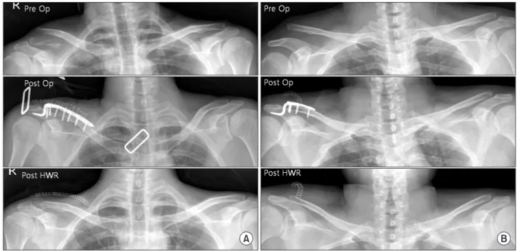
Fig. 4. (A) The radiograph shows preoperative and immediate postoperative, immediate post hardware removal of the lateral end fracture of the clavicle. (B) The radiograph shows preoperative and immediate post-operative, immediate post hardware removal of acromioclavicular dislocation.
Results
Radiologic Results
In acromioclavicular dislocation, all cases except one showed a maintenance of reduction after implant removal. The mean CC distance on the operated shoulder assessed at the final follow-up was 7 mm (range, 1.5–16 mm), and the mean CC distance on contralateral shoulder at the final follow-up was 6.2 mm (range, 2.1–9.0 mm). The difference of mean distance between the operated and contralateral sides was 0.8 mm. This did not show a statistically significant difference (p=0.19). In lateral end fracture of the clavicle, all cases except one showed a bony union. The mean time to union was 4.2 months (range, 3–6 months) (Fig. 4).
Clinical Results
The mean CMS and ASES assessed for acromioclavicular dislocation were 51.5 ± 12.3 and 55.3 ± 13.2, respectively, just before implant removal and 83.2 ± 11.8 and 92.4 ± 12.3, respectively, at the final follow-up. The mean CMS and ASES as- sessed for lateral end fracture of the clavicle were 58.2 ± 14.6 and 50.7 ± 18.2, respectively, just before implant removal and 75.1 ± 5.7, 90.3 ± 10.7, respectively, at the final follow-up.
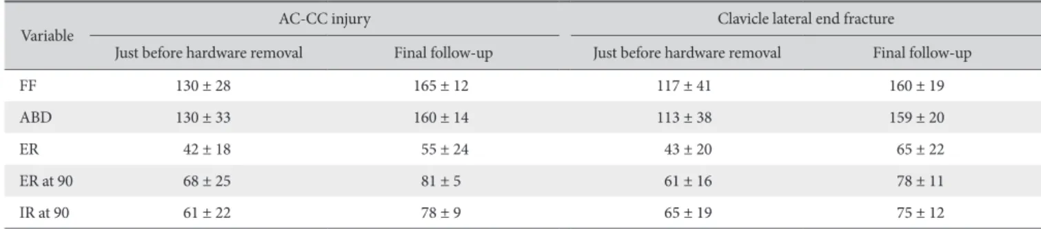
The functional scores were improved, and this improvement showed a statistical significance in acromioclavicular dislocation (p<0.001, p<0.001) and lateral end fracture of the clavicle (p<0.001, p<0.001) (Table 1, 2). The ROM just before implant
removal and final follow-up in acromioclavicular dislocation and the lateral end fracture of the clavicle are listed in Table 3. The ROM was increased in all direction, and this increase showed statistical significance.
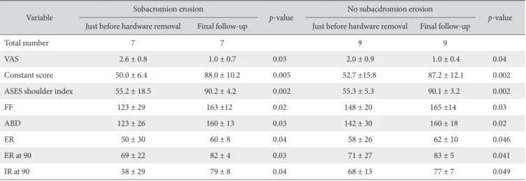
The functional scores (CMS and ASES) in accordance with subacromial erosion in acromioclavicular dislocation and lateral end fracture of the clavicle are listed in Table 4, 5. There was a statistically significant improvement of functional scores in acro- mioclavicular dislocation and lateral end fracture of the clavicle, regardless of the presence or absence of subacromial erosion.
Complications
In acromioclavicular dislocation patients, reduction loss after implant removal had developed in 1 case (6.3%), fixation failure in 1 case (6.3%), superficial infection with erythema and serous discharge in 1 case (6.3%), stiffness just before implant removal in 3 cases (18.8%), and subacromial erosion in 9 cases (56.3%).
In patients with lateral end fracture of the clavicle, nonunion had developed in 1 case (5.0%), fixation failure in 2 cases (10.0%), stiffness just before implant removal in 7 cases (35.0%), and subacromial erosion in 11 cases (55.0%).
Discussion
The hook plate was introduced as a treatment for acromio- clavicular dislocation and lateral end fracture of the clavicle in the 1970s. This method provides significant security with the
Table 1. Functional Score Just before Implant Removal and Final Follow-up in Acromioclavicular Dislocation
Functional score Just before
implant removal Final follow-up p-value
CMS 51.5 ± 12.3 83.2 ± 11.8 <0.001
ASES shoulder index 55.3 ± 13.2 92.4 ± 12.3 <0.001 Values are presented as mean ± standard deviation.
CMS: Constant-Murley score, ASES: American Shoulder and Elbow Sur- geons.
Table 2. Functional Score Just before Implant Removal and Final Follow-up in Lateral End Fracture of the Clavicle
Functional score Just before
implant removal Final follow-up p-value
CMS 58.2 ± 14.6 75.1 ± 5.7 <0.001
ASES shoulder index 50.7 ± 18.2 90.3 ± 10.7 <0.001 Values are presented as mean ± standard deviation.
CMS: Constant-Murley score, ASES: American Shoulder and Elbow Sur- geons.
Table 3. Range of Motion Just before Implant Removal and Final Follow-up
Variable AC-CC injury Clavicle lateral end fracture
Just before hardware removal Final follow-up Just before hardware removal Final follow-up
FF 130 ± 28 165 ± 12 117 ± 41 160 ± 19
ABD 130 ± 33 160 ± 14 113 ± 38 159 ± 20
ER 42 ± 18 55 ± 24 43 ± 20 65 ± 22
ER at 90 68 ± 25 81 ± 5 61 ± 16 78 ± 11
IR at 90 61 ± 22 78 ± 9 65 ± 19 75 ± 12
Values are presented as mean ± standard deviation.
AC-CC: acromioclavicular coracoclavicular, FF: forward flexion, ABD: abduction, ER: external rotation, ER at 90: external rotation at 90°, IR at 90: internal rota- tion at 90°.
use of screws, letting rotational stability and its subacromial fixation allowing horizontal stability.6) Moreover, the profound blood supply is guaranteed by minimal contact surface. Securing internal fixation without direct damage to the ACJ makes early shoulder joint exercise possible. Fixation with the hook under the acromion gives vertical stability,2) preventing a breakout of the plate from the clavicle and minimizing the risk of malunion and pseudarthrosis, as well as relieving fracture site stress.7) Sev- eral previous studies in ther literature presented good functional results of hook plate fixation in acromioclavicular dislocation and lateral end fracture of the clavicle.8) Our study showed good clinical results after hook plate fixation in acromioclavicular dislocation and lateral end fracture of the clavicle. Moreover, radiologic evaluation showed good results. CC interval in acro-
mioclavicular dislocation at the final follow-up showed that no statistically significant difference compared with contralateral side. All cases except one showed a maintenance of reduction after implant removal. In lateral end fracture of the clavicle, all cases except one showed bony union, and the mean union time after the operation was 4.1 months.
Due to the strong fixation of the hook plate, the early ROM exercise is recommended after operation. Even though the ROM interval after operation is different depending on the surgeon, about 2 to 3 weeks after operation are reported.3,9-11) Same as other studies, we usually started ROM exercise after 2 weeks postoperatively. However, the ROM just before implant removal was not good even though bony union and reduction were maintained.
Table 4. The Functional Scores and Range of Motion according to Subacromial Erosion in Acromioclavicular Dislocation
Variable Subacromion erosion
p-value No subacdromion erosion
p-value Just before hardware removal Final follow-up Just before hardware removal Final follow-up
Total number 7 7 9 9
VAS 2.6 ± 0.8 1.0 ± 0.7 0.03 2.0 ± 0.9 1.0 ± 0.4 0.04
Constant score 50.0 ± 6.4 88.0 ± 10.2 0.005 52.7 ±15.8 87.2 ± 12.1 0.002
ASES shoulder index 55.2 ± 18.5 90.2 ± 4.2 0.002 55.3 ± 5.3 90.1 ± 3.2 0.002
FF 123 ± 29 163 ±12 0.02 148 ± 20 165 ±14 0.03
ABD 123 ± 26 160 ± 13 0.03 142 ± 30 160 ± 18 0.02
ER 50 ± 30 60 ± 8 0.04 58 ± 26 62 ± 10 0.046
ER at 90 69 ± 22 82 ± 4 0.03 71 ± 27 83 ± 5 0.041
IR at 90 58 ± 29 79 ± 8 0.04 68 ± 13 77 ± 7 0.049
Values are presented as number only or mean ± standard deviation.
VAS: visual analogue scale, ASES: American Shoulder and Elbow Surgeons, FF: forward flexion, ABD: abduction, ER: external rotation, ER at 90: external rotation at 90°, IR at 90: internal rotation at 90°.
Table 5. The Functional Scores and Range of Motion according to Subacromial Erosion in Lateral End Fracture of the Clavicle
Variable Subacromion erosion
p-value No subacromion erosion
p-value Just before hardware removal Final follow-up Just before hardware removal Final follow-up
Total number 11 11 9 9
VAS 0.8 ± 0.6 0.5 ± 0.3 0.2 0.9 ± 0.3 0.6 ± 0.4 0.4
Constant score 70.5 ± 8.9 81.0 ± 7.8 0.04 72.7 ± 8.3 83.2 ± 6.5 0.02
ASES shoulder index 73.4 ± 16.9 87.9 ± 11.6 0.02 78.4 ± 12.7 91.4 ± 7.32 0.012
FF 105 ± 40 150 ± 26 0.013 131 ± 38 172 ± 3 0.04
ABD 96 ± 32 140 ± 40 0.03 133 ± 36 167 ± 12 0.02
ER 42 ± 23 70 ± 12 0.015 44 ± 18 74 ± 15 0.014
ER at 90 61 ± 18 76 ± 9 0.03 61 ± 15 77 ± 13 0.026
IR at 90 65 ± 21 77 ± 9 0.03 66 ± 17 78 ± 11 0.023
Values are presented as number only or mean ± standard deviation.
VAS: visual analogue scale, ASES: American Shoulder and Elbow Surgeons, FF: forward flexion, ABD: abduction, ER: external rotation, ER at 90: external rota- tion at 90°, IR at 90: internal rotation at 90°.
Moreover, there were 3 patients who showed a ROM below 100 degree of abduction and forward flexion before implant removal in acromioclavicular dislocation (18.8%) and 7 cases in the lateral end fracture of the clavicle (35.0%). We cannot de- fine the cause of this limited ROM in our study. With reviewed the literature, rigid and static stabilization of AC joint by plate does not reproduce the primary dynamic unit of lateral clavicle, which contributes significantly to the freedom of motion in the shoulder. In addition, known shortcomings included by hook plate include bony erosion, shoulder impingement, and rotator cuff damage, which could lead to persistent pain and restricted ROM in combination or singly. Therefore, many previous stud- ies recommended implant removal after confirming stabilization during the follow-up.3,5,9) According to Renger et al.,12) after fixa- tion hook plate, 68% of patients had a complication of limited ROM and pain on ROM. In our study, we removed the hook plate at the mean postoperative period of 3.9 months (range, 3–5 months) in acromioclavicular dislocation and 4.2 months (range, 2–6 months) in lateral end fracture of the clavicle. After implant removal, the ROM showed an improvement. The ROM after implant removal and before implant removal showed a statistically significant difference. In addition to the ROM, the functional scores also showed improved results, as compared between before implant removal and final follow-up.
According to Davut et al, after hardware removal at postop- erative 6 months, 28 patients with displaced lateral end fracture of the clavicle that used a hook plate showed good results. CMS was 97, DASH score was 3.5 in the mean follow-up of 5.4 years.13) According to Kienast et al, after hardware removal, the acromioclavicular dislocation patients that used a hook plate showed good results. CMS was 92.4 in the mean follow-up of 3 years.14)
In our study, patients with acromioclavicular dislocation and lateral end fracture of the clavicle who used a hook plate showed good results and increased functional score after hardware removal. Although we cannot suggest whether the improved result is due to implant removal itself or is part of the natural healing process, implant removal after stabilization dur- ing the follow-up period has been confirmed to provide some advantages to the functional aspects.
In addition to the stiffness as a complication of hook plate, several other complications have been reported, such as sub- acromial erosion, impingement, rotator cuff tear, bursitis, non- union, and infection. Subacromial erosion is reported as one of the most common complications. The use of a hook plate could easily pierce the subacromial bursa, and in some specimens, the tip end of the hook had reached the acromial undersurface.
According to Tiren et al.,13) subacromial bursitis and subacromial erosion on an x-ray are signs of a mismatch between the plate and the anatomy of the patients. In addition, stress concentra- tion at the hooks and altered joint kinematics may cause ero-
sion in the acromion. Gao et al.15) reported that 37.5% (9/24) of acromioclavicular dislocation patients demonstrated subacro- mial erosion in the final radiographs. In our study, subacromial erosion was developed in 56.3% of patients (9/16) with acro- mioclavicular dislocation and in 55.0% of patients (11/20) with lateral end fracture of the clavicle just before implant removal.
However, regardless of the presence and/or absence of sub- acromial erosion, the functional score showed an improvement in acromioclavicular dislocation and lateral end fracture of the clavicle, as compared between just before implant removal and final follow-up.
There are some limitations to consider while interpreting our results. First, our study design is retrospective in nature. Patients who were enrolled in this study did not represent the entire population of those who had been treated by a hook plate due to acromioclavicular dislocation and lateral end fracture of the clavicle. Second, a comparison between patients with plate re- moval and without plate removal was not carried out. Therefore, whether the stiffness and lower functional score before implant removal were caused by hook design, and impingement due to plate is not definitely concluded. Further study is necessary.
Third, subacromial erosion was only evaluated by simple radio- logic findings. Therefore, we could not quantify and classify the degree of subacromial erosion. Further studies using radiologic evaluation, such as computed tomography, might be neces- sary to quantify and classify the erosion, leading us to better understand the reason for the erosion and the relationship with implant design and acromial shape. Finally, the evaluation time point was not consistent. The implant removal time was not the same after the initial treatment. However, we tried to remove the implant 4 months after the operation.
Conclusion
Hook plate fixation of acromioclavicular dislocation and lat- eral end fracture of the clavicle resulted in good results. Although the functional score was not promising before implant removal, the functional score was improved after implant removal. Sub- acromial erosion was a common finding after hook plate fixation in both acromioclavicular dislocation and lateral end fracture of the clavicle. However, the presence or absence of subacromial erosion did not affect the functional improvement at the final follow-up.
References
1. van der Meijden OA, Gaskill TR, Millett PJ. Treatment of clavi- cle fractures: current concepts review. J Shoulder Elbow Surg.
2012;21(3):423-9.
2. Muramatsu K, Shigetomi M, Matsunaga T, Murata Y, Tagu- chi T. Use of the AO hook-plate for treatment of unstable
fractures of the distal clavicle. Arch Orthop Trauma Surg.
2007;127(3):191-4.
3. Kim YS, Lee HM, Jang HG. Surgical treatment of unstable distal clavicle fractures: comparison of transacromial pin fixation and hook plate fixation. Clinics Shoulder Elbow. 2013;16(2):123- 9.
4. Tambe AD, Motkur P, Qamar A, Drew S, Turner SM. Fractures of the distal third of the clavicle treated by hook plating. Int Orthop. 2006;30(1):7-10.
5. Drijfhout van Hooff CC, Haverlag R, Willems WJ. Evaluation of the use of the hook plate in Neer type 2 lateral clavicle frac- tures and rockwood types 3-5 acromioclavicular joint disloca- tions. Eur J Trauma Emerg Surg. 2013;39(6):619-26.
6. Martinoli C, Bianchi S, Prato N, et al. US of the shoulder: non- rotator cuff disorders. Radiographics. 2003;23(2):381-401;
quiz 534.
7. Wickham MQ, Wyland DJ, Glisson RR, Speer KP. A biome- chanical comparison of suture constructs used for coracocla- vicular fixation. J South Orthop Assoc. 2003;12(3):143-8.
8. Alhamam NM, Bella IH, Uddin FZ, Al-Afaleq MA, Al-Afaleq SA, Al-Khalifa FK. Clinical outcomes of hook-plate fixation in the tratemtn of unstable distal clavicular fractures and acromioclavicular joint dislocations. Saudi J Med Med Sci.
2015;3(1):50-3.
9. Kumar N, Sharma V. Hook plate fixation for acute acromiocla-
vicular dislocations without coracoclavicular ligament recon- struction: a functional outcome study in military personnel.
Strategies Trauma Limb Reconstr. 2015;10(2):79-85.
10. Jeon HS, Woo YK, Hwang SH, Suh SP, Kang SG, Lee JK. Analy- sis of treatment results of unstable distal clavicle fractures using hook plate. J Korean Orthop Assoc. 2014;49(5):374-80.
11. Kim KC, Shin HD, Cha SM, Jeon YS. Hook plate fixation for unstable distal clavicle fracture: a prospective study. Clin Shoulder Elbow. 2011;14(1):6-12.
12. Renger RJ, Roukema GR, Reurings JC, Raams PM, Font J, Verleisdonk EJ. The clavicle hook plate for Neer type II lateral clavicle fractures. J Orthop Trauma. 2009;23(8):570-4.
13. Tiren D, van Bemmel AJ, Swank DJ, van der Linden FM. Hook plate fixation of acute displaced lateral clavicle fractures: mid- term results and a brief literature overview. J Orthop Surg Res.
2012;7:2. doi: 10.1186/1749-799X-7-2.
14. Kienast B, Thietje R, Queitsch C, Gille J, Schulz AP, Meiners J.
Mid-term results after operative treatment of rockwood grade III-V acromioclavicular joint dislocations with an AC-hook- plate. Eur J Med Res. 2011;16(2):52-6.
15. Gao YS, Zhang YL, Ai ZS, Sun YQ, Zhang CQ, Zhang W.
Transarticular fixation by hook plate versus coracoclavicular stabilization by single multistrand titanium cable for acute Rockwood grade-V acromioclavicular joint dislocation: a case- control study. BMC Musculoskelet Disord. 2015;16:360.