INTRODUCTION
Current evidence indicates that chronic subclinical inflam- mation may play an important role in the genesis and pro- gression of atherosclerosis, and many recent reports have ex- plored the association between C-reactive protein (CRP) lev- els and atherosclerosis. CRP is present in the acute reaction phase of inflammation, and elevated levels of serum CRP are related to a poorer outcome in patients with cardiovas- cular diseases (1, 2).
Several studies have suggested that CRP is a predictor of subsequent cardiovascular events in healthy adults (3). Recen- tly, high-sensitivity C-reactive protein (hs-CRP) has emerged as a useful biomarker for vascular inflammation associated with atherosclerosis. While a significant amount of data has been amassed about vascular disease in adults, limited data exists concerning the low grade inflammation in obese chil- dren. Because atherosclerosis frequently begins in childhood, we assessed the association of two markers of early atheroscle- rosis, serum hs-CRP level and atherosclerotic risk factors with carotid intima-media wall thickness (IMT) and brachial flow-mediated dilation (FMD) in obese children.
MATERIALS AND METHODS Subjects
The study subjects were 83 children. Thirty-eight children were obese (14-16 yr of age, male), and 45 were sex/age-ma- tched healthy control children. All were nonsmokers with- out any regular medication and had no family history of pre- mature vascular disease. None of the children had symptoms of infection during the 2 weeks before the study.
Anthropometric measurement and clinical examination
Body height and weight were measured in light clothes using a portable standiometer. Body mass index (BMI) was calculated as weight divided by the square of the height (kg/
m2). In children, the 95th percentile for BMI in the 2000 CDC growth chart is 21-23 (4), so we used BMI >23 as our prospectively defined criterion for obesity, and BMI <21 was defined as the non-obese control level.
Eui Jung Roh, Jae Woo Lim, Kyoung Og Ko, Eun Jung Cheon
Department of Pediatrics, College of Medicine, Konyang University, Daejeon, Korea
Address for correspondence Eun Jung Cheon, M.D.
Department of Pediatrics, College of Medicine, Konyang University, 685 Gasoowon-dong, Seo-gu, Daejeon 302-241, Korea
Tel : +82.42-600-9230, Fax : +82.42-600-9090 E-mail : [email protected]
*This study was supported by a grant from the Korea Research Foundation (KRF-2005-E00208).
192
A Useful Predictor of Early Atherosclerosis in Obese Children: Serum High-sensitivity C-reactive Protein
Childhood obesity seems to contribute to the development of vascular inflammation and the progression of arterial wall changes. High-sensitivity C-reactive protein (hs- CRP) has recently emerged as a useful biomarker for vascular inflammation asso- ciated with atherosclerosis. The objectives of this study were to evaluate the asso- ciation of the serum hs-CRP level with ultrasonic findings of early atherosclerosis, carotid intima-media wall thickness (IMT) and brachial flow-mediated dilation (FMD), in obese children. Thirty eight obese children and 45 sex/age-matched healthy con- trol children were recruited. Serum CRP levels were measured by the high-sensi- tive latex turbidimetric immunoassay, and we measured carotid IMT and brachial FMD using high-resolution B-mode ultrasonography. Obese children had signifi- cantly higher hs-CRP levels (1.40±±0.74 mg/L vs. 0.55±±0.49 mg/L, p<0.01), as well as increased IMT (0.52±±0.09 mm vs. 0.41±±0.07 mm, p<0.01) and impaired FMD (7.35±±7.78% vs. 20.34±±16.81%, p<0.01) compared to healthy controls.
Serum hs-CRP correlated positively with IMT (r=0.413, p<0.05) and inversely with FMD (r=-0.350, p<0.05) in the obesity group. Measurement of the serum hs-CRP level is a simple, cheap, and highly reproducible assay and correlates with IMT and FMD in obese children. Thus, it would be a useful marker for evaluating and esti- mating the degree of atherosclerosis in children.
Key Words : C-reactive Protein; Intima-media Thickness; Flow-mediated Dilation; Obesity
Received : 25 April 2006 Accepted : 16 August 2006
Serum lipids and apolipoprotein
Venous blood samples were taken in the morning after overnight fasting (10-12 hr). Serum total cholesterol (TC), low density lipoprotein cholesterol (LDL-C), high density lipoprotein cholesterol (HDL-C), and triglyceride (TG) con- centrations were measured by standard enzymatic methods with the use of Boehringer Mannheim GmbH and a fully automatic analyzer. Serum apolipoprotein A-1 (Apo A-1), apolipoprotein B (Apo B), and apolipoprotein E (Apo E) were measured by immunonephelometry (Behring Nephelome- ter II, Dade Behring, Inc., Newark, DE, U.S.A.).
High-sensitivity C-reactive protein
A BN II Nephelometer Analyzer (Dade Behring, Inc.) was used to measure serum hs-CRP values using a high sensitiv- ity latex-enhanced immunonephelometric assay method. The detection limit of this assay was 0.17 mg/dL. Because the 90th percentile of normal CRP distribution is 0.3 mg/dL, we included only patients with CRP values below 0.3 mg/
dL to avoid the influence of acute infection.
Ultrasound studies
All ultrasound studies were performed in the morning on fasting subjects with the use of Hewlett-Packard Sonos 5500 and a 3-11 MHz linear array transducer (Philips, Andover, Massachusetts, U.S.A.). All ultrasound scans were performed by an experienced vascular operator who was unaware of the children’s clinical details. Blood pressure was measured from the brachial artery of the nondominant arm 3 times during the ultrasound study with the use of a standard sphygmo- manometer, and the mean value of these 3 measurements was used for the analyses. The brachial artery evaluations and carotid artery evaluations were performed by using standard techniques for the study of children.
Brachial artery evaluation
Brachial artery diameter was measured during reactive hyperemia from B-mode ultrasound images at rest. The sub- jects lay quietly for 10 min before the first scan. The brachial artery was scanned in a longitudinal section 5 to 15 cm above the elbow. Depth and gain settings were set to optimize ima- ges of the lumen-arterial wall interface, and the operating parameters were kept constant during the study. A resting scan was performed, and the arterial flow velocity was mea- sured using a Doppler signal. Increased flow was then induced by inflation of a pneumatic tourniquet placed around the forearm (distal to the scanned part of the artery) to a pressure of 250 mmHg for 5 min, followed by release. Subsequent scans were taken continuously for 60 sec after cuff deflation.
We also included a repeated flow velocity recording for the first 15 sec after the cuff was released. Vessel diameter was
measured by an experienced reader who were blind to the study subjects’ laboratory data. The arterial diameter was measured at a fixed distance from an anatomic marker with the use of ultrasonic calipers. Measurements were taken from the anterior to the posterior M line at end diastole, incident with the R wave on a continuously recorded ECG. The maxi- mal proportional dilatation from baseline (FMD) was assessed.
Carotid artery analysis
Carotid artery IMT measurements were done according to a predetermined, standardized scanning protocol for the right carotid artery. Briefly, the proximal part of the carotid bulb was identified and the segments of the common carotid arteries 1 to 2 cm proximal to the bulb were scanned. The image was focused on the posterior wall, and the resolution box function was used to magnify the arterial far wall. All scans were digitally stored for subsequent offline analysis.
One end-diastolic frame (captured adjacent to the R wave on a continuously recorded ECG) for each interrogation angle was selected and analysed for mean IMT. Five measurements for far wall IMT were taken manually for each image using ultrasonic calipers, and the average values of these readings were used for the analyses.
Statistical analyses
SPSS version 11.0 was used for all statistical calculations.
Results were expressed as mean±standard deviation. Com- parisons between two groups were performed by the inde- pendent student-t test. Univariate and multivariate regres- sion analyses were used to dclineate the relationships between arterial changes and CRP. Due to the skewed distribution of hs-CRP levels, CRP values were logarithmically transformed prior to regression analyses. For all analyses, probability (p) values below 0.05 were considered statistically significant.
RESULTS Clinical characteristics
Height and weight were significantly higher in the obesity group than in the control group. BMI was 29.40±3.18 kg/
m2in the obesity group and was 18.43±1.00 kg/m2in the control group (Table 1). Obese children had significantly higher systolic blood pressure (SBP) (133.05±8.84 mmHg vs. 115.16±8.62 mmHg), diastolic blood pressure (DBP) (84.95±9.02 mmHg vs. 75.16±6.92 mmHg), hs-CRP levels (0.10±0.07 mg/dL vs. 0.06±0.04 mg/dL, p<0.01) and significantly higher LDL-C, TG, Apo B and lower HDL- C than the control group (Table 2). TC and Apo E were higher and Apo A-1 was lower in the obesity group than in the con- trol group, but these differences were not statistically signif- icant.
Ultrasonic findings
Obese children were also found to have increased IMT (0.52
±0.09 mm vs. 0.40±0.07 mm, p<0.001) and impaired FMD (7.35±7.78% vs. 20.34±16.81%, p<0.001) com- pared to healthy controls (Table 3).
Relationships between IMT, FMD and atherosclerotic risk factors
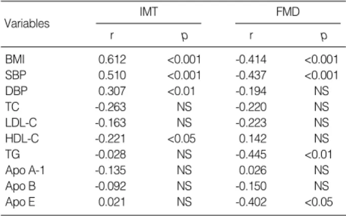
IMT correlated with BMI (r=0.612, p<0.001), SBP (r=
0.510, p<0.001), DBP (r=0.307, p<0.05), and HDL-C (r=
-0.221, p<0.05). FMD correlated with BMI (r=-0.414, p<
0.001), SBP (r=-0.437, p<0.001), TG (r=-0.445, p<0.05), and Apo E (r=-0.402, p<0.05) (Table 4).
Relationships between hs-CRP and atherosclerotic risk factors
Log [CRP]showed a positive correlation with BMI (r=
0.470, p<0.001), SBP (r=0.209, p<0.05), DBP (r=0.230, p<0.05), Apo E (r=0.308, p<0.01), and TG (r=0.288, p<
0.05) by simple regression (Table 5).
Relationships between hs-CRP and ultrasonic findings
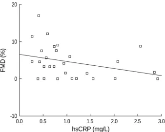
Serum hs-CRP correlated positively with IMT (r=0.413, p<0.05) and inversely with FMD (r=-0.350, p<0.05) in the obesity group (Fig. 1, 2).
Values are mean±standard deviation.
BMI, body mass index; BP, blood pressure; p, p-value.
Obesity (n=38) Control (n=45) p
Age (yr) 14.76±0.8 14.00±0.00 NS
Height (cm) 164.47±9.0 155.14±7.34 <0.01 Weight (kg) 80.05±13.30 44.53±5.13 <0.01 BMI (kg/m2) 29.40±3.18 18.43±1.00 <0.01 Systolic BP (mmHg) 133.05±8.84 115.16±8.62 <0.01 Diastolic BP (mmHg) 84.95±9.02 75.16±6.92 <0.01 Table 1.Comparison of characteristics between obesity and control groups
Values are mean±standard deviation.
IMT, intima-media thickness; FMD, flow-mediated dilation; p, p-value.
Obesity (n=38) Control (n=45) p
IMT (mm) 0.52±0.09 0.41±0.07 <0.01
FMD (%) 7.35±7.78 20.34±16.81 <0.01
Table 3.Comparison of ultrasonic findings between obesity and control groups
IMT, intima-media thickness; FMD, flow-mediated dilation; BMI, body mass index; SBP, systolic blood pressure; DBP, diastolic blood pres- sure; PP, pulse pressure; TC, total cholesterol; LDL-C, low density lipop- rotein cholesterol; HDL-C, high density lipoprotein cholesterol; TG, triglyc- eride; Apo A-1, apolipoprotein A-1; Apo B, apolipoprotein B; Apo E, apo- lipoprotein E; r, correlation coefficient; p, p-value.
IMT
r p
Variables FMD
r p
BMI 0.612 <0.001 -0.414 <0.001
SBP 0.510 <0.001 -0.437 <0.001
DBP 0.307 <0.01 -0.194 NS
TC -0.263 NS -0.220 NS
LDL-C -0.163 NS -0.223 NS
HDL-C -0.221 <0.05 0.142 NS
TG -0.028 NS -0.445 <0.01
Apo A-1 -0.135 NS 0.026 NS
Apo B -0.092 NS -0.150 NS
Apo E 0.021 NS -0.402 <0.05
Table 4.Correlation of IMT and FMD with BMI, BP, lipid profile, and apolipoprotein concentrations
BMI, body mass index; SBP, systolic blood pressure; DBP, diastolic blood pressure; PP, pulse pressure; TC, total cholesterol; LDL-C, low density lipoprotein cholesterol; HDL-C, high density lipoprotein choles- terol; TG, triglyceride; Apo A-1, apolipoprotein A-1; Apo B, apolipopro- tein B; Apo E, apolipoprotein E; r, correlation coefficient; p, p-value.
hs-CRP
r p
Variables
BMI 0.470 <0.001
SBP 0.209 <0.05
DBP 0.230 <0.05
TC 0.094 NS
LDL-C 0.104 NS
HDL-C -0.123 NS
TG 0.308 <0.01
Apo A-1 -0.182 NS
Apo B 0.017 NS
Apo E 0.288 <0.01
Table 5.Correlation of log hs-CRP with BMI, BP, lipid profile, and apolipoprotein concentrations
Values are mean±standard deviation.
TC, total cholesterol; LDL-C, low density lipoprotein cholesterol; HDL-C, high density lipoprotein cholesterol; TG, triglyceride; Apo A-1, apolipop- rotein A-1; Apo B, apolipoprotein B; Apo E, apolipoprotein E, hs-CRP;
high-sensitivity C-reactive protein; p, p-value.
Obesity (n=38) Metabolic
parameters Control (n=45) p
TC (mg/dL) 193.46±38.20 180.38±39.84 NS HDL-C (mg/dL) 63.20±15.92 72.58±17.06 <0.05 LDL-C (mg/dL) 114.09±29.90 99.71±29.91 <0.05 TG (mg/dL) 148.63±78.83 96.49±54.97 <0.01 Apo A1 (mg/dL) 74.17±34.88 78.76±36.66 NS Apo B (mg/dL) 37.89±20.0 30.00±12.38 <0.05
Apo E (mg/dL) 3.54±1.31 3.42±1.71 NS
hs-CRP (mg/L) 1.40±0.74 0.55±0.49 <0.01 Table 2.Comparison of metabolic parameters between obesity and control groups
DISCUSSION
The present study showed that elevated serum CRP levels were significantly associated with endothelial dysfunction and increased carotid artery IMT in obese children. While there have been many previous studies relating CRP and cardiovascular risk factors (5, 6), the association of CRP with subclinical atherosclerotic findings has been examined pri- marily in adults, with few studies in obese children (7, 8).
Historically, CRP has been measured in clinical laborato- ries by immunoturbidimetric and immunonephelometric assays designed to detect active inflammation and infection.
The dynamic range of these assays is from 3 mg/L (90th per- centile of the general population) to well over 200 mg/L. Al- though such assays are automated and reproducible, they have a relatively low detection limit of 3 to 8 mg/L and thus do not have appropriate sensitivity in the range required for the determination of cardiovascular risk (9). To achieve the desired limit of quantification, hs-CRP assays have been developed that can reliably measure hs-CRP concentrations as low as 0.15 mg/L (<2.5th percentile of the general population). When measured with a high-sensitivity assay, CRP levels appear to have a predictive value for cardiovascular morbidity and mor- tality. Moreover, baseline levels of CRP predict the risk of future myocardial infarction, stroke, and peripheral atheroscle- rosis, even after adjustment for other known cardiovascular risk factors (10). We employed this method in the present study, and we found that obese children had significantly higher hs-CRP levels than control subjects. Further studies will be required to determine cut-off values for hs-CRP in obese children.
High-resolution ultrasound is a reliable, noninvasive method for detecting early structural and functional atherosclerotic changes in the arterial wall. Increased carotid IMT is a struc-
tural marker of early atherosclerosis that correlates with vas- cular risk factors and is related to the severity and extent of coronary artery disease. FMD of the brachial artery is a marker of endothelial function that can be assessed by measuring arte- rial diameter responses to increased flow (11). Brachial artery FMD occurs mainly as the result of endothelial release of nitric oxide and correlates with coronary endothelial function (12).
Both increased IMT and impaired FMD have been detected in young children who have risk factors for atherosclerosis such as familial hypercholesterolemia (13) and diabetes (14).
In the present study, obese children were also found to have increased IMT and impaired FMD compared to the healthy controls. In addition, IMT was positively correlated with BMI, SBP, and DBP, and negatively correlated with HDL-C concentrations. Zhu et al. reported a similar result in 43 obese children (15), and Kim et al. showed that IMT corre- lated with DBP and homocysteine concentrations (16).
Woo et al. have confirmed that the degree of endothelial dysfunction correlated with BMI as assessed by multivariate analysis (17). In the current study, FMD was shown to cor- relate with BMI, SBP, and TG and Apo E concentrations.
Traditional risk factors for cardiovascular disease include aging, hypertension, dyslipidemia, smoking, and diabetes.
The contribution of lipid accumulation to atherosclerotic disease is well known, but laboratory and experimental evi- dence indicates that chronic inflammatory processes also play an important role in the development of atherosclerosis (18).
In order to effectively prevent the incidence of cardiovascu- lar events, it is important to weigh the influence of each risk factor on the cardiovascular system. In addition, it should be borne in mind that various cardiovascular factors are not independent of one another but often have direct or indirect interactions (19). In this respect, we investigated the relation- ship of inflammatory markers with other risk factors. Elevat-
FMD (%)
20
10
0
-10
0.0 0.5 1.0 1.5 2.0 2.5 3.0
hsCRP (mg/L)
Fig. 1.Linear correlation of high-sensitivity C-reative protein (hs- CRP) and flow-mediated dilation (FMD) in the obesity group (r=
-0.327, p<0.05, hs-CRP=6.472-1.89*FMD).
IMT (mm)
0.7
0.6
0.5
0.4
0.3
0.2
0.0 0.5 1.0 1.5 2.0 2.5 3.0
hsCRP (mg/L)
Fig. 2.Linear correlation of high-sensitivity C-reative protein (hs- CRP) and intima-media thickness (IMT) in the obesity group (r=
0.437, p<0.05, hs-CRP=0.377+0.042*IMT).
ed serum CRP levels were positively associated with elevat- ed BMI, confirming previous observations in both children (19) and adults (20). The mechanisms underlying this asso- ciation with BMI or obesity might be as follows; the adipose tissue is a source of cytokines such as tumor necrosis factor-a (TNF-a) and interleukin-6 (IL-6), and these cytokines stim- ulate the production of acute-phase proteins such as C-reac- tive protein in the liver (21). Not unexpectedly, then, we found that serum CRP levels were positively associated with BMI. Because we have not checked proinflammatory cyto- kines such as TNF-a and IL-6, we can not verify the eleva- tion of these cytokines as a consequence of obesity.
In addition to BMI, CRP was also found to be closely cor- related with SBP, DBP, and ApoE and TG concentrations.
Previous studies reported that CRP levels are significantly positively correlated with TG, total ratio of serum cholesterol to serum HDL cholesterol (22), fibrinogen levels, heart rate, SBP (19), smoking, and white blood cell count (5) and neg- atively correlated with HDL-C levels (19, 22). Several stud- ies have examined the relationship between CRP and carotid IMT in adults. Hak et al. found a highly significant relation- ship between markers of obesity and CRP in healthy, mid- dle-aged women, but failed to find a significant relationship between CRP and subclinical atherosclerosis of the carotid system (20). Sitzer et al. performed a large scale study that showed the serum CRP level is linearly positively correlated with IMT in apparently healthy adults (3). Hayaishi et al.
reported that the hs-CRP level is positively associated with carotid IMT in young patients with type 1 diabetes (23).
Several studies have also shown an association between ele- vated CRP concentrations and endothelial dysfunction in adult patients with coronary artery disease (24) and with type 2 diabetes (25). In contrast to the significant amount of data that has accumulated from adult individual, limited data exists concerning CRP and subclinical atherosclerosis in children.
Jarvisalo et al. reported that, in healthy children, elevated serum CRP levels were significantly associated with decreased endothelial vasodilatory function and increased IMT (26).
Our results are consistent with the Mangge’s report (27) that serum hs-CRP correlated positively with IMT and inversely with FMD in the obesity group. In other words, if hs-CRP is increased in asymptomatic obese children, we can assume the existence of and advanced subclinical atherosclerosis, in- creased IMT, and decreased endothelial vasodilatory function without performing an ultrasound.
In summary, we found a highly significant correlation bet- ween the serum CRP concentration and the early stage of atherosclerosis in obese children. Although ultrasonography allows visualization of early subclinical stages of atherosclero- sis in obese children, the measurement of the serum hs-CRP level is simpler and cheaper than ultrasonography, is highly reproducible, and well correlates with IMT and FMD in obese children. Thus, hs-CRP would be a useful screening marker for evaluating and estimating the degree of atherosclerosis
in children.
REFERENCES
1. Haverkate F, Thompson SG, Pyke SD, Gallimore JR, Pepys MB.
Production of C-reactive protein and risk of coronary events in sta- ble and unstable angina. European Concerted Action on Thrombosis and Disabilities Angina Pectoris Study Group. Lancet 1997; 349:
462-6.
2. Liuzzo G, Biasucci LM, Gallimore JR, Grillo RL, Rebuzzi AG, Pepys MB, Maseri A. The prognostic value of C-reactive protein and serum amyloid A protein in severe unstable angina. N Engl J Med 1994;
331: 417-24.
3. Kuller LH, Tracy RP, Shaten J, Meilahn FN. Relation of C-reactive protein and coronary heart disease in the MRFIT nested case-con- trol study. Multiple Risk Factor Intervention Trial. Am J Epidemiol 1996; 144: 537-47.
4. Ogden DL, Kuczmarski RJ, Flegal KM, Mel Z, Guo S, Wel R, Grum- mer-Strawn LM, Gurtin LR, Roche AF, Johnson CL. Centers for Disease Control and Prevention 2000 growth charts for the United States: improvements to the 1977 National Center for Health Statis- tics version. Pediatrics 2002; 109: 46-60.
5. Saito M, Ishimitsu T, Minami J, Ono H, Ohrui M, Matsuoka H. Rela- tions of plasma high-sensitivity C-reactive protein to traditional car- diovascular risk factors. Athoerosclerosis 2003; 167: 73-9.
6. Ryu SY, Lee YS, Park J, Kang MG, Kim KS. Relations of plasma high-sensitivity C-reactive protein to various cardiovascular risk fac- tors. J Korean Med Sci 2005; 20: 379-83.
7. Sitzer M, Markus HS, Mendall MA, Liehr R, Knorr U, Steinmetz H.
C-reactive protein and carotid intimal medial thickness in a commu- nity population. J Cardiovasc Risk 2002; 9: 97-103.
8. Hashimoto H, Kitagawa K, Hougaku H, Shimizu Y, Sakaguchi M, Nagai Y, Iyama S, Yamanishi H, Matsumoto M, Hori M. C-reactive protein is an independent predictor of the rate of increase in early carotid atherosclerosis. Circulation 2001; 104: 63-7.
9. Rifai N, Ridker PM. High-sensitivity C-reactive protein: a novel and promising marker of coronary heart disease. Clinical Chemistry 2001;
47: 403-11.
10. Rohde LE, Hennekens CH, Ridker PM. Survey of C-reactive protein and cardiovascular risk factors in apparently healthy men. Am J Car- diol 1999; 84: 1018-22.
11. Jarvisalo MJ, Raitakari M, Toikka JO, Putto-Laurila A, Rontu R, Laine S, Lehtimaki T, Ronnemaa T, Viikari J, Raitakari OT. Endothe- lial dysfunction and increased arterial intima-media thickness in chil- dren with type 1 diabetes. Circulation 2004; 109: 1750-5.
12. Joannides R, Haefeli WE, Linder L, Richard V, Bakkali EH, Thuillez C, Luscher TF. Nitric oxide is responsible for flow-dependent dilata- tion of human peripheral conduit arteries in vivo. Circulation 1995;
92: 1314-9.
13. Celermajer DS, Sorensen KE, Gooch VM, Spiegelhalter DJ, Miller OI, Sullivan ID, Lloyd JK, Deanfield JE. Non-invasive detection of endothelial dysfunction in children and adults at risk of atheroscle- rosis. Lancet 1992; 340: 1111-5.
14. Singh TP, Groehn H, Kazmers A. Vascular function and carotid inti- mal-medial thickness in children with insulin-dependent diabetes mellitus. J Am Coll Cardiol 2003; 41: 661-5.
15. Zhu W, Huang X, He J, Li M, Neubauer H. Arterial intima-media thickening and endothelial dysfunction in obese chinese children. Eur J Pediatr 2005; 164: 337-44.
16. Kim YJ, Shim YH, Yoo JH, Lee K, Hong YM. Measurement of the common carotid artery by ultrasound as a predictor of atheroscle- rosis in obese adolescents. Korean J Pediatr 2005; 48: 745-52.
17. Woo KS, Chook P, Yu CW, Sung RY, Qiao M, Leung SS, Lam CW, Metreweli C, Celermajer DS. Overweight in children is associated with arterial endothelial dysfunction and intima-media thickening.
Int J Obes 2004; 28: 852-7.
18. Ross R. Atherosclerosis-an inflammatory disease. New Engl J Med 1999; 340: 115-26.
19. Cook DG, Mendall MA, Whincup PH, Carey IM, Ballam L, Morris JE, Miller GJ, Strachan DP. C-reactive protein concentration in chil- dren: relationship to adiposity and other cardiovascular risk factors.
Atherosclerosis 2000; 149: 130-50.
20. Hak AE, Stehouwer CD, Bots ML, Polderman KH, Schalkwijk CG, Westendorp IC, Hofman A, Witteman JC. Association of C-reactive protein with measures of obesity, insulin resistance, and subclinical atherosclerosis in healthy, middle aged women. Arterioscler Thromb Vasc Biol 1999; 19: 1986-91.
21. Bassuk SS, Rifai N, Ridker PM. High-sensitivity C-reactive protein:
clinical importance. Curr Probl Cardiol 2004; 29: 439-93.
22. Wu DM, Chu NF, Shen MH, Chang JB. Plasma C-reactive protein levels and their relationship to anthropometric and lipid character- istics among children. J Clin Epidemol 2003; 56: 94-100.
23. Hayaishi-Okano R, Yamasaki Y, Katakami N, Ohtoshi K, Gorogawa S, Kuroda A, Matsuhisa M, Kosugi K, Nishikawa N, Kajimoto Y, Hori M. Elevated C-reactive protein associates with early-stage caro- tid atherosclerosis in young subjects with type 1 diabetes. Diabetes Care 2002; 25: 1432-8.
24. Fichtlscherer S, Rosenberger G, Walter DH, Breuer S, Dimmeler S, Zeiher AM. Elevated C-reactive protein levels and impaired endothe- lial vasoreactivity in patients with coronary artery disease. Circula- tion 2000; 102: 1000-6.
25. Tan KC, Chow WS, Tam SC, Ai VH, Lam CH, Lam KS. Atorvas- tatin lowers C-reactive protein and improves endothelium-dependent vasodilation in type 2 diabetes mellitus. J Clin Endocrinol Metab 2002; 87: 563-8.
26. Jarvisalo MJ, Harmoinen A, Hakanen M, Paakkunainen U, Viikari J, Hartiala J, Lehtimaki T, Simell O, Raitakari OT. Elevated serum C-reactive protein levels and early arterial changes in healthy chil- dren. Arterioscler Thromb Vasc Biol 2002; 22: 1323-8.
27. Mangge H, Schauenstein K, Stroedter L, Griesl A, Maerz W, Bor- kenstein M. Low grade inflammation in juvenile obesity and type 1 diabetes associated with early signs of atherosclerosis. Exp Clin En- docrinol Diabetes 2004; 112: 378-82.