INTRODUCTION
The term synovial sarcoma refers to the morphology that resembles developing synovium (1). Eighty five to 95% of synovial sarcomas occur in the extremities near the large joints.
Only 5-15% of synovial sarcomas affect the head and neck, mediastinum, abdominal wall, and retroperitoneum (1-4).
Primary retroperitoneal synovial sarcoma is extremely rare and has poor prognosis (6). There are 19 cases of retroperi- toneal synovial sarcoma in the English literature.
CASE REPORT
A 36-yr-old woman was admitted to our hospital with epi- gastric pain radiating to the back, which had persisted for 1 week. On physical examination there was tenderness in the epigastric region. The pulse rate was 74 per minute and blood pressure was 110/80 mmHg. Complete blood cell count re- vealed hemoglobin 8.7g/dL with mean corpuscular volume of 76.5 fL and white blood count 6,700 cells/ L with 73.6%
of polymorphonuclear leukocytes.
Ultrasonogram showed a solid and cystic retropancreatic mass (Fig. 1A). Computed tomography (CT) showed a 10- cm low attenuating retropancreatic mass with a mild contrast enhancement. It extended from the portocaval space of the hepatic hilum to just above the right renal vein. The margin
of the mass was relatively well-defined with surrounding struc- tures by the fat plain but the border between the mass and the vena cava was somewhat blurred (Fig. 1B). Duodeno- graphy showed a widening of the duodenal C-loop by the mass but there was no passage disturbance (Fig. 1C). Celiac arteriography showed avascular mass, which displaced the surrounding vessels without tumor staining.
At operation, the tumor adhered to the inferior vena cava and caudate lobe of the liver. Gross inspection of the resected specimen revealed an 8-cm lobulated mass in dark brownish color. The cut surface of the mass showed a grayish tan, fish flesh appearance with hemorrhage (Fig. 1D).
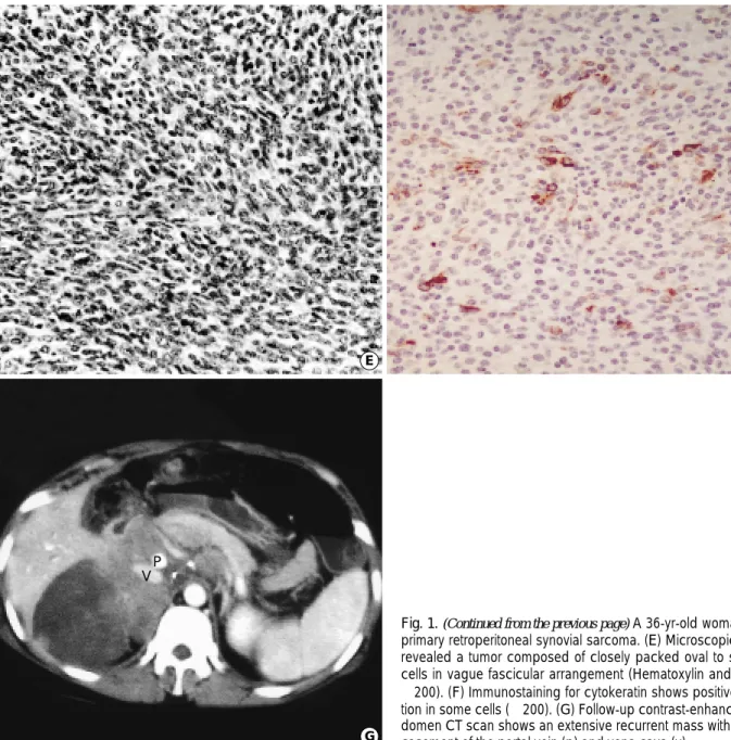
Histologic examination demonstrated a monophasic fibrous pattern consisting of closely packed round to oval spindle- shaped cells with hyperchromatic nuclei and small amount of eosinophilic cytoplasm without evidence of epithelial dif- ferentiation (Fig. 1E). Immunohistochemically the tumor cells showed strong positivity for vimentin and focally weak positivity for cytokeratin (Fig. 1F), but were negative for epi- thelial membrane antigen and S-100 protein. Mitoses were frequently seen. Final diagnosis was a poorly differentiated primary retroperitoneal synovial sarcoma.
Five months from the operation, she was readmitted due to low back pain and jaundice. Follow-up CT scan showed extensive tumor recurrence at the original tumor bed as well as liver metastasis (Fig. 1G).
Hotaek Song, Byung Hee Koh,
On Koo Cho, Hyunchul Rhim, Yongsoo Kim, Eun Kyung Hong*, Yong Wook Park�
Department of Diagnostic Radiology, College of Medicine, Hanyang University; Department of Pathology*, National Cancer Center, Koyang;
Department of Pathology�, College of Medicine, Hanyang University, Seoul, Korea
Address for correspondence Byung Hee Koh, M.D.
Department of Diagnostic Radiology, Hanyang University Hospital, 17 Haengdang-dong, Sungdong-gu, Seoul 133-792, Korea Tel : +82.2-2290-9159, Fax : +82.2-2293-2111 E-mail : [email protected]
419 J Korean Med Sci 2002; 17: 419-22
ISSN 1011-8934
Copyright � The Korean Academy of Medical Sciences
Primary Retroperitoneal Synovial Sarcoma
: A Case Report
A case of a 36-yr-old woman with retroperitoneal synovial sarcoma is described.
Her presenting symptom was epigastric pain that radiating to the back. On radi- ologic study, bulky retropancreatic soft tissue mass was detected which showed cystic and solid components. At operation, complete resection of the tumor was not possible because of the adhesion to the vena cava and the liver. During the follow-up, extensive tumor recurrence and liver metastasis were revealed. Prima- ry retroperitoneal synovial sarcoma is a very rare malignant tumor with high mor- tality and recurrence rates. Retroperitoneal synovial sarcoma usually appears as a nonspecific soft tissue mass that do not have specific imaging features dif- ferentiating it from other mesenchymal tumors. However general radiologic find- ings and anatomic location of the tumor may help the diagnosis. In addition, syn- ovial sarcoma should be included in the differential diagnosis of retroperitoneal soft tissue mass detected in young adults.
Key Words : Tomography, X-ray Computed; Retroperitoneal Neoplasms; Sarcoma, Synovial
Received : 2 March 2001 Accepted : 3 August 2001
420 H. Song, B.H. Koh, O.K. Cho, et al.
DISCUSSION
The exact nature of differentiation in synovial sarcoma is controversial. Potential origins of the synovial sarcoma include normal synovium, arthrogenous mesenchyma, and primitive pluripotent mesenchyma. Usually synovial sarcoma occurs in the vicinity of the joint capsules, bursae, and tendon sheaths.
But the occurrences of the tumor in various extraarticular sites such as the mediastinum, abdominal wall, retroperito-
neum, intraperitoneum, and esophagus have been reported (2-4). Synovial sarcoma is most prevalent in adolescents and young adults between 15 and 40 yr of age.
A primary retroperitoneal sarcoma has been defined as a tu- mor arising in the retroperitoneal space with an origin of me- sodermal structures exclusive of bony, renal, visceral, adrenal, and pancreatic tissues (6).
Retroperitoneal synovial sarcoma usually appears as a non- specific soft tissue mass that does not have specific imaging
Fig. 1.A 36-yr-old woman with primary retroperitoneal synovial sarcoma. (A) Transverse scan of ultrasonogram at the level of celiac axis shows a lobulated solid and cystic mass displacing the pancreas (anteriorly arrows). (B) Delayed contrast-enhanced CT scan at the level of celiac axis shows a low-attenuating mass (arrows) with anterior displacement of pancreas (p) and lateral displacement vena cava (v). Obliteration of fat plane between mass and vena cava is noted. (C) Duodenography shows widening of duodenal C-loop. (D) The cut surface of the lobulated mass shows a grayish tan, fish flesh appearance with hemorrhage (Fig. 1 continued next).
A B
C D
V P
Primary Retroperitoneal Synovial Sarcoma 421
features differentiating it from other mesenchymal tumors (2, 3, 8). However radiologic findings and anatomic location of the tumor may help the diagnosis. In addition, synovial sarcoma should be considered when retroperitoneal soft tis- sue mass is found in young adults. On CT, these tumors are hypo-dense and may show an irregular enhancement in the periphery with a poor enhancement in the central area, reflect- ing the necrotic, cystic, and hemorrhagic changes (8). In the region of 30% of cases show intratumoral calcification and extensive calcification suggest a favorable prognosis (3, 8). CT is still recommended as the best imaging method for assess- ing the local extent of the primary tumor and is a useful tool in the planning of appropriate therapy as well as evaluation of tumor response to ongoing treatment (8).
Histologically there are two types of synovial sarcomas:
biphasic and monophasic. Biphasic type is an admixture of epithelial cells and spindle cells. Monophasic type is com- posed of either only epithelial cells or spindle cells (1-4). Both types have a mortality rate of 40%. The known poor pro- gnostic factors are frequent mitotic figures (more than 10 or 15 mitoses per 10 HPF) and extensive tumor necrosis, and least favorable for the poorly differentiated (small cell) type.
Favorable factors are young age of the patient (15 yr or youn- ger) and tumor size smaller than 5 cm, and distal rather than proximal location in the extremities (5). Pathologically, major differential diagnosis of monophasic fibrous synovial sarcoma includes hemangiopericytoma, malignant peripheral nerve sheath tumor (MPNST, malignant schwannoma), and fibro-
Fig. 1.(Continued from the previous page) A 36-yr-old woman with primary retroperitoneal synovial sarcoma. (E) Microscopic study revealed a tumor composed of closely packed oval to spindle cells in vague fascicular arrangement (Hematoxylin and Eosin.
×200). (F) Immunostaining for cytokeratin shows positive reac- tion in some cells (×200). (G) Follow-up contrast-enhanced ab- domen CT scan shows an extensive recurrent mass with an en- casement of the portal vein (p) and vena cava (v).
E F
G V
P
422 H. Song, B.H. Koh, O.K. Cho, et al.
sarcoma. Hemangiopericytoma shows abundant vasculature, with no spindle cell and negativity for cytokeratin. MPNST may show close resemblance to the monophasic fibrous type of synovial sarcoma, but there is no evidence that synovial sarcoma arises in a large nerve or in a patient with neurofi- bromatosis. Immunohistochemically MPNST shows focal weak positivity for S-100 and negativity for cytokeratin. Fi- brosarcoma composed of long spindle cells and shows arrange- ment of the fibroblasts in distinct intersecting fascicles (her- ringbone pattern) (7).
Surgical ablation remains the mainstay of management of retroperitoneal sarcomas, but complete resection rate of is ap- proximately 50% (6). The recurrence rate ranged from 28%
to 36% even with adequate surgical and adjunctive therapies (3).
REFERENCES
1. Cotran RS, Kumar V, Robbins SL. Pathologic basis of disease, 5th ed.
Philadelphia: W.B. Saunders 1994: 1261-9.
2. Shmookler BM. Retroperitoneal synovial sarcoma. A report of four cases. Am J Clin Pathol 1982; 77: 669-73.
3. Ko SF, Chou FF, Huang CH, Ng SH, Wan YL, Lee TY, Lin JW, Chen WJ. Primary synovial sarcoma of the gastrocolic ligament. Br J Radiol 1998; 71: 438-40.
4. Fetsch JF, Meis JM. Synovial sarcoma of the abdominal wall. Can- cer 1993; 72: 469-77.
5. Kransdort MJ. Malignant soft-tissue tumors in a large referral popu- lation: distribution of diagnoses by age, sex, and location. Am J Roent- genol 1995; 164: 129-34.
6. Ziarn BH, Makley JT, Carter JR. Primary retroperitoneal sarcomas:
common symptoms, common diagnoses, uncommon disease. Clin Orthop 1996; 331: 277-82.
7. Enzinger FM, Weiss SW. Soft tissue tumors, 3rd ed. St. Louis: Mosby 1995: 759-86.
8. Israels SJ, Chan HS, Daneman A, Weitzman SS. Synovial sarcoma in childhood. Am J Roentgenol 1984; 142: 803-6.