pISSN 2288-9272 eISSN 2383-8493 J Oral Med Pain 2016;41(2):61-71 http://dx.doi.org/10.14476/jomp.2016.41.2.61
Comparison between the Subjective Evaluation and the Objective Evaluation of the Effect of Pain Control in the Masticatory Muscle Pain
Dong-Keun Kim, Chi-Hyuk Ahn, Mi-Jin Hwang, Yeon-Hee Lee, Soo-Kyung Kang, Q-Schick Auh, Jung-Pyo Hong, Yang-Hyun Chun
Department of Orofacial Pain and Oral Medicine, Kyung Hee University School of Dentistry, Seoul, Korea
Received May 4, 2016 Revised May 24, 2016 Accepted June 9, 2016
Purpose: This study was designed to evaluate the comparison between the subjective and the objective evaluation of pain control effect in masticatory muscle pain depending on time and dose change.
Methods: The patients were recruited to this study and diagnosed according to the Research Diagnostic Criteria for Temporomandibular Disorders (RDC/TMD). Experimental group were di- vided into three groups; saline injection group (n=10), morphine 1.5 mg injection group (n=10), and morphine 3.0 mg injection group (n=10). Evaluation list was the subjective pain evalua- tion (visual analogue scale, McGill pain questionnaire) and the objective pain evaluation (pres- sure pain threshold [PPT], pressure pain tolerance [PTO]). The subjective and the objective pain evaluation were performed at the times of just before injection, 10 minutes, 30 minutes, 1 hour, 24 hours, and 48 hours after injection. Then, data were statistically analyzed.
Results: The results were as follows: 1) There is no statistically significant difference be- tween the results of the subjective and the objective pain evaluation with regard to the short- term (within 1 hour) analgesic effect of morphine sulfate. 2) However, after 1 hour of injec- tion, while the subjective pain evaluation score still decreased, the objective pain evaluation didn’t show significant changes in PPT and PTO (1 hour, p<0.05; 24 hours, p<0.01; 48 hours, p<0.001). 3) In comparison to changes in the dose, the McGill pain questionnaire was the most statistically effective method among the subjective pain evaluations (1.5 mg, p<0.05; 3 mg, p<0.01).
Conclusions: Therefore, it was revealed that the subjective pain evaluation was more effective to evaluate long-term pain control, and that the McGill pain questionnaire could be an effec- tive way to evaluate pain control depending on dose changes. It requires further investigations with time and dose extension.
Key Words: Masticatory muscle pain; Morphine; Objective pain evaluation; Subjective pain evaluation
Correspondence to:
Yang-Hyun Chun
Department of Orofacial Pain and Oral Medicine, Kyung Hee University School of Dentistry, 26 Kyungheedae-ro, Dongdaemun-gu, Seoul 02447, Korea Tel: +82-2-958-9443
Fax: +82-2-958-9478 E-mail: [email protected]
JOMP
Journal of Oral Medicine and PainCopyright Ⓒ 2016 Korean Academy of Orofacial Pain and Oral Medicine. All rights reserved.
CC This is an open-access article distributed under the terms of the Creative Commons Attribution Non-Commercial License (http://creativecommons.org/licenses/by-nc/4.0/),
INTRODUCTION
Pain is a more or less localized sensation of discomfort, distress, or agony, resulting from the stimulation of special- ized nerve endings by the medical dictionary definition.
1)It serves as a protective mechanism insofar as it induces the sufferer to remove or withdraw from the source. And pain
is an unpleasant sensory and emotional experience associ- ated with actual or potential tissue damage or described in terms of such damage by the International Association for the Study of Pain (IASP) definition of pain.
2)The subjective experience of pain arises by four dis-
tinct processes which consist of transduction, transmission,
modulation and perception.
3)Transduction is the process
by which noxious stimuli lead to electrical activity in the appropriate sensory nerve endings. Transmission refers to the neural events that carry the nociceptive input into the Central Nervous System (CNS) for proper processing.
Modulation refers to the ability of the CNS to control the pain-transmitting neurons. And if nociceptive input reaches the cortex, perception occurs; this immediately initiates a complex interaction among neurons in the higher center of the brain.
Four terms related with pain—nociception, pain, suffering, and pain behavior—are differently used.
3)Nociception refers to the noxious stimulus originating from the sensory recep- tor. Pain is an unpleasant sensation perceived in the cortex, usually as a result of incoming nociceptive input. The term suffering refers to still another phenomenon: how the hu- man reacts to the perception of pain. Pain behavior refers to the individual’s audible and visible actions that communi- cate his suffering to others.
The patient subjectively feels and objectively expresses pain. Difference between the subjective and the objective pain evaluations generally exists. It therefore makes clini- cians difficult to exactly select and decide contents, dura- tion and the final goal of treatment. Such difference also can be varied even depending on the duration of pain con- trol, types of drug and dose change.
This study was designed to evaluate the comparison be- tween the subjective and the objective evaluation of pain control in masticatory muscle pain with time and by dose change.
MATERIALS AND METHODS
1. Subjects
The subjects participated in this study were volunteers among outpatients of Department of Oral Medicine, Kyung Hee University Dental Hospital (Seoul, Korea) during the pe- riod from July to August in 2014. This study was conduct- ed in Department of Oral Medicine, Kyung Hee University Dental Hospital after receiving approval from Institutional Review Board of Kyung Hee University Dental Hospital.
The participants were informed about the details of this experiment and signed the consent paper after reading it.
All participants are limited that visual analogue scale (VAS)
is over 50 and ages between 20 to 55 years who were di- agnosed to axis I; group Ia, myofascial pain according to the Research Diagnostic Criteria for Temporomandibular Disorders (RDC/TMD).
The subjects who had systemic musculoskeletal pain, systemic arthritis, malignant tumor, hypertension, diabe- tes mellitus, and cardiovascular disease were excluded.
Pregnant women and chronic analgesic or psychiatric drug users were also excluded.
2. Methods
One researcher randomly divided the subjects into three groups with 10 people each who had diagnosed according to the RDC/TMD. Another researcher injected 0.2 mL drug with 27 G subcutaneous needle and 1.0 mL disposable sy- ringe (Sofjec; Hwajin Medical, Cheonan, Korea) into each subject for 10 seconds, and injection point was the most painful area on unilateral masticatory muscle in palpation.
The other researcher randomly ordered the sequence of injected drugs. As a result, double blind procedure about the injected drug was performed by both researchers and subjects.
Initially classified subject groups were morphine sulfate (15 mg/1 mL; BC World Pharm, Seoul, Korea) 3.0 mg injec- tion group (n=10), morphine sulfate (15 mg/1 mL; BC World Pharm) 1.5 mg injection group (n=10), and saline (NaCl 9 mg/1 mL; JW Pharmaceutical, Seoul, Korea) injection group (n=10).
Each group was evaluated for both subjective and objec- tive pain at just before the injection, 10 minutes, 30 min- utes, 1 hour, 24 hours, and 48 hours after the injection.
3. Pain Evaluation
1) Subjective pain evaluation
The methods used to evaluate the subjective pain in this study were VAS and McGill pain questionnaire (MPQ) test.
For the VAS test, subjects were asked to mark their pain with marking pen (namepen; Monami, Yongin, Korea) on 100 mm straight line according to pain extent; start point of 100 mm straight line with no pain, end point with the strongest pain that they could imagine. The result was con- verted into numbers according to the percentage.
For the MPQ method, the MPQ in Korean version was
applied to evaluate the patient’s pain. The patient’s subjec- tive pain was digitalized and the data of the questionnaire was calculated by average of total score, then the values were recorded and calculated for the VAS and the MPQ.
2) Objective pain evaluation
The methods used to evaluate the objective pain were pressure pain threshold (PPT) test and pressure pain toler- ance (PTO) test.
The PPT test used in this study was to estimate the PPT around the most painful masticatory area before and after the injection with the pressure pain measuring instrument (Wagner Instruments, Greenwich, CT, USA), and then the estimation of pressure pain was converted into number.
The PTO was applied to the same area and used the same pressure pain measuring instrument to calculate the pain limit of same pressure, and its estimation was also convert- ed into number.
We kept the patient’s masticatory system as relaxed po- sition as possible without tooth contact to use the pressure pain measuring instrument. The pressure was applied to muscle vertically with 11 mm diameter probe with 30 kPa/s velocity; the measured kgf value was divided by area of the probe and converted to kPa value. The subjects were asked to raise their left hand at the moment that they felt the first pain and intolerable pain, and then the values were record- ed and calculated for the PPT and the PTO.
4. Statistical Analysis
The results from two subjective pain evaluation tests and two objective pain evaluation tests were analyzed by the repeated measures ANOVA (analysis of variance) test using descriptive statistics and Greenhouse-Geisser method for the short-term pain evaluation and one-way ANOVA test and Dunnett’s multiple comparison test for the long-term objec- tive pain evaluation. At first, we verified the effects; within subject effects, between subject effects and interaction ef- fects. When there were effects, we tried post hoc through multiple comparisons using Tukey HSD (honestly signifi- cant differences) method.
All the statistics significance level were p<0.05 and study scores were analyzed with PASW Statistics version 18.0 (IBM Co., Armonk, NY, USA).
RESULTS
1. Short-term Pain Evaluation
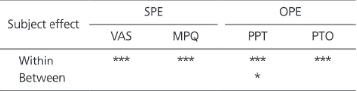
The p-value summary of post hoc multiple comparisons by Tukey HSD in the short-term subjective pain evalua- tion and the objective pain evaluation was in Table 1. VAS (p<0.001) and MPQ (p<0.001) in the short-term subjec- tive pain evaluation were significantly different and PPT (p<0.001) and PTO (p<0.001) in the short-term objective pain evaluation were significantly different.
Therefore, the difference between the subjective pain evaluation and the objective pain evaluation were no sta- tistically significant in the short-term pain evaluation with time.
2. Long-Term Subjective Pain Evaluation
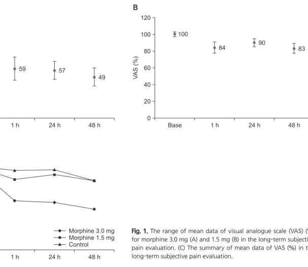
The mean data of VAS (%) for morphine 3.0 mg injec- tion group in the long-term subjective pain evaluation were shown in Fig. 1A. Those for morphine 1.5 mg injection group in the long-term subjective pain evaluation were in Fig. 1B. The summary of mean data of VAS (%) in the long- term subjective pain evaluation was in Fig. 1C.
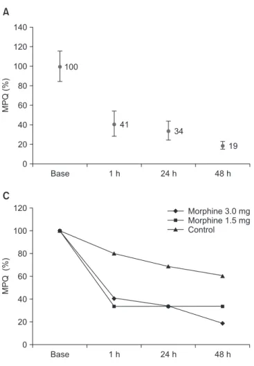
The mean data of MPQ (%) for morphine 3.0 mg injec- tion group in the long-term subjective pain evaluation were shown in Fig. 2A. The mean data of MPQ (%) for morphine 1.5 mg injection group in the long-term subjective pain evaluation were in Fig. 2B. The summary of mean data of MPQ (%) in the long-term subjective pain evaluation was in Fig. 2C.
The p-value summary by one-way ANOVA test and Dunnett’s multiple comparison test in the long term subjec- tive pain evaluation was in Table 2.
Table 1. The p-value summary in the short-term SPE and OPE
aSubject effect SPE OPE
VAS MPQ PPT PTO
Within *** *** *** ***
Between *
SPE, subjective pain evaluation; OPE, objective pain evaluation; VAS, visual analogue scale; MPQ, McGill pain questionnaire; PPT, pressure pain threshold; PTO, pressure pain tolerance.
a
Post hoc multiple comparisons by Tukey honestly significant differences (HSD).
*p<0.05, ***p<0.001.
The morphine 3.0 mg injection group was more signifi- cantly different than the morphine 1.5 mg injection group in 1 hour (p<0.05), 24 hours (p<0.01), and 48 hours (p<0.01) in VAS. Twenty-four hours after the injection (p<0.01) and 48 hours after the injection (p<0.01) were more significant- ly different than 1 hour after the injection (p<0.05) in the morphine 3.0 mg injection group.
The morphine 3.0 mg injection group was more sig- nificantly different than the morphine 1.5 mg injection group in 24 hours (p<0.01) and 48 hours (p<0.001) in MPQ. Twenty-four hours after the injection (p<0.01) were more significantly different then 1 hour after the injection (p<0.05). Forty-eight hours after the injection (p<0.001) were more significantly different than 24 hours after the in- jection (p<0.01) in the morphine 3.0 mg injection group.
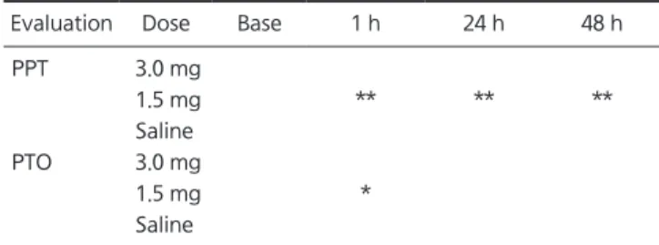
3. Long-Term Objective Pain Evaluation
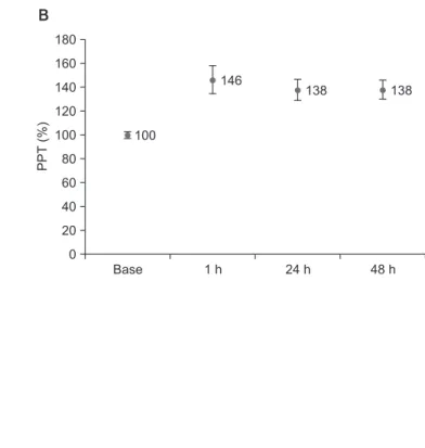
The mean data of PPT (%) for morphine 3.0 mg injec- tion group in the long-term objective pain evaluation were in Fig. 3A. The mean data of PPT (%) for morphine 1.5 mg
injection group in the long-term objective pain evaluation were in Fig. 3B. The summary of mean data of PPT (%) in the long-term objective pain evaluation was in Fig. 3C.
The mean data of PTO (%) for morphine 3.0 mg injec- tion group in the long term objective pain evaluation were in Fig. 4A. The mean data of PTO (%) for morphine 1.5 mg injection group in the long term objective pain evaluation were in Fig. 4B. The summary of mean data for conversion to PTO (%) in the long term objective pain evaluation was in Fig. 4C.
The p-value summary by one-way ANOVA test and Dunnett’s multiple comparison test in the long-term objec- tive pain evaluation were in Table 3.
The morphine 1.5 mg injection group was more signifi- cantly different than the morphine 3.0 mg injection group in 1 hour (p<0.01), 24 hours (p<0.01), and 48 hours (p<0.01) in PPT. The morphine 1.5 mg injection group was more significantly different than the morphine 3.0 mg injection group in 1 hour (p<0.05) in PTO.
120
100
80
60
40
20
0
Base 1 h 24 h 48 h
100
59 57
49
120
100
80
60
40
20
0
Base 1 h 24 h 48 h
100
84
90
83
120
100
80
60
40
20
0
Base 1 h 24 h 48 h
Morphine 3.0 mg 1.5 mg Control
Morphine
A
VAS(%)
B
VAS(%)
C
VAS(%)
Fig. 1. The range of mean data of visual analogue scale (VAS) (%)
for morphine 3.0 mg (A) and 1.5 mg (B) in the long-term subjective
pain evaluation. (C) The summary of mean data of VAS (%) in the
long-term subjective pain evaluation.
DISCUSSION
Pains are perceived after activation of first afferent nerves by peripheral stimulations generating electrical impulses that transmitted to central nervous system through serial neuro-pathway. Pain-developing stimulations, such as tis- sue damages or inflammation, release pain neurotransmit- ters. These neurotransmitters activate receptors located on
cell membrane, which cause excitatory action potential.
Peripheral pain receptors play an important role in initiat- ing pain pathway. Thus, wide and varied studies on periph- eral tissue are underway so as to regulate pain by modulat- ing peripheral pain receptors. A typical drug to control pain with its analgesic effect is opioid. Opioids are still the most powerful drugs for severe pain but their use is hampered by side effects such as respiratory depression, nausea, consti- pation, addiction and tolerance.
4)However, since peripher- ally-acting opioid agonists discovered, peripheral analgesic effect of opioid without central side effects has been an- ticipated.
5,6)Opioid receptors are synthesized at dorsal root ganglion and migrate along the neuronal axon to peripher- al and central nerve terminals.
7-9)It has an analgesic effect on post-operative pain or chronic pain.
10)Especially, its pe- ripheral analgesic effect is very strong under inflammatory state.
11)There are studies of applying opioid during oral sur- gery so that it can reduce post-operative pain.
12)Morphine sulfate (5 mg or 10 mg) was applied in temporomandibular
A B
MPQ(%)
C
140
100 80 60 40 20 0
Base 1 h 24 h 48 h
100
41
34
19 120
140
100 80 60 40 20 0
Base 1 h 24 h 48 h
100
34 34
120
34
120
100
80
60
40
20
0
Base 1 h 24 h 48 h
Morphine 3.0 mg 1.5 mg Control
Morphine
MPQ(%)MPQ(%)
Fig. 2. The range of mean data of McGill pain questionnaire (MPQ) (%) for morphine 3.0 mg (A) and 1.5 mg (B) in the long-term subjective pain evaluation. (C) The summary of mean data of MPQ (%) in the long-term subjective pain evaluation.
Table 2. The p-value summary in the long-term subjective pain evaluation
aEvaluation Dose Base 1 h 24 h 48 h
VAS 3.0 mg * ** **
1.5 mg Saline
MPQ 3.0 mg * ** ***
1.5 mg * * *
Saline
VAS, visual analogue scale; MPQ, McGill pain questionnaire.
a
One-way ANOVA test and Dunnett’ s multiple comparison test.
*p<0.05, **p<0.01, ***p<0.001.
joint (TMJ) to reduce pain.
13)The study of Chun et al.,
14)which was about the influence of terminal AMPA receptor to the muscle nociception and c-fos activation, proposed that GluR1 and GluR2, the AMPA receptor subunits, were developed in the trigeminal gan- glion neurons and masseter afferent nerve cell bodies. As a result, acute muscle pain is partially mediated by AMPA re- ceptors located in the terminal, and when several terminal glutamate receptor subunits are blocked it might reduce the muscle pain and central nerve activation more effectively.
In the study of Ro et al.,
15)the animal model of hypertonic saline (HS) infusion protocol causes peripheral release of glutamate, and that blockade of peripheral NMDA receptors significantly reduces HS-induced nociceptive behavior and central neuronal activation.
Chun and Ro
16)suggested that intramuscular capsaicin in rat masseter muscle significantly induced increase of tri- geminal caudalis (Vc) neuron response and that the block- ade of peripherally localized mGluR5 could effectively
attenuate muscular hypersensitivity.
The study of Yoo et al.
17)was to evaluate the pain con- trol by morphine injection to patient’s masticatory mus- cle. For this study, patients with masticatory muscle pain were recruited and diagnosed according to the RDC/TMD.
Experimental group were divided into three groups; saline injection group (n=10), lidocaine injection group (n=10), and morphine injection group (n=10). Evaluation list was the subjective pain evaluation (VAS, MPQ, and pain draw- ing) and the objective pain evaluation (PPT and PTO) and evaluation time were 0, 10, 30, 60 minutes after injection, and then data were statistically analyzed. In this study, the subjective pain evaluation and the objective pain evalua- tion were significantly different within group (p<0.001). The subjective pain drawing evaluation (p<0.001) were signifi- cantly different between groups. The objective PPT evalua- tion (p=0.025) were significantly different between groups.
The morphine injection group (p=0.001) were more signifi- cantly different than the saline injection group in the pain
160
100 80 60 40 20 0
Base 1 h 24 h 48 h
100
127
111
120 116
140
180
100 80 60 40 20 0
Base 1 h 24 h 48 h
100
146
138 120
140 138 160
160
100 80 60 40 20 0
Base 1 h 24 h 48 h
140 120
A B
C
PPT(%)
Morphine 3.0 mg 1.5 mg Control
Morphine
PPT(%) PPT(%)
Fig. 3. The range of mean data of pressure pain threshold (PPT) (%)
for morphine 3.0 mg (A) and 1.5 mg (B) in the long-term objective
pain evaluation. (C) The summary of mean data of PPT (%) in the
long-term objective pain evaluation.
drawing evaluation. Therefore, it was considered that the morphine injection was effective to control masticatory muscle pain within 60 minutes.
The study of Ko et al.
18)was to evaluate the role of peri pheral opioid receptors in masticatory muscle pain control. For this study, patients with masticatory muscle pain were recruited and diagnosed according to the RDC/TMD. Experimental group were divided into four groups; saline injection group
(n=10), lidocaine injection group (n=10), morphine 1.5 mg injection group (n=10), and morphine 3 mg injection group (n=10). Evaluation was performed by the subjective pain evaluation (VAS, MPQ, and pain drawing) and the objective pain evaluation (PPT and PTO), and evaluation time was 0, 1, 24, 48 hours after injection. Data were statistically ana- lyzed. The results were as follows: The subjective pain eval- uation were significantly different in morphine 3 mg group at 48 hours after injection (VAS, p<0.01; MPQ, p<0.001;
pain drawing, p<0.05). The objective pain evaluation were significantly different in morphine 1.5 mg group at 1 hour after injection (PPT, p<0.01; PTO, p<0.05). The morphine 3 mg group were more significantly different than lidocaine group and morphine 1.5 mg group in the MPQ evalua- tion (1 hour, p<0.01; 24 hours, p<0.01; 48 hours, p<0.001).
Therefore, it was revealed that the morphine 3 mg injection was effective to control masticatory muscle pain within 48 hours and more effective than lidocaine injection.
The study of Bae et al.
19)was to evaluate the sex differences
100
80
60
40
20
0
Base 1 h 24 h 48 h
100
111
96 120
104
100 80 60 40 20 0
Base 1 h 24 h 48 h
100
125 121
160
119 140
120
100 80 60 40 20 0
Base 1 h 24 h 48 h
140 120
A B
PTO(%)
C
PTO(%)PTO(%)
Morphine 3.0 mg 1.5 mg Control
Morphine
Fig. 4. (A) The range of mean data of pressure pain tolerance (PTO) (%) for morphine 3.0 mg (A) and 1.5 mg (B) in the long-term objective pain evaluation. (C) The summary of mean data of PTO (%) in the long-term objective pain evaluation.
Table 3. The p-value summary in the long-term objective pain evaluation
aEvaluation Dose Base 1 h 24 h 48 h
PPT 3.0 mg
1.5 mg ** ** **
Saline
PTO 3.0 mg
1.5 mg *
Saline
PPT, pressure pain threshold; PTO, pressure pain tolerance.
a