Endobronchial carcinoid tumor is the most com- mon malignant pulmonary tumor of the childhood tumors, accounting for 50 to 80% of childhood tumors. Endobronchial carcinoid tumor is generally accompanied by symptoms such as cough, hemopt- ysis, wheezing, weight loss, dyspnea, and chest pain, as well as by radiological symptoms including ate- lectasis, sclerosing lesion, infiltration, and lump patterns. This case report is about a patient whose asthma had been treated for several years but was not properly regulated, and who showed a unilateral overinflation in the chest x-ray that was diagnosed as an endobronchial carcinoid tumor after addi-
tional tests.
CASE
A 14-year old, male, child patient who had been undergoing treatment for asthma in another hospi- tal for three years due to repeated coughing, wheez- ing, and dyspnea visited our institution. The patient was hospitalized due to the exacerbation of his coughing, wheezing, and dyspnea, and underwent treatment using a bronchodilator, steroids, and a leukotriene receptor antagonist. However, since the
Kosin Medical Journal 2017;32:221-226.
https://doi.org/10.7180/kmj.2017.32.2.221 KMJ
Case Report
A 14-year-old boy with endobronchial carcinoid tumor presenting with asthma
Yunmo Gu, Youngjin Hwang, Sungwon Kim
Department of Pediatrics, Busan St. Mary’s Hospital, Busan, Korea
Introduction: Bronchial carcinoid tumors seldom occur in children, sometimes mistaken for a minor disease and diagnosed slowly.
Materials and Methods: We report on a patient who diagnose tumors slowly because confused with asthma.
Results: This case describes a 14-year-old boy, presenting with asthma-like symptoms throughout 3 years.
He was treated as asthma but wax and wane. Chest x-ray showed an hyperlucent left lung, so we rechecked high resolution computed tomography (HRCT) for unilateral hyperinflation diseases diagnosis. It was found 1×1㎝ nodule in left main bronchus. We did bronchoscopy and discovered a round mass in the left bronchus, 2∼3㎝ away from carina. In the biopsy, it was bronchial carcinoid tumor, so we resected tumor.
Discussion: Because symptoms of bronchial carcinoid tumors are various, it can often be misdiagnosed firstly.
It is confused with asthma, pneumonia and foreign body. An additional examination were necessary when respiratory symptoms persist.
Key Words: Bronchial, Carcinoid, Tumor
Corresponding Author: Sung Won Kim. Department of Pediatrics, Busan St. Mary's Hospital, 25-14, Yongho-ro 232 beon-gil, Nam-gu, Busan 48575, Korea
Tel: +82-51-933-7981, Fax: +82-51-936-7531, E-mail: [email protected]
Received:
Revised:
Accepted:
Aug. 18, 2015 Aug. 28, 2015 Oct. 26, 2015
improvement of the symptoms through the treat- ment was slow, the peak expiratory flow rate (PEFR) was kept as low as 200 to 220 L/ml (normally higher than 325.6).
Allergy tests showed serum eosinophil count 190 /uL (reference value, 0 to 350 /uL), total IgE 75.57 U/ml (reference value, 0.5 to 393 U/ml), and eosino- phil cationic protein (ECP) 9.82 /uL (reference value, 0 to 15/uL), all of which were in the normal ranges.
The ImmunoCAP lab tests showed a high European mite level of 16.5 kUA/L (reference value, 0 to 3.5) and a high American mite level of 29.5 kUA/L (reference value, 0 to 3.5).
In the tuberculosis tests, the result of the tuber- culin test performed using purified protein de- rivatives (PPD) was less than 0.5 ㎜ (reference value, 1 cm or less). The sputum test result was negative.
There was neither weight loss nor contact with a tuberculosis patient, nor night sweats.
Before administration of a bronchodilator, the pulmonary function tests showed forced expiratory volume in 1second (FEV1) 52% (reference value, 80%
or higher), forced vital capacity (FVC) 66% (reference value, 80% or higher), and FEV1/FVC 0.73 (reference value, 0.8 or higher) with respiratory obstruction.
After the administration of a bronchodilator, the test results were FEV1 59%, FVC 70%, and FEV1/FVC 77, and the increase of FEV1 by 13% (reference value, 12% or higher) indicated airway hyper- reponsiveness (Fig. 1).
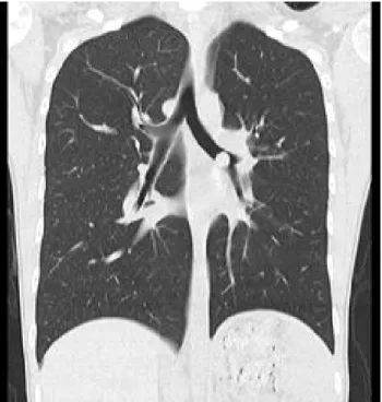
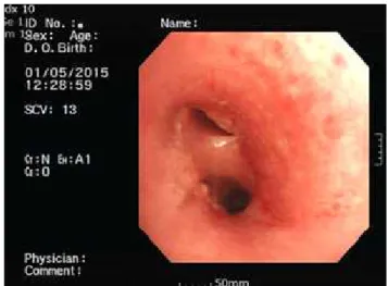
The chest x-ray photography showed a pulmo- nary overinflation in both lungs, and the radio- lucency of the left lung was significantly higher than that of the right lung (Fig. 2). To verify the obstruction of the upper bronchus suspected by the unilateral increase of the radiolucency, chest high resolution computed tomography (HRCT) was performed. The HRCT showed a nodule of 1×1 ㎝ in size which blocked the left main bronchus in a check valve type (Fig. 3). The bronchoscopy showed a round lump at the left main bronchus about 2 to 3 ㎝ from the tracheal bifurcation (Fig. 4). The biopsy verified a typical carcinoid tumor. Thus, a thor- acotomy was performed to solve the bronchial ob- Fig. 1. Spirometry shows obstructive lung disease with hypersensitivity of air way.
endobronchial carcinoid tumor presenting with asthma
struction by the lump (sleeve lobectomy and left bronchial terminal anastomosis) (Fig. 5).
The coughing and wheezing of the child patient were improved after the operation. The patient has been relatively healthy since his discharge, and has not undergone any treatment for asthma or pneumonia.
DISCUSSION
Primary pulmonary tumor is very rare in children.
Bronchial carcinoid tumor is the most common pri- mary pulmonary tumor in children, accounting for 50 to 80% of pulmonary tumors in children or adolescents.1 The ratio is about 1 to 2% in adults, but is particularly high among children during
puberty.2
The most common symptoms of bronchial carci- noid tumor are cough, hemoptysis, wheezing, weight loss, dyspnea, and chest pain.3 Most of the tumors are found in the proximal part of the respira- tory tract, where obstruction or hemorrhage is found as a symptom. Sometimes, cough, wheezing, he- moptysis, chest pain, or recurrent pneumonia may be repeatedly found in the same region.4
About 75% of bronchial carcinoid tumors can be found through chest x-ray radiography. The central lesion is a mucosal obstruction of atelectasis, while the peripheral lesion is mostly symptomless but may be found as an incidental solitary pulmonary nodule.5
Chest CT, which is the most useful method of diagnosis, shows the range and location of the tumor Fig. 2. Chest x-ray shows both lung hyperinflation with
Left lung hyperlucent lung field.
Fig. 3. High resolution computed tomography (HRCT) shows an endobronchial nodule in Left main brohcus (1 x 1 cm).
as well as the relation with the mediastinum.
Bronchial carcinoid tumor of the typical type is accompanied by a mediastinal adenopathy in 5 to 20% of cases.6 The case reported herein did not show a mediastinal adenopathy.
Bronchoscopy is implemented to confirm the di- agnosis, and biopsy is possible in 3/4 of the patients.
Biopsy is allowed in most of the patients because of the low risk of severe hemorrhage.7
Other diagnostic methods include magnetic reso- nance imaging, somatostatin receptor scintigraphy, and positron emission tomography, all of which are rarely employed as a practical diagnostic method.
Carcinoid tumors are caused in peptide-amine secreting neuroendocrine cells transported from the neural crest. Carcinoid tumors are mostly found in the digestive organs, followed in order of fre- quency by the lungs, thymus, and ovaries.8
The diagnostic rate was low in the past, but with the development of medical technologies such as
HRCT and endoscopy early diagnosis is now increasing. The patient of this case report had been undergoing treatment for asthma, but was diag- nosed with a bronchial carcinoid tumor through an auxiliary diagnostic instrument.
Bronchial carcinoid tumors are histologically classified into typical carcinoma, atypical carcino- ma, large cell neuroendocrine carcinoma, and small cell neuroendocrine carcinoma.9 The patient of this case report had the typical carcinoma. The typical carcinoma usually remains in Stage 1, while the atypical carcinoma often moves on to Stage 2 or further.
Differential diagnosis should be performed with diseases showing unilateral hyperlucent lung in chest x-ray radiography, most of which are found at the pulmonary parenchyma, respiratory tracts, pulmonary blood vessels, thorax, and mediastinum.
Representative diseases include Swayer-James or Macleod syndrome, congenital lobar emphysema, and pulmonary vascular abnormalities.
Fig. 4. Bronchoscopy before surgery shows a tumor obstructed left main bronchus from carina.
Fig. 5. Bronchoscopy after surgery shows bronchus resected a tumor.
endobronchial carcinoid tumor presenting with asthma
Swayer-James or Macleod syndrome, which is a postinfectious obliterative bronchiolitis following infection with adenovirus, respiratory syncytial vi- rus, or measles, causes abnormal growth of the lungs with unilateral hyperlucent lung. The disease may be accompanied by chronic cough, pneumonia, and wheezing.
Congenital lobar emphysema is often found with severe dyspnea as well as local pulmonary ob- struction in the neonatal period. This disease may be accompanied by various symptoms, from mild tachypnea to wheezing and severe dyspnea. The infiltrated pulmonary lobe loses the function due to the overinflation.
The pulmonary vascular abnormalities showing unilateral hyperlucent lung in chest x-ray radiog- raphy include unilateral pulmonary agenesis, anomalous origin of the left pulmonary artery, and Scimitar syndrome (pulmonary venolobar syn- drome). Unilateral pulmonary agenesis is found in the neonatal period with movement of the media- stinum due to the volume decrease of the infiltrated lung and overinflation of the opposite lung.
Anomalous origin of the left pulmonary artery may affect the right main bronchus by causing over- inflation of the right lung or atelectasis. Scimitar syndrome may cause overinflation of the left lung due to hypoplasia of the right lung.
Congenital cystic adenomatoid malformation, which is also a disease that should be differentiated, causes a harmartoma instead of normal pulmonary tissue. The disease may be diagnosed by prenatal ultrasonography, but if missed, hyperlucent lung
is found by chest x-ray radiography or CT at the part where a pulmonary tissue has not been formed.
The disease may be accompanied by dyspnea, re- peated pneumonia, or pneumothorax.10
REFERENCES
1. Lal DR, Clark I, Shalkow J, Downey RJ, Shorter NA, Klimstra DS, et al. Primary epithelial lung malignancies in the pediatric population. Pediatr Blood Cancer 2005;45:683-6.
2. Hauso O, Gustafsson BI, Kidd M, Waldum HL, Drozdov I, Chan AKC, et al. Neuroendocrine tu- mor epidemiology: contrasting Norway and North America. Cancer 2008;113:2655-64.
3. Wang LT, Wilkins EW Jr, Bode HH. Bronchial carcinoid tumors in pediatric patients. chest 1993;103:1426-8.
4. Eyssartier E, Ang P, Bonnemaison E, Gibertini I, Diot P, Carpentier E, et al. Characteristics of Endobronchial Primitive Tumors in Children.
Pediatric Pulmonol 2014;49:121-5.
5. Skuladottir H, Hirsch FR, Hansen HH, Olsen JH.
Pulmonary neuroendocrine tumors: incidence and prognosis of histological subtypes. A pop- ulation-based study in Denmark. Lung Cancer 2002;37:127-35.
6. Divisi D, Crisci R. Carcinoid tumors of the lung and multimodal therapy. Thorac Cardiovasc Surg 2005;53:168-72.
7. Fink G, Krelbaum T, Yellin A, Bendayan D, Saute M, Glazer M, et al. Pulmonary carcinoid: pre-
sentation, diagnosis, and outcome in 142 cases in Israel and review of 640 cases from the literature. Chest 2001;119:1647-51.
8. Kulke MH, Mayer RJ. Carcinoid tumors. N Engl J Med 1999;340:858-68.
9. Travis WB. The concept of pulmonary neuro- endocrine tumors. In: Travis WB, Brambilla E, Muller-Hermeli HK, Harris CC, editors. Pathology
and genetics of tumours of lung, pleura, thymus, and heart. Lyon: IARC Press; 2004. p.19-20.
10. Steven RB. Glenna BW. Emphysema and Overinflation. In: Robert MK, Bonita FS, Joseph WS, Nina FS, Richard EB, editors. Nelson Textbook of Pediatrics. 19th ed. Philadelphia:
Elsevier saunders; 2011. p.1460-1461.