성인에서 자기 공명 영상을 이용한 족관절 형태 계측
단국대학교 의과대학 정형외과학교실 유문집⋅김유진
Ankle Morphometry in Adults using MRI
Moon-Jib Yoo, M.D., You-Jin Kim, M.D.
Department of Orthopaedic Surgery, Dankook University College of Medicine, Cheonan, Korea
=Abstract=
Purpose: To provide basic data on anatomy of the ankle joint in adults using magnetic resonance imaging as well as to compare measurements with available data of corresponding dimension in the different sizes of the total ankle replacements design (HINTEGRAⓇ).
Materials and Methods: Magnetic resonance (MR) sagittal and coronal images from one hundred-forty nine were studied. Post traumatic, arthritic, or grossly deformed ankles at any reason were excluded. Lengths, widths of the main parts of this articulation and also the radius of curvature of talar dome were measured in the MR images.
Statistical analysis was performed on these measurements.
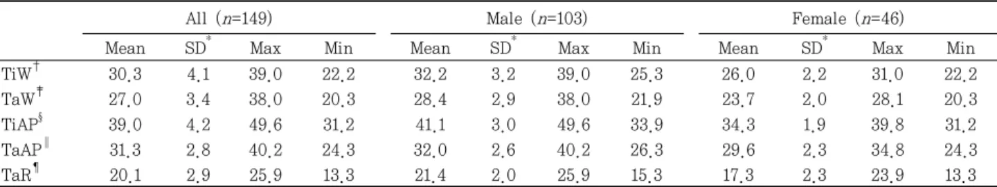
Results: On MR coronal image data, average tibial width was 30.0±4.1 mm and talar width was 27.0±3.4 mm. On sagittal image date, average anteroposterior length of the distal tibia was 39.0±4.2 mm, average anteroposterior length of the talus was 31.3±2.8 mm, and average sagittal radius of curvature of talar dome was 20.1±2.9 mm. All measurements in male were significantly larger than in female (p<0.01), but a significant difference according to age was not found.
Conclusion: Ankle morphometric data define a basis of size, particularly useful in the design of ankle prostheses.
Key Words: Ankle, Morphometry, MRI
∙ Address for correspondence You-Jin Kim, M.D.
Department of Orthopaedic Surgery, Dankook University College of Medicine, 16-5 Anseo-dong, Cheonan-si, Choongnam, 330-715, Korea
Tel: +82-41-550-6612 Fax: +82-41-550-3950 E-mail: [email protected]
서 론
진행된 관절염의 치료에서 관절 기능의 보호와 동통 해소를 동시에 유지하기 위하여 시행하는 관절 치환술은 고
관절과 슬관절에서 성공적으로 많이 시행되고 있으나 족관 절에서는 시행 초기에 나온 나쁜 결과4)로 인하여 최근까지 널리 시행되지 못하였다. 족관절 전치환술의 실패는 주로 족관절의 안정성과 운동성의 부적절한 회복, 인대의 안정화 역할에 대한 인식의 부재 그리고 형태에 대한 이해 부족7)으 로 인한 것이었다. 그리하여 본 저자들은 자기 공명 영상을 통하여 족관절의 형태 계측을 시행하고 이를 인공 족관절 삽입물의 크기와 비교하기로 하였으며, 이러한 연구는 환자 에 따라 가장 적합한 삽입물을 선택하는 데 도움이 되며 한 국인 체격에 맞는 인공 족관절의 설계 및 제작을 위한 기초 자료를 제공할 수 있으리라 생각된다.
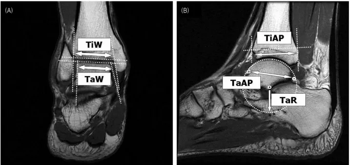
(A) (B)
Figure 1. Measurement of ankle morphometry. (A) In the coronal image, the tibial width (TiW) is the distance of the two intersections of the two lines that are drawn fitting the internal profiles of the two malleoli and the line that is drawn fitting the top of the tibial mortise.
The talar width (TaW) is the distance of the two intersections of the two lines that are drawn fitting the medial and lateral talar articular profiles and the line that is drawn fitting the top of talar articular profile. (B) In the sagittal image, the tibial thickness (TiAP) is the distance between the most anterior point of the tibial anterior profile and the corresponding point along the tibial anterio-posterior axis on the posterior tibial profile. The talar length (TaAP) is the length of the segment connecting to the posterior and the most anterior point of the trochlear tali segittal arc. The sagittal radius of the trochlear tali (TaR) arc is the radius of the circumference fitting the points of the trochear tali arc profile.
대상 및 방법
2002년 10월부터 2007년 3월까지 본원에 내원하여 족관 절 자기 공명 영상을 시행한 환자 중 족관절에 영향을 미칠 외상력이 있거나 관절염의 변화가 시작된 경우와 다른 어떤 원인에서든 변형이 있는 경우를 제외한 149예를 대상으로 하였다. 대상에 포함된 149명의 연령은 20세에서 78세까지 로 평균 40.1세였고, 남자가 103명, 여자가 46명이었다.
족관절의 형태 계측은 자기 공명 영상(1.5T signa ex- cite+, GE, USA, 2002)으로 얻은 이미지를 사용하였으며, 자기 공명 영상은 환자가 앙와위로 누운 상태에서 부하를 가하지 않고 족부의 중립위와 족관절을 15도 족저 굴곡시킨 상태에서 촬영하였다. 측정 방법은 족관절 자기 공명 영상의 시상면과 관상면에서 각각의 측정치를 0.1 mm 단위로 측정 하였다. 관상면에서는 경골의 폭(TiW)과 거골의 폭(TaW) 을 측정하였으며, 경골의 중간면을 지나는 관상면에서 내외 측 과의 안쪽 윤곽의 두 선과 경골 원위부 관절면의 선과 만 나는 두 점 사이의 길이를 구하여 경골의 폭으로 정의하였 고, 거골의 폭은 내외측 거골 관절면의 윤곽의 두 선과 거골 관절면 상단의 선이 만나는 두 점 사이의 길이를 구하여 그 측정치로 정의하였다. 시상면에서는 경골 전후 길이(TiAP)
와 거골 전후 길이(TaAP), 거골 반지름(TaR)을 측정하였 으며, 경골 원위부 관절 중간면을 지나는 시상면 사진을 이용하여 계측하였다. 경골 전후 길이는 경골의 전방에 있는 최대 전방점에서 경골 전후 축을 따라 경골 후방에 있는 점 사이의 거리로 정의하였으며, 거골 전후 길이와 거 골 반지름은 거골 활차 호(arc)의 최대 전방에서 최대 후방 까지의 거리와 거골 활차 호로 이루어지는 원의 반지름으로 각각 정의하였다(Fig. 1). 각 계측치의 평균과 표준편차, 최 대값, 최소값을 구하였으며 남녀간에 따른 족관절 형태 계측의 차이를 보기 위하여 남녀군으로 구분하여 비교하였 다. 연령에 따른 차이가 있는지를 보기 위하여 30세 미만과 30-39세, 40-49세, 50-59세, 60세 이상 군으로 나누어 계측치를 비교하였다. 통계학적 분석은 SPSS 10.0을 이용 하여 남성과 여성의 형태 차이가 있는지 여부를 알기 위해 student t-test를, 연령별 차이를 알기 위해 ANOVA test를 적용하였다.
결 과
자기 공명 영상에서의 족관절 형태 계측 결과는 경골의 폭이 30.0±4.1 mm, 거골의 폭이 27.0±3.4 mm, 경골 전후
Table 1. Mean, Standard Deviation, Maximum, Median Values (mm) for all 5 Measurements of the Ankle Joint
All (n=149) Male (n=103) Female (n=46)
Mean SD* Max Min Mean SD* Max Min Mean SD* Max Min
TiW† 30.3 4.1 39.0 22.2 32.2 3.2 39.0 25.3 26.0 2.2 31.0 22.2
TaW‡ 27.0 3.4 38.0 20.3 28.4 2.9 38.0 21.9 23.7 2.0 28.1 20.3
TiAP§ 39.0 4.2 49.6 31.2 41.1 3.0 49.6 33.9 34.3 1.9 39.8 31.2
TaAP∥ 31.3 2.8 40.2 24.3 32.0 2.6 40.2 26.3 29.6 2.3 34.8 24.3
TaR¶ 20.1 2.9 25.9 13.3 21.4 2.0 25.9 15.3 17.3 2.3 23.9 13.3
*SD, standard deviation; †TiW, the tibia width; ‡TaW, the talar width; §TiAP, the tibia thickness; ∥TaAP, the talar length;
¶TaR, the sagittal radius of the trochlear tail.
Table 2. Data Measurement (mm) according to Age
Age (years) TiW* TaW† TiAP‡ TaAP§ TaR∥
20-29 (n=40) 30.6±3.8 27.0±2.8 38.3±4.1 30.8±3.2 20.1±2.7
30-39 (n=35) 30.7±3.5 27.2±2.9 39.7±3.0 31.7±3.0 20.7±2.7
40-49 (n=37) 29.8±4.4 26.8±4.2 38.6±4.3 31.3±2.4 19.9±2.5
50-59 (n=25) 30.3±4.6 27.4±3.8 40.1±5.3 31.4±2.4 20.1±3.6
60- (n=12) 29.2±4.7 26.1±3.8 38.7±4.5 31.7±2.9 19.7±3.2
*TiW, the tibia width; †TaW, the talar width; ‡TiAP, the tibia thickness; §TaAP, the talar length; ∥TaR, the sagittal radius of the trochlear tail.
길이는 39.0±4.2 mm, 거골의 전후 길이는 31.3±2.8 mm 그리고 거골 반지름 20.1±2.9 mm이였다. 계측치는 성별과 연령에 따라서 넓은 범위의 계측치를 보였으며, 전체 149예 에서는 거골의 폭이 최대 계측치와 최소 계측치의 차이가 17.7 mm로 가장 넓은 큰 차이를 보였다. 남성의 경우도 역 시 거골의 폭이 가장 넓은 범위의 계측치를 보였다. 하지만, 여성의 경우는 거골 반지름이 최대 계측치 23.9 mm와 최소 계측치 13.3 mm로 가장 큰 차이를 보이는 것으로 나타났다.
성별에 따른 계측치의 차이는 계측치 모두에서 통계적으 로 남성에서 큰 수치를 보이는 것으로 나타났다(p<0.01) (Table 1).
본 연구의 대상 중에 20세에서 29세의 군은 40명, 30세 에서 39세의 군은 35명, 40세에서 49세의 군은 37명, 50세 에서 59세의 군은 25명 그리고 60세 이상의 군은 12명으로 일원 배치 분산 분석을 이용하여 각 연령군별 계측 값을 비 교한 결과에는 유의한 차이를 보이지 않았다(Table 2).
고 찰
족관절 병변의 고전적인 치료 방법인 족관절 유합술은 Alberth에 의해 1978년 처음 기술된 이후 진행된 족관절 질 환의 보편적인 치료법으로 알려져 있다1,8,13). 그러나 족관절 유합술의 문제점으로 족관절 운동 소실, 오랜 안정 가료로 인한 거골하 관절과 중거골 관절의 강직, 가관절증과 긴
유합 기간 등이 있다.
이러한 족관절 유합술과 더불어 족관절 관절염의 수술적 치료 방법인 족관절 전치환술은 1970년대 초반 처음으로 시도되었다. 그러나, 족관절 전치환술은 시행 초기에 많은 부작용등으로 인하여 족관절의 해부학적 특징과 더욱 유사 한 디자인이 개발되어 향상된 임상 결과를 보고하기 시작한 1980년 후반과 1990년대까지는 널리 시행되지 못하였고
2,3,6,9,14), 족관절 유합술이 족관절 관절염의 주된 수술적
치료로서 사용되었다. 초기 족관절 전치환술의 실패 요인 으로는 시멘트의 사용과 과도한 구속 또는 비구속 형태 (over or under constraint), 창상 치유, 삽입물의 해리, 족 관절 삼각인대 및 외측 인대의 중요성에 대한 인식의 부족 으로 인한 것이었다. 1세대 디자인의 단점을 보완하기 위 하여 개발되어진 2세대 디자인은 족배 굴곡과 족저 굴곡 뿐 아니라 전후방 또는 내외측 활주를 가능하게 하는 디자 인인 반구속 형태(semi-constrained)와 비시멘트 삽입물로 되어있으며, 대표적인 형태로는 Buechel-Pappas, STAR, Agility형이 있다.
2000년 현재 33가지의 서로 다른 인공 족관절이 있으며4), 이후 최소한 8개의 인공 족관절, 즉 Salto (Tornier SA, Saint Ismier, France), HINTEGRA (Newdeal SA, Lyon), Mobility (Depuy), Ramses (France MBA), TNK (Kyocera Corporation, Japan), OSG (Corin), AES (Biomet) and BOX (Finsbury Orthopaedics)이 개발되어 사용되고 있다.
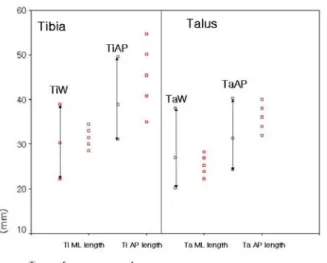
Figure 2. Diagram shows that values of the total ankle replacement design (HINTEGRA) superimposed to corresponding measurement from the present study. The sizes made available by replacement design are reported along the horizontal axis, for the tibial (Ti) and talar (Ta) components, and for the antero-posterior (AP), medio-lateral (ML) dimensions.
TNK형을 제외한 7개의 인공 족관절은 Buechel-Pappas 족관절에 의해 처음 사용되었던 세 부분으로 구성된 형태 (three part mobile bearing)를 사용하고 있다. 이 중 최 근 국내에 소개되어 많이 사용되고 있는 HINTEGRA형은 2000년 5월 처음 환자에게 치환되었으며, Hintermann 등5)은 HITEGRA 인공 족관절 122예의 평균 18.5개월 (1-3년) 추시에서 91.8%의 생존률을 보고하였다.
족관절 전치환술의 수술시 슬관절이나 고관절과 달리 관 절을 탈구시키지 않고 치환술을 시행하므로, 수술 시야가 제한되어 환자의 족관절에 비해 작은 대치물이 삽입되는 경 향이 있다. 또한 다양한 크기의 삽입물이 제작되지 않았기 때문에 개개인에게 가장 적합한 삽입물을 선택하는데 어려 움이 있으며, 삽입물로 덮히지 않은 부분에 이소성 골화 또는 골극이 발생하여 충돌에 의한 통증을 유발한다12). 그 러므로 인공 족관절 디자인 특히, 수술 기구와 삽입물의 크기는 매우 중요한데 이를 위해 다수 개체수에 대한 족관절 형태 계측치에 대한 통계학적 분석이 필요하다. 이에 본 저 자들은 자기 공명 영상을 통하여 성인의 족관절 형태 계측을 시행하였다. 본 연구 결과 경골의 폭은 30.0±4.1 mm, 거골 의 폭은 27.0±3.4 mm, 경골 전후 길이는 39.0±4.2 mm, 거골의 전후 길이는 31.3±2.8 mm 그리고 거골 반지름 20.1±2.9 mm이였다. Stagni 등11)은 기본적인 관상면과 시 상면 방사선 사진을 이용한 반자동 방사선학적 계측 방법10) 에 의한 족관절 형태 계측에서 경골의 폭은 31.9±3.5 mm, 거골의 폭은 30.4±3.3 mm, 경골의 최대 두께(maximal
tibial thickness)는 41.4±3.9 mm, 거골 활차 길이(tro- chlea tali length)는 41.7±4.4 mm 그리고 거골 활차 호의 반지름(sagittal radius of the trochlea tali arc)은 23.4±
3.1 mm으로 보고 하였다. 계측치의 정의에 대한 차이는 있 으나 이를 본 연구 결과와 비교하여 볼 때 본 연구에서 거골 의 전후 길이를 제외한 나머지의 계측치에서 2-3 mm 정도 작은 것으로 조사되었으며, 이는 서구인에 비해 작은 족관 절 형태를 나타내는 것으로 사료된다. 본 연구에서 측정한 거골의 전후 길이는 거골 활차 길이에 비해 10.1 mm의 차 이를 보였는데 이는 거골의 활차가 중앙에서 후방으로 들어 가는 형태를 보이므로 자기 공명 영상의 시상면에서 측정한 본 연구와 방사선 사진을 기본으로 하는 연구의 차이일 것 으로 사료된다.
본 저자들은 이 계측치와 현재 국내에 많이 사용되어 지고 있는 HINTEGRA 인공 족관절 삽입물의 규격마다 계측치와 상응하는 크기를 본 연구의 계측치와 비교하였 다(Fig. 2). 거골의 전후 길이와 경골의 전후 길이의 계측치 가 삽입물에 비해 작았는데, 이는 위에서 기술 한 바와 같이 자기 공명 영상의 시상면의 측정으로 인하여 실제 길이보다 저평가되었다는 것을 감안하더라도, 족관절 전치환술을 시행 할 경우 돌출이 발생할 수 있을 것이라 생각된다. 이에 본 저자들은 한국인의 족관절과의 적응도를 높이기 위해서 작은 전후 길이의 삽입물이 필요할 것으로 생각하였다.
본 연구의 제한점으로는 사다리꼴 모양의 원위 경골과 거골에 대해 전후 길이와 폭만의 이차원적인 계측을 하였던 점과 역학적인 연구 없이 단지 형태학적인 면만을 연구한 점 등이 있었으나, 본 연구를 통해 족관절 형태 계측치의 평균값과 분포도를 얻을 수 있었고, 부족하나마 기존 제품 과의 비교를 통해 삽입물의 적응도를 높이기 위하여 개선 해야 할 점을 연구함으로서 향후 한국형 인공 족관절 개발에 기초 자료를 제공할 수 있으리라 생각된다.
결 론
자기 공명 영상을 통한 족관절의 형태 계측에서 거골의 폭이 최대와 최소의 차가 17.7 mm로 가장 넓은 범위의 계측치를 보였으며, 남성의 경우 역시 거골의 폭이 여성의 경우에서는 거골의 반지름이 가장 큰 범위의 계측치를 보이 는 것으로 나타났다. 성별에 따른 계측치는 남성이 여성에 비해 유의하게 큰 것으로 나타났으며, 연령군에 따른 차이는 없었다.
REFERENCES
1. Bae DK, Kim KT and Kim JW: Total ankle arthroplasty -report of 6 cases-. J Korean Orthop Assoc, 27: 34-40, 1992.
2. Buechel FF Sr, Buechel FF Jr and Pappas MJ: Ten-year evaluation of cementless Buechel-Pappas meniscal bearing total ankle replacement. Foot Ankle Int, 24: 462-472, 2003.
3. Buechel FF Sr, Buechel FF Jr and Pappas MJ: Twenty-year evaluation of cementless mobile bearing total ankle re- placements. Clin Orthop Relat Res, 424: 19-26, 2004.
4. Giannini S, Leardini A and O’Connor JJ: Total ankle replacement: review of the designs and of the current status.
Foot Ankle Surg, 6: 77-88, 2000.
5. Hintermann B, Valderrabano V, Dereymaeker G and Dick W: The HINTEGRA ankle: rationale and short-term results of 122 consecutive ankles. Clin Orthop Relat Res, 424: 57- 68, 2004.
6. Knecht SI, Estin M, Callaghan JJ, et al: The Agility total ankle arthroplasty. Seven to 16-year follow-up. J Bone Joint Surg, 86-A: 1161-1171, 2004.
7. Morrey BF and Wiedeman GP: Complications and long term results of ankle arthrodeses following trauma. J Bone
Joint Surg, 62-A: 777-784, 1980.
8. Leardini A: Geometry and mechanics of the human ankle complex and ankle prosthesis design. Clin Biomech, 16:
706-709, 2001.
9. Pyevich MT, Saltzman CL, Callaghan JJ and Alvine FG:
Total ankle arthroplasty : a unique design. Two to 12-year follow-up. J Bone Joint Surg, 80-A: 1410-1420, 1998.
10. Stagni R, Leardini A, Catani F and Cappelllo A: A new semi-automated measurement technique based on X-ray pictures for ankle morphometry. J Biomech, 37: 1113-1118, 2004.
11. Stagni R, Leardini A, Ensini A and Cappello A: Ankle mor- phometry evaluated using a new semi-automated technique based on X-ray pictures. Clin Biomech, 20: 307-311, 2005.
12. Stamatis ED and Myerson MS: How to avoid specific complications of total ankle replacement. Foot Ankle Clin, 7: 765-789, 2002.
13. Stauffer RN and Segal NM: Total ankle arthroplasty: four years’ experience. Clin Orthop Relat Res, 160: 217-221, 1981.
14. Wood PL and Deakin S: Total ankle replacement. The results in 200 ankles. J Bone Joint Surg, 85-B: 334-341, 2003.