Letter to the Editor
Vol. 26 No. 5, 2014 659
Received April 3, 2013, Revised September 9, 2013, Accepted for publication October 13, 2013
Corresponding author: Sung-Yul Lee, Department of Dermatology, Soon Chun Hyang University Hospital Cheonan, 31 Suncheonhyang 6-gil, Dongnam-gu, Cheonan 330-721, Korea. Tel: 82-41-570-2270, Fax: 82-41-570-2783, E-mail: [email protected]
This is an Open Access article distributed under the terms of the Creative Commons Attribution Non-Commercial License (http://
creativecommons.org/licenses/by-nc/3.0) which permits unrestricted non-commercial use, distribution, and reproduction in any medium, provided the original work is properly cited.
effects3,4. The other advantages of using this pinhole technique with ablative lasers are that is easier to use and less expensive than the treatment modality involving pulsed dye laser, as ablative lasers are usually available in most dermatology clinics.
In conclusion, the pinhole method using the erbium : YAG laser could be beneficial for treating CALMs, which have been proven difficult to treat with other methods.
Furthermore, the pinhole method using the CO2 laser can be used an effective treatment alternative for telangiec- tasia.
ACKNOWLEDGMENT
We thank Eun Jin Yeon for editing the manuscript.
REFERENCES
1. Whang SW, Lee KY, Cho SB, Lee SJ, Kang JM, Kim YK, et al.
Burn scars treated by pinhole method using a carbon dioxide laser. J Dermatol 2006;33:869-872.
2. Cho SB, Lee SJ, Kang JM, Kim YK, Kim TY, Kim DH. The treatment of burn scar-induced contracture with the pinhole method and collagen induction therapy: a case report. J Eur Acad Dermatol Venereol 2008;22:513-514.
3. Yang JH, Han SS, Won CH, Chang SE, Lee MW, Choi JH, et al. Treatment of elastosis perforans serpiginosa with the pinhole method using a carbon dioxide laser. Dermatol Surg 2011;37:524-526.
4. Lee SM, Kim YJ, Chang SE. Pinhole carbon dioxide laser treatment of secondary anetoderma associated with juvenile xanthogranuloma. Dermatol Surg 2012;38:1741-1743.
http://dx.doi.org/10.5021/ad.2014.26.5.659
A Case of Primary Cutaneous Scar Infection Caused by Aspergillus niger
Euy-Hyun Chung, Sung-Yul Lee
Department of Dermatology, Soon Chun Hyang University College of Medicine, Cheonan, Korea
Dear Editor:
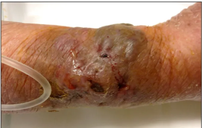
Here, we report the case of a 78-year-old healthy woman who presented with a 7.5×8-cm erythematous eschar-like crusted indurated plaque and pustules with purulent discharges on a chicken-pecked scar on the right forearm that developed 2 month prior to her visit (Fig. 1). The patient was diagnosed with type 2 diabetes and hyper- tension 12 years earlier. She was afebrile and otherwise healthy.
Skin biopsy was performed, including staining with hema- toxylin and eosin, and Gomori methenamine silver (GMS) for histologic, bacteriologic, and mycologic examination.
The histologic sections stained with hematoxylin and eosin exhibited numerous dichotomously branching and septate hyphae in the granulated tissue. Meanwhile, GMS staining showed dark brown/black-colored hyphae walls (Fig. 2).
Cultures from the skin biopsy specimens and exudates on Sabouraud’s agar at 37oC repeatedly exhibited rapidly
Letter to the Editor
660 Ann Dermatol
Fig. 2. Septate hyphae with dichotomous branching in the dermis (Gomori methenamine silver, ×400).
Fig. 1. Erythematous eschar-like crusted indurated plaque and pustules with purulent discharges on the right forearm.
growing colonies consistent with Aspergillus niger. On the basis of these findings, the patient was diagnosed with primary cutaneous aspergillosis due to A. niger. The patient was treated with oral terbinafine hydrochloride 250 mg/day for 3 months, which resulted in complete healing of the lesion 12 weeks after treatment initiation.
Cutaneous aspergillosis can occur as either a primary or secondary infection1,2. The initial infection is clinically characterized by macules, papules, nodules, plaques, or hemorrhagic bullae, which may progress into necrotic ul- cers covered by a heavy black eschar3.
The diagnosis of most cutaneous aspergillosis infections generally requires the biopsy of a skin lesion for both culture and histopathology. Fungal isolates from culture media are identified on the basis of colony morphology, color, and sporulation. Meanwhile, histopathologic exami- nation with routine stains variably demonstrates Aspergillus hyphae. GMS stain clearly shows hyphae, because the hyphal cell walls stained black, whereas the tissue back- ground stained green (Fig. 2). Aspergillus hyphae should have acute-angle branching and frequent septations. The fruiting bodies of Aspergillus are rarely observed in tissue samples unless there is an overwhelming burden of organisms at the site. Therefore, although a tentative diagnosis of aspergillosis can be made on the basis of histopathologic GMS staining, a definitive diagnosis requires the identification of Aspergillus grown in culture.
The recommended treatment for primary cutaneous aspergillosis includes voriconazole, itraconazole, and ampho- tericin B. However, recent studies suggest a significant portion of Aspergillus species may be resistant to conven-
tional treatment4. In the present case, oral administration of terbinafine 250 mg/day was effective.
Primary cutaneous aspergillosis caused by A. niger in a healthy patient is extremely rare. To our knowledge, only few cases have been reported worldwide, including only 1 case in Korea5. In summary, we present a unique and interesting case of primary cutaneous aspergillosis caused by A. niger.
ACKNOWLEDGMENT
This work was supported in part by the Soon Chun Hyang University Research Fund.
REFERENCES
1. Allo MD, Miller J, Townsend T, Tan C. Primary cutaneous aspergillosis associated with Hickman intravenous catheters.
N Engl J Med 1987;317:1105-1108.
2. Carlile JR, Millet RE, Cho CT, Vats TS. Primary cutaneous aspergillosis in a leukemic child. Arch Dermatol 1978;114:
78-80.
3. van Burik JA, Colven R, Spach DH. Cutaneous aspergillosis.
J Clin Microbiol 1998;36:3115-3121.
4. Woodruff CA, Hebert AA. Neonatal primary cutaneous asper- gillosis: case report and review of the literature. Cutaneous aspergillosis. J Clin Microbiol 1998;36:3115-3121.
5. Kang JD, You DO, Youn NH, Park SD. A case of primary cutaneous infection of the burn scar by Aspergillus niger.
Korean J Med Mycol 2002;7:97-101.