Received May 18, 2010, Revised January 5, 2011, Accepted for publication January 5, 2011
Corresponding author: Bong Seok Shin, M.D., Department of Dermato- logy, Chosun University Medical School, 588 Seoseok-dong, Dong-gu, Gwangju 501-717, Korea. Tel: 82-62-220-3130, Fax: 82-62-220-3215, E-mail: [email protected]
This is an Open Access article distributed under the terms of the Creative Commons Attribution Non-Commercial License (http://
creativecommons.org/licenses/by-nc/3.0) which permits unrestricted non-commercial use, distribution, and reproduction in any medium, provided the original work is properly cited.
CASE REPORT
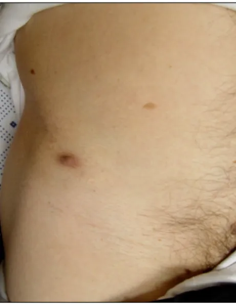
Fig. 1. Brown-colored cutaneous nodule on the linear scar of right girdle area.
Cutaneous Extraskeletal Osteosarcoma on the Scar of a Previous Bone Graft
Sang Gon Park, M.D.1, Ji Young Song, M.D., In Guk Song, M.D., Min Sung Kim, M.D., Bong Seok Shin, M.D.
Departments of Dermatology, 1Internal Medicine, Hemato-oncology, School of Medicine, Chosun University, Gwangju, Korea
Extraskeletal osteosarcoma (ESOS) is a very rare malignant tumor of mesenchymal origin. It is rarer than osseous osteosarcoma and there are very few reports of the skin being a primary site. Most reported cutaneous ESOS were accompanied with metastasis in other organs. A 56-year-old man presented with a painful, 1.5×0.8 cm sized, brown- colored nodule on the right girdle area for 3 months. The histologic findings revealed a tumor that was confined to the dermis without connection to the subcutaneous tissue. In addition, there were large amounts of thin and lace-like bony trabeculae and osteoid with neoplastic cells in a highly pleomorphic sarcomatous stroma. (Ann Dermatol 23(S2) S160∼S164, 2011)
-Keywords-
Cutaneous, Extraskeletal, Osteosarcoma, Scar
INTRODUCTION
Extraskeletal osteosarcoma (ESOS) is defined as a malignant mesenchymal neoplasm that produces osteoid, bone, and/or chondroid material, and is located in the soft tissues without attachment to the bone or periosteum1. ESOS occurs rarely, with an incidence of 4∼5% of
osteosarcoma2,3 and 1% of soft tissue sarcoma4. Up to 13% of cases have been reported with association to previous trauma or irradiation, but the etiological correlation of mechanical injury is difficult to assess1,5. It presents as a progressively enlarging soft tissue mass, which is painful in about one third of patients. The duration of symptoms vary from a few weeks to several months with a mean of 6.5 months4. Approximately 10%
of reported ESOSs are superficially located in soft tissue.
ESOS in skin develops de novo or by metastasis and is exceedingly rare. Herein, we describe a 56-year-old patient that presented with a nodule on a scar in the right girdle area, with a review of the literature.
CASE REPORT
A 56-year-old man presented with a painful, slowly
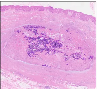
Fig. 2. Extension of mid- to lower dermis by solid tumor (H&E,
×40).
Fig. 4. Microscopic finding of the periphery of the tumor: plump osteoid in a stroma of malignant tumor cells and osteoclast-like giant cells are observed (H&E, ×200).
Fig. 3. Microscopic finding of central portion of the tumor: a large amount of thin and lace-like bony trabeculae and osteoid with intervening malignant osteoblasts are seen (H&E, ×100).
growing nodule located on the right girdle area (Fig. 1).
On physical examination, a firm, non-ulcerated, 1.5×0.8 cm sized, brown-colored exophytic nodule was observed on a linear scar of the right girdle area. The lesion presented 3 months ago, and had been preceded by intermittent pricking pain without specific skin lesions.
Ten years ago, the patient went to a local physician with intermittent left foot pain. X-ray showed an osteolytic lesion in the fourth and fifth metatarsal bone of his left foot. He had undergone curettage and autogenous bone graft that was taken from the right iliac crest. Pathologic interpretation was osteoblastoma. He showed positive
results for several weeks, but then developed pain and swelling on the distal portion of his left leg. X-ray showed progression of the osteolytic lesion and soft tissue swelling in his left foot. He had an additional biopsy for evaluation, which revealed osteosarcoma. Following that, an ampu- tation was performed below the left knee. Postoperatively, he was not given adjuvant chemotherapy, radiotherapy or trauma in the tumor area.
The tumor was excised with a clinical diagnosis of dermatofibroma. Microscopically, the tumor was confined to the dermis without connection to subcutaneous tissue (Fig. 2). Large amounts of thin and lace-like bony trabeculae and osteoid formed by neoplastic cells were observed in a highly pleomorphic sarcomatous stroma (Fig. 3). The neoplastic bone and osteoid exhibited an oteoblastic rim (Fig. 4). Immnunohistochemically, tumor cells were immunoreactive for vimentin, but negative for S-100 protein, desmin, and cytokeratin.
Beyond histopathological examination, physical findings and laboratory tests were within normal limits, and positron emission tomography-computed tomography (PET- CT) revealed no other bone involvement. The final diagnosis was primary cutaneous osteoblastic ESOS, which developed on previously traumatized skin as an operation scar. The patient improved without relapse or metastases for 18 months.
DISCUSSION
ESOS, initially described by Wilson6 in 1941, is a malignant mesenchymal neoplasm arising in soft tissues
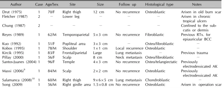
Table 1. Review of reported cases of primary cutaneous extraskeletal osteosarcomas
Author Case Age/Sex Site Size Follow up Histological type Notes
Drut (1975) 1 70/F Right thigh 12 cm No recurrence Osteoblastic Arisen in old burn scar Fletcher (1987) 2 − Lower leg − − − Arisen in chronic
tropical ulcers
Chung (1987) 2 − − − − − Confined to the sub- cutis or dermis Reyes (1989) 1 62/M Temporoparietal 5×3 cm No recurrence Fibroblastic Previous RTx. for
epiauricular BCC Kuo (1992) 1 51/F Popliteal area 3×3 cm − Osteo/fibroblastic − Kobos (1995) 1 78/M Shoulder 1×1 cm Local recurrence Osteoblastic − Kircik (1995) 1 83/F Frontal/parietal Large Lung metastasis − Previous trauma Pillay (2000) 1 56/F Scalp 8 cm Neck metastasis Osteo/fibroblastic − Santos-Juanes (2004) 1 96/F Temple 4×3 cm No recurrence Osteo/telangiectatic Previously
electrodessicated AK Massi (2006)9 1 84/M Scalp 2×2 cm No recurrence Osteoblastic Previously
electrodessicated AK Salamanca (2008)10 1 60/M Right thigh 9×6×5 cm Lung metastasis Chondroblastic −
Song (2009) 1 56/M Right girdle area 1.5×0.8 cm No recurrence Osteoblastic Arisen in operation scar Several articles do not mention about the information of precise tumor’s histological type but we can infer from the descriptions of histology and the figures. F: female, M: male, RTx.: radiotherapy, BCC: basal cell carcinoma, AK: actinic keratosis, Osteo: osteoblastic.
with no connection to skeletal structures, and composed of cells producing osteoid, bone, and/or chondroid material. Based on the predominant matrix, ESOS is divided microscopically into six distant subtypes, which include fibroblastic, osteoblastic, chondroblastic, oste- oclastic or giant cell, telangiectatic, and small cell osteosarcoma1. ESOS usually occurs in patients over 40-years-old (mean age 47.5 years) with equal gender incidence; in contrast to primary osseous osteosarcoma its peak incidence in the 2nd decade of life. ESOS most commonly affects lower extremities, and in particular the thighs and other sites include the upper extremities, shoulder and retroperitoneum. A majority of cases of ESOS have been found in deep soft tissue7, but have rarely been confined to the subcutis or even the dermis2,5,8. The clinical course of ESOS is aggressive, and tumor- related mortality is 75% within 5 years of diagnosis. Tumor size (<5 cm versus ≥5 cm) appears to be the only reliable prognostic variable. However, histological subtypes are regarded by some as another variable of clinical course, and chondroblastic type has a slightly favorable outcome compared to the rest.
Cutaneous ESOS usually develops by de novo or meta- stasis. Skin as a primary site is rare and only 13 cases of primary cutaneous osteosarcoma have been reported to date (Table 1)5,7,9,10. With an exception of 2 undocu- mented cases, it occurred either on the cephalic area (5 cases) or in the extremities (lower leg 5 cases, and shoulder 1 case). The age distribution of primary cutaneous ESOS ranged from 51 to 96-years-old with a mean age of 70
years and most of them had a relatively short duration of less than one year. In most cases that occurred on the cephalic area, they arose at the site of a preceding insult, such as electrodessication (2 cases), radiotherapy (1 case) and trauma (1 case), whereas there was no previous insult in 6 cases that occurred in the extremities. Three cases that were over 5 cm in size were metastasized to the lung within a few months after initial diagnosis, but had no distinct histological types that were clearly related to the prognosis.
Bone tumor metastasis almost exclusively spreads hematogenously, and rarely by lymphatic dissemination, or direct dissemination to overlying skin of the primary lesion, or implantation11. A metastasis of bone tumor mani- fested by pulmonary involvement in the early stage and secondarily by involvement of other sites, such as distant bone, brain, lymph node, and other retroperitoneal sites12,13.
Cutaneous metastasis, whether from osteosarcoma or ESOS, appears to be exceedingly rare, as there are only 11 cases reported in the literature (Table 2)11-20. In most cases of skin metastasis, solitary or multiple lesions appeared, except in 2 undocumented cases, and all solitary lesions developed in the cephalic area.
The average age of metastatic cutaneous ESOS is 39.5 years lower than that of primary cutaneous ESOS, as it spreads to the skin from the osteosarcoma that is developed at an early age. Until cutaneous metastasis develops, they progress relatively fast at a mean of 12 months and are accompanied with distant metastasis to
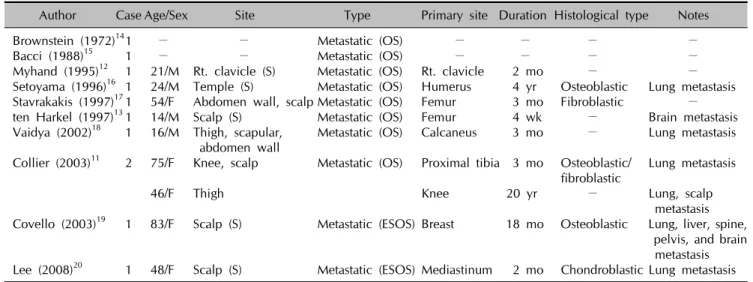
Table 2. Review of reported cases of metastatic cutaneous extraskeletal osteosarcomas
Author Case Age/Sex Site Type Primary site Duration Histological type Notes Brownstein (1972)141 − − Metastatic (OS) − − − − Bacci (1988)15 1 − − Metastatic (OS) − − − − Myhand (1995)12 1 21/M Rt. clavicle (S) Metastatic (OS) Rt. clavicle 2 mo − − Setoyama (1996)16 1 24/M Temple (S) Metastatic (OS) Humerus 4 yr Osteoblastic Lung metastasis Stavrakakis (1997)171 54/F Abdomen wall, scalp Metastatic (OS) Femur 3 mo Fibroblastic − ten Harkel (1997)131 14/M Scalp (S) Metastatic (OS) Femur 4 wk − Brain metastasis Vaidya (2002)18 1 16/M Thigh, scapular, Metastatic (OS) Calcaneus 3 mo − Lung metastasis
abdomen wall
Collier (2003)11 2 75/F Knee, scalp Metastatic (OS) Proximal tibia 3 mo Osteoblastic/ Lung metastasis fibroblastic
46/F Thigh Knee 20 yr − Lung, scalp
metastasis Covello (2003)19 1 83/F Scalp (S) Metastatic (ESOS) Breast 18 mo Osteoblastic Lung, liver, spine,
pelvis, and brain metastasis Lee (2008)20 1 48/F Scalp (S) Metastatic (ESOS) Mediastinum 2 mo Chondroblastic Lung metastasis Several articles do not mention about the information of precise tumor’s histological type but we can infer from the descriptions of histology and the figures. F: female, M: male, Rt: right, S: solitary, OS: osteosarcoma, ESOS: extraskeletal osteosarcoma.
the lung or brain. It seems that there is no interrelationship between histological type and prognosis.
Due to the loss of data, we could not identify whether the histological type between the previous osseous oste- osarcoma and the present cutaneous ESOS was identical or not. However, the previous tumor was completely removed and recurrence or other metastatic lesions were not observed. In addition, the interval of tumor deve- lopment was quite long (10 years) compared to reported metastatic cases and no metastatic lesion on PET-CT was observed. Therefore, we diagnosed that this case was a primary cutaneous ESOS occurring on the operation scar.
However, we could not completely rule out a metastatic ESOS because there was one report11, in which metastatic ESOS developed after 20 years of the primary osteosar- coma.
When making a differential diagnosis, bone forming soft tissue tumors such as myositis ossificans and sarcomas with osseous metaplasia including fibrosarcoma, malig- nant fibrous histocytoma, synovial sarcoma, epithelioid fibrosarcoma, and liposarcoma should be excluded. Other ossified tumors originating in the skin should also be considered. Osteoma cutis shows mature ossification without atypia or mitosis. Metaplastic ossification may also be seen in benign or malignant cutaneous tumors including pilomatricoma, intradermal nevus, desmoplastic malignant melanoma, basal cell carcinoma, and chond- roid syringoma. In our case, the clinical and histo- pathological features supported a diagnosis of cutaneous ESOS.
Close long-term follow up will be needed for our patient including regular physical examinations and radiological
studies because of less favorable osteoblastic histo- pathologic type, although the patient had a tumor less than 5 cm and no evidence of another distant metastasis was found, particularly in the lung.
REFERENCES
1. Weiss SW, Goldblum JR. Osseous soft tissue tumor. Soft tissue tumor. 5th ed. St. Louis: CV Mosby, 2008:1051-1059.
2. Lidang Jensen M, Schumacher B, Myhre Jensen O, Steen Nielsen O, Keller J. Extraskeletal osteosarcomas: a clinico- pathologic study of 25 cases. Am J Surg Pathol 1998;22:
588-594.
3. Sordillo PP, Hajdu SI, Magill GB, Golbey RB. Extraosseous osteogenic sarcoma: a review of 48 patients. Cancer 1983;
51:727-734.
4. Allan CJ, Soule EH. Osteogenic sarcoma of the somatic soft tissues. Clinicopathologic study of 26 cases and review of literature. Cancer 1971;27:1121-1133.
5. Chung EB, Enzinger FM. Extraskeletal osteosarcoma. Cancer 1987;60:1132-1142.
6. Wilson H. Extraskeletal ossifying tumors. Ann Surg 1941;113:
95-112.
7. Kobos JW, Yu GH, Varadarajan S, Brooks JS. Primary cuta- neous osteosarcoma. Am J Dermatopathol 1995;17:53-57.
8. Bane BL, Evans HL, Ro JY, Carrasco CH, Grignon DJ, Benjamin RS, et al. Extraskeletal osteosarcoma: a clinico- pathologic review of 26 cases. Cancer 1990;65: 2762-2770.
9. Massi D, Franchi A, Leoncini G, Maio V, Dini M. Primary cutaneous osteosarcoma of the scalp: a case report and review of the literature. J Cutan Pathol 2007;34:61-64.
10. Salamanca J, Dhimes P, Pinedo F, Gómez de la Fuente E, Pérez Espejo G, Martínez-Tello FJ. Extraskeletal cutaneous chondroblastic osteosarcoma: a case report. J Cutan Pathol 2008;35:231-235.
11. Collier DA, Busam K, Salob S. Cutaneous metastasis of osteosarcoma. J Am Acad Dermatol 2003;49:757-760.
12. Myhand RC, Hung PH, Caldwell JB, James WD, Sau P, Hargis JB. Osteogenic sarcoma with skin metastases. J Am Acad Dermatol 1995;32:803-805.
13. ten Harkel AD, Hogendoorn PC, Beckers RC, Sprij AJ, Taminiau AH, Van der Woude HJ, et al. Skin metastases of osteogenic sarcoma: a case report with review of the literature. J Pediatr Hematol Oncol 1997;19:266-267.
14. Brownstein MH, Helwig EB. Metastatic tumors of the skin.
Cancer 1972;29:1298-1307.
15. Bacci G, Avella M, Picci P, Briccoli A, Dallari D, Campanacci M. Metastatic patterns in osteosarcoma. Tumori 1988;74:421-427.
16. Setoyama M, Kanda A, Kanzaki T. Cutaneous metastasis of
an osteosarcoma. A case report. Am J Dermatopathol 1996;
18:629-632.
17. Stavrakakis J, Toumbis-Ioannou E, Alexopoulos A, Rigatos GA. Subcutaneous nodules as initial metastatic sites in osteosarcoma. Int J Dermatol 1997;36:606-609.
18. Vaidya S, Jones KP, Fisher C. Letter to the editor: osteogenic sarcoma-cutaneous metastases. Med Pediatr Oncol 2002;38:
453-454.
19. Covello SP, Humphreys TR, Lee JB. A case of extraskeletal osteosarcoma with metastasis to the skin. J Am Acad Dermatol 2003;49:124-127.
20. Lee WJ, Lee DW, Chang SE, Lee MW, Choi JH, Moon KC, et al. Cutaneous metastasis of extraskeletal osteosarcoma arising in the mediastinum. Am J Dermatopathol 2008;30:
629-631.