Korean J Clin Microbiol Vol. 15, No. 2, June, 2012 http://dx.doi.org/10.5145/KJCM.2012.15.2.70
Primary Cutaneous Cryptococcosis in a Patient with Iatrogenic Cushing’s Syndrome: A Case Report
and Review of the Literature
Young Jin Ko, Mi Hyun Hong, Chul-Min Park, Hee-Won Moon, Mina Hur, Yeo-Min Yun
Department of Laboratory Medicine, Konkuk University School of Medicine and Medical Center, Seoul, Korea
Cryptococcus neoformans (C. neoformans) is a ubiq- uitous yeast-like fungus that has been a common op- portunistic human pathogen, especially in immuno- compromised patients. Although skin lesions due to C. neoformans are found in 10-15% of patients with systemic cryptococcosis, primary cutaneous crypto- coccosis without systemic infection is rare and now considered a distinct clinical entity. We report a case of primary cutaneous cryptococcosis in a patient with iatrogenic Cushing’s syndrome. A 73-year-old woman presented with pain and discharge from lesions on her left forearm. The patient had been treated with oral corticosteroids for 20 years, and as a result had developed iatrogenic Cushing’s syndrome. A skin fragment of the ulcer was cultured, and the encapsu-
lated fungus were isolated and identified as C. neo- formans using a Vitek2 system (Vitek2 ID-YST, bio- Mérieux, France) and API 20C (bioMérieux). Concu- rrent blood and urine cultures were negative for growth. At first, she was treated with antibiotics due to suspicion of cellulitis. After surgical resection and treatment with systemic and oral fluconazole, her wound was improved with scar. Primary cutaneous cryptococcosis should be considered when skin le- sions are not responsive to antibiotics and accurate identification is important for proper treatment.
(Korean J Clin Microbiol 2012;15:70-73)
Key Words: Cryptococcus neoformans, Cushing’s synd- rome, Primary cutaneous cryptococcosis
Received 19 October, 2011, Revised 24 November, 2011 Accepted 12 December, 2011
Correspondence: Hee-Won Moon, Department of Laboratory Medicine, Konkuk University School of Medicine, 120-1 Neungdong-ro, Gwangjin-gu, Seoul 143-729, Korea. (Tel) 82-2-2030-5583, (Fax) 82-2-2030-5587, (E-mail) [email protected]
70 서 론
Cryptococcus neoformans는 환경에 널리 존재하는 효모균으 로 비둘기 배설물과 같은 조류 분비물에 오염된 토양과 부패된 나무, 과일, 채소, 먼지 등으로부터 발견되며 주로 면역억제 환 자에서 기회감염을 일으킨다[1-4]. 주된 침입 경로는 호흡기이 며, 일차적으로 폐에 병변을 일으키고 혈행성으로 중추 신경계, 피부 등의 다른 장기를 침범한다[5]. 피부 침범은 전신성 크립 토콕쿠스증(systemic cryptococcosis) 환자의 약 10-15%에서 볼 수 있으며, 구진, 결절, 농포, 반상출혈, 육아종, 농양 등 다양한 병변을 나타낸다[6]. 전신적 질환 없이 피부 병변만 나타나는 일차성 피부 크립토콕쿠스증(Primary cutaneous cryptococcosis) 은 드물고, 최근에는 별도의 질환군으로 생각되고 있다[7]. 이 렇게 전신적 크립토콕쿠스증이 동반되지 않는 경우, 피부 병변 에서 정확한 동정이 없이는 봉와직염 등으로 오인되어, 부적절 한 치료를 시행할 수 있다[8]. 저자들은 의인성 쿠싱 증후군 (iatrogenic Cushing’s syndrome) 환자에서 C. neoformans에 의
한 일차성 피부 크립토콕쿠스증을 경험하여, 문헌 고찰과 함께 보고하는 바이다.
증 례
73세 여자 환자가 내원 3일 전부터 발생한 좌측 팔에 발생한 직경 5×5 cm 크기의 삼출성 궤양과 동통을 주소로 내원하였다 (Fig. 1A). 환자는 특별히 피부에 큰 외상을 입은 병력은 없었 으나 가벼운 긁힘이 있었다고 하였고, 50년간 농사일을 하며 농촌에 거주 중이었다. 과거력 상 류마티스 관절염으로 20-30 년간 경구 스테로이드를 복용한 기왕력이 있었고 이로 인해 의 인성 쿠싱 증후군이 발생하였으며, 당뇨, 고혈압, 골관절염이 동반되어 있었다. 수개월 전 요추 압박골절 수술 후 거동이 불 편하여 둔부에 욕창으로 인한 죽은 조직 제거술과 편측 V-Y 전진 피판 연조직 덮개술을 받았다. 가족력상 특이 사항은 없 었다. 내원 당시 체온 37.1oC, 혈압 101/73 mmHg, 맥박 91회/
분, 호흡수 20회/분이었고, 검사 소견으로 일반 혈액 검사에서 백혈구 7,730/μL (호중구 60.3%, 림프구 31.4%, 단구 8.0%, 호 염기구 0.3%), 혈색소 10.3 g/dL, 혈소판 464,000/μL로 경도의 빈혈이 있었으며, 적혈구침강속도는 55 mm/h, C 반응성 단백 은 3.87 mg/dL로 증가해 염증이 있음을 시사하였다. 진단 시
Young Jin Ko, et al. : Primary Cutaneous Cryptococcosis in Cushing’s Syndrome
71
Fig. 1. Skin lesion due to C. neofor- mans on the patient’s left forearm before (A) and after (B) debridement and antifungal treatment for 10 days.
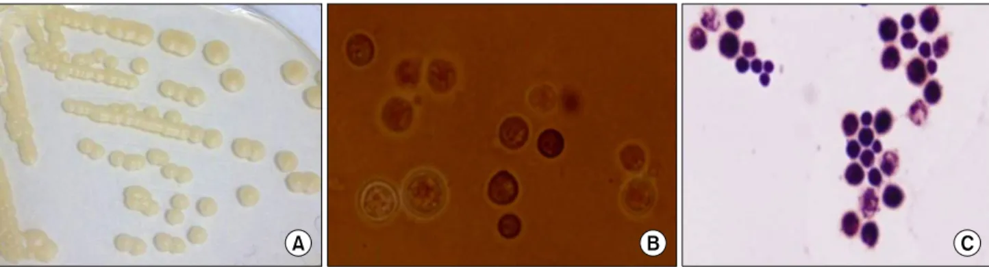
Fig. 2. Mucoid and creamy colonies of C. neoformans on Sabouraud dextrose agar (A). Photomicrograph of an India ink preparation (B), and Gram staining (C), illustrating the irregular-sized, encapsulated, spherical yeast cells of C. neoformans (×1,000). After subculture, capsules became thinner than initial preparation in India ink stain.
Table 1. Clinical characteristics of primary cutaneous cryptococcosis cases reported in Korea
Sex/Age Site Skin finding Underlying condition Treatment References
F/78 F/63 F/57 F/73
Forearm Forearm Forebead &
perioral area Forearm
Ulcer Ulcer Ulcer Ulcer
Iatrogenic Cushing’s syndrome Long-term steroid use
Non-insulin-dependent diabetes mellitus Iatrogenic Cushing’s syndrome
Itraconazole
Fluconazole and debridement Fluconazole
Fluconazole and debridement
16 17 18 Present study
시행한 기저 혈청 cortisol 농도는 4.84 μg/dL, 기저 혈청 ACTH 농도는 16.97 pg/mL, 급속 ACTH 자극 검사 후 혈청 cortisol 농도는 11.98 μg/dL로 의인성 쿠싱 증후군에 적합한 소견을 보였다. 화학검사에서 총단백질, 알부민, 칼슘이 약간 감소되었고, 요검사에서 단백뇨, 케톤뇨, 빌리루빈뇨 소견을 보 였다. 흉부 X-선 검사상 오래된 좌측 흉수 소견 외에는 이상 소 견은 없었다. 궤양 변연부의 조직에서 얻은 검체를 혈액한천 배지에 접종하여 배양한 결과 24시간 후 희고 작은 집락이 관 찰되었고, 집락의 일부를 채취하여 Sabouraud dextrose agar에 계대배양하여 효모균 집락을 관찰할 수 있었다. 집락은 점성이 있고 크림색을 띠었으며, India ink 염색에서 협막이 있는 둥근 형태의 포자가 관찰되었다(Fig. 2). 효모균은 urease를 생성하였 으며, Vitek2 system (Vitek2 ID-YST, bioMérieux, Marcy
l’Etoile, France)에서 C. neoformans probability 99%로 동정되 었고, API 20C (bioMérieux)를 이용한 20종류의 효소생성능 시 험에도 48시간 후 glucose, 2-keto-D-gluconate, xylose, adonitol, galactose, sorbitol, N-acetyl-D-glucosamine, cellobiose, maltose, sucrose, trehalose, melezitose, raffinose 반응이 양성이었고, 72 시간 후 inositol, Methyl-D-glucoside 양성으로 C. neoformans 를 probability 99.9%로 동정할 수 있었다. 함께 시행한 혈액 배 양 및 뇨 배양에서는 균이 검출되지 않았으며, 뇌척수액 배양 은 시행하지 않았다. 초기에는 세균성 봉와직염으로 판단하였 고, 배양 및 결과 확인 과정이 신속하게 진행되지 못하여, 10일 간 cefazedone, ciprofloxacin, clindamycin 치료를 하였다. 원발 성 피부 크립토콕쿠스증으로 진단된 후, 치료로 내원 당일과 11일째 외과적 변연 절제술을 2회 시행하였으며(Fig. 1B) 10일
72
Korean J Clin Microbiol 2012;15(2):70-73간의 fluconazole (400 mg/day) 정주요법과 이후 3개월간의 경 구 투약 요법(400 mg/day)을 지속하여 병변은 반흔을 남기고 치유되었다.
고 찰
C. neoformans는 환경에 널리 분포하며, 인체 감염은 일차적 으로 호흡기를 통해 흡입하는 것으로 되어 있으며, 면역기능의 감소가 병을 일으키는 데 중요하여 후천성 면역 결핍증, 악성 림프종, 유육종증, 결체조직 질환 등을 가진 사람들이나 오랫동 안 스테로이드나 면역억제제를 투여 받은 환자에서 잘 발생한 다[9]. 임상적으로는 숙주의 반응에 따라 90%는 폐에 국한되 나, 소수에서는 혈행성으로 전신에 퍼진다. 임상적으로 가장 많 이 침범되는 기관은 중추신경계로 전체 효모균증의 70-80%를 차지하며 폐와 중추신경계 이외에도 간, 신장, 전립선, 피부, 뼈 등을 침범한다[10]. 봉와직염의 소견을 보이는 경우 종종 세균 감염으로 오진되어 항생제 치료를 받기도 하며 본 증례의 경우 도 효모균증으로 진단되기 전 발열이 지속되어 항균제 치료를 받았고 검사실과 임상과에서 피부 병변 배양에 대한 경각심이 부족하여 배양 및 확인과정이 지연되었다.
피부 크립토콕쿠스증은 대부분이 전신성 질환에서 속발되며 원발성으로 피부에만 발생하는 경우는 매우 드물다. Neuville 등[7]은 원발성 피부 크립토콕쿠스증으로 진단하기 위한 기준 을 제시하였고 이에 따르면 병변이 피부에만 국한되어 있어야 하며, 진단 후 최소한 4주간의 추적기간 동안에 전신 침범이 없 어야 한다고 하였다. 또한 병변 부위는 외부 노출이 되는 부위 로서 병변 부위의 피부 손상 기왕력이 있거나, 피부손상을 일 으키기 쉬운 직업이나 취미가 있고, 농촌에 거주하는 경우 진 단의 근거가 된다[7]. 원발성 질환과 이차성 질환의 피부 병변 도 차이가 있는데 이차성의 경우 다발성이며 여기저기 퍼져있 는 경우가 많고, 배꼽모양 구진(umbilicated papule)이 많지만 원발성은 상완처럼 노출된 부위에 단독 병변이며, 봉와직염이 나 궤양이 흔하다[7]. 본 환자의 경우 발열과 다른 병변의 국소 봉와직염을 동반하였으나, 혈액 배양 및 뇨배양 모두 음성이었 으며, 호흡기 및 중추신경계를 비롯한 전신감염의 증상 및 증 후가 없었고 발병 후부터 치료기간 동안 병변이 피부에만 국한 되어 있었던 점 등으로 원발성 피부질환으로 진단할 수 있었 다.
원발성 피부 감염은 피부의 작은 외상을 통한 균의 직접 침 투에 의하여 발생한다고 알려져 있으나 정확한 감염경로는 아 직도 논란이 있다. 본 증례의 경우 환자는 농촌에서 거주하며 농사일을 50년간 해왔기 때문에 환경적인 원인에 의한 감염 가 능성이 있다. 원발성 피부감염도 대개의 경우 면역 기능이 저 하된 사람에서 기회 감염으로 발생되며 국외 보고들의 경우 기 저질환으로 HIV 감염[11], 종양[12], 이식[13], 스테로이드 제
제를 사용한 경우 등에서 발생했다[7]. 동정의 경우 조직소견으 로만 진단하는 경우도 있었으나, 정확하게 동정을 시행하는 것 이 필요할 것으로 생각되며, 추후 질환의 임상적 의의를 연구 하는 데도 필수적이라고 생각된다. 본 증례는 생화학적 반응 소견으로 충분히 C. neoformans로 동정할 수 있었다. 국내에서 의 기존 보고로는 주로 집락의 성상 및 병리학적 소견으로 진 단한 경우가 많고, 일부 보고에서 효모균이 관찰되는 경우 C.
neoformans로 간주하는 경우도 있었다[14,15]. 생화학적 동정 으로 진단한 C. neoformans에 의한 원발성 피부감염 증례를 고 찰해보면, 본 증례와 마찬가지로 위험요인으로 부신피질 호르 몬제의 장기간 사용이 많았으며, 모두 고령의 여성인 공통점이 있었으나 [16-18] 더 많은 증례 분석이 필요하겠다(Table 1). 또 한 최근 보고에 따르면 건강한 사람에서도 원발성 피부감염이 보고되어[19], 면역억제환자뿐 아니라 건강인에서도 이러한 피 부병변이 보일 경우 원발성 크립토콕쿠스증을 의심하여 배양 등의 검사를 통해 정확한 동정을 할 필요가 있다.
결론적으로 항균제에 잘 치유되지 않는 피부 병변의 경우 원 발성 피부 크립토콕쿠스증의 가능성도 고려해야 하며, 검사실 에서도 전신적 감염이 아닌 피부병변이라도 신속한 동정 및 보 고가 중요하다. 국내에서는 장기간 스테로이드 제제를 사용한 고령에서 주로 발생했으므로, 이러한 경우 더욱 경각심을 가질 필요가 있고, 동시에 전신적 감염 여부의 면밀한 관찰이 필요 할 것이다.
참 고 문 헌
1. Ellis DH and Pfeiffer TJ. Ecology, life cycle, and infectious propagule of Cryptococcus neoformans. Lancet 1990;336:923-5.
2. Levitz SM. The ecology of Cryptococcus neoformans and the epidemiology of cryptococcosis. Rev Infect Dis 1991;13:1163-9.
3. Ruiz A, Fromtling RA, Bulmer GS. Distribution of Cryptococcus neoformans in a natural site. Infect Immun 1981;31:560-3.
4. Kumlin U, Olsen B, Granlund M, Elmqvist LG, Tärnvik A.
Cryptococcosis and starling nests. Lancet 1998;351:1181.
5. Fitzpatrick TB, ed. Dermatology in general medicine. 4th ed. New York; McGraw-Hill, Health Professions Division, 1993.
6. Anderson DJ, Schmidt C, Goodman J, Pomeroy C. Cryptococcal disease presenting as cellulitis. Clin Infect Dis 1992;14:666-72.
7. Neuville S, Dromer F, Morin O, Dupont B, Ronin O, Lortholary O; French Cryptococcosis Study Group. Primary cutaneous crypto- coccosis: a distinct clinical entity. Clin Infect Dis 2003;36:337-47.
8. Probst C, Pongratz G, Capellino S, Szeimies RM, Schölmerich J, Fleck M, et al. Cryptococcosis mimicking cutaneous cellulitis in a patient suffering from rheumatoid arthritis: a case report. BMC Infect Dis 2010;10:239.
9. Hernandez AD. Cutaneous cryptococcosis. Dermatol Clin 1989;7:
269-74.
10. Powderly WG. Cryptococcal meningitis and AIDS. Clin Infect Dis 1993;17:837-42.
11. Gatti M, Di Silverio A, Cespa M, Mosca M. Primary unusual cutaneous cryptococcosis in an HIV former drug-abuser patient.
Young Jin Ko, et al. : Primary Cutaneous Cryptococcosis in Cushing’s Syndrome
73
Mycoses 1997;40:101-2.
12. Romano C, Taddeucci P, Donati D, Miracco C, Massai L. Primary cutaneous cryptococcosis due to Cryptococcus neoformans in a woman with non-Hodgkin's lymphoma. Acta Derm Venereol 2001;
81:220-1.
13. Zorman JV, Zupanc TL, Parac Z, Cucek I. Primary cutaneous cryptococcosis in a renal transplant recipient: case report. Mycoses 2010;53:535-7.
14. Kim YJ, Seo SJ, Ro BI. A case of primary cutaneous cryptoco- ccosis misdiagnosel as skin tuberculosis. Korean J Med Mycol 2003;8:16-20.
15. Shin DH, Kim KS, Lee JM, Choi JS, Kim KH. Primary cutaneous
cryptococosis. Ann Dermatol 1999;11:27-9.
16. Kim HJ, Min HG, Lee ES. Two cases of cutaneous cryptoccosis mimicking cellulitis. Korean J Med Mycol 1998;3:190-4.
17. Kang HY, Kim NS, Lee ES. Primary cutaneous cryptococcosis treated with fluconazole. Korean J Dermatol 2000;38:838-40.
18. Park JH, Ryoo YW, Lee KS. Primary cutaneous cryptococcosis successfully treated with fluconazole. Ann Dermatol 2000;12:
148-51.
19. Pau M, Lallai C, Aste N, Aste N, Atzori L. Primary cutaneous cryptococcosis in an immunocompetent host. Mycoses 2010;53:
256-8.
=국문초록=
의인성 쿠싱 증후군 환자에서 발생한 일차성 피부 크립토콕쿠스증:
증례보고 및 문헌 고찰
건국대학교 의학전문대학원 진단검사의학교실 고영진, 홍미현, 박철민, 문희원, 허미나, 윤여민
C. neoformans는 환경에 널리 존재하는 효모균으로 주로 면역억제 환자에서 기회감염을 일으킨다. 피부 침범은 전신성 크립토콕쿠스증 환자의 약 10-15%에서 볼 수 있으며, 전신적 질환 없이 피부 병변만 나타나는 일차성 피부 크립토콕쿠스 증은 드물고, 최근에는 별도의 질환군으로 생각되고 있다. 저자들은 의인성 쿠싱 증후군 환자에서 C. neoformans에 의한 일차성 피부 크립토콕쿠스증을 경험하여 보고하는 바이다. 73세 여자 환자가 좌측 팔의 삼출성 궤양과 동통을 주소로 내원하였다. 환자는 20년 동안 경구 스테로이드를 복용하였고 그 결과, 의인성 쿠싱 증후군이 발생하였다. 궤양 변연부의 조직에서 얻은 검체를 배양한 결과 효모균 집락을 관찰할 수 있었다. 효모균은 urease를 생성하였으며, Vitek2 system (Vitek2 ID-YST, bioMérieux, Marcy l’Etoile, France) 및 API 20C (bioMérieux)에서 C. neoformans로 동정되었다. 함께 시행 한 혈액 배양 및 뇨 배양에서는 균이 검출되지 않았다. 세균성 봉와직염으로 판단해 항균제 치료를 하였으나 배양 결과 가 보고된 후 원발성 피부 크립토콕쿠스증으로 진단하고, 치료로 외과적 변연 절제술과 fluconazole 정주요법 및 경구 투약 요법을 지속하여 병변은 반흔을 남기고 치유되었다. 결론적으로 항균제에 잘 치유되지 않는 피부 병변의 경우 원발 성 피부 크립토콕쿠스증의 가능성도 고려해야 하며, 정확히 동정하여 적절한 치료를 하는 것이 중요하다. [대한임상미생 물학회지 2012;15:70-73]
교신저자 : 문희원, 143-729, 서울시 광진구 능동로 120-1 건국대학교 의학전문대학원 진단검사의학교실 Tel: 02-2030-5583, Fax: 02-2030-5587 E-mail: [email protected]