Blood Res2017;52:316-39. bloodresearch.or.kr
324 Letters to the Editor
tionships between circulating leukocytes and infection in pa- tients with acute leukemia. Ann Intern Med 1966;64:328-40.
4. Knoll GA, MacDonald I, Khan A, Van Walraven C. Mycopheno- late mofetil dose reduction and the risk of acute rejection after renal transplantation. J Am Soc Nephrol 2003;14:2381-6.
5. Rerolle JP, Szelag JC, Le Meur Y. Unexpected rate of severe leuco- penia with the association of mycophenolate mofetil and valgan- ciclovir in kidney transplant recipients. Nephrol Dial Transplant 2006;22:671-2.
6. Jaffe E, Arber DA, Campo E, Harris NL, Quintanilla-Fend L.
Hematopathology. 2nd ed. St. Louis, MO: Elsevier, 2016.
7. Dhote R, Simon J, Papo T, et al. Reactive hemophagocytic syn- drome in adult systemic disease: report of twenty-six cases and literature review. Arthritis Rheum 2003;49:633-9.
8. Pal R, Monaghan SA, Hassett AC, et al. Immunomodulatory de- rivatives induce PU.1 down-regulation, myeloid maturation ar- rest, and neutropenia. Blood 2010;115:605-14.
9. Zacharias N, Gallichio MH, Conti DJ. Graft-versus-host disease after living-unrelated kidney transplantation. Case Rep Transplant 2014;2014:971426.
10. Venton G, Crocchiolo R, Fürst S, et al. Risk factors of Ganciclovir- related neutropenia after allogeneic stem cell transplantation: a retrospective monocentre study on 547 patients. Clin Microbiol Infect 2014;20:160-6.
11. Page AV, Liles WC. Granulocyte colony-stimulating factor, granulocyte-macrophage colony-stimulating factor, and other immunomodulatory therapies for the treatment of infectious dis- eases in solid organ transplant recipients. Curr Opin Organ Transplant 2008;13:575-80.
12. Shoup M, Weisenberger JM, Wang JL, Pyle JM, Gamelli RL, Shankar R. Mechanisms of neutropenia involving myeloid matu- ration arrest in burn sepsis. Ann Surg 1998;228:112-22.
13. Henter JI, Horne A, Aricó M, et al. HLH-2004: Diagnostic and therapeutic guidelines for hemophagocytic lymphohistiocytosis.
Pediatr Blood Cancer 2007;48:124-31.
A case of primary plasma cell leukemia exhibiting hemophagocytic plasma cells relapsed with multiple cutaneous plasmacytoma
TO THE EDITOR: Cutaneous infiltration in multiple myelo- ma (MM) is an extremely rare condition with poor prognosis that accounts for only approximately 0.6% of patients with MM according to a recent large-scale study [1]. Primary plasma cell leukemia (PCL) constitutes only 2%–5% of all myeloma cases, with a higher proportion of light-chain-only cases presenting as PCL rather than as MM. Typically, the immunophenotype of PCL differs from that of multiple mye- loma (MM) in that it lacks aberrant CD56 expression and more frequently shows an abnormal karyotype [2].
Hemophagocytosis in the bone marrow is a characteristic feature of some aggressive disorders, such as hemophago- cytic syndrome by histiocytes, but more rarely by myelo- blasts, in acute myeloid leukemia. Furthermore, this charac- teristic is extremely rare among plasma cells, particularly in PCL, and only a few reports on such cases have been published [3-8]. Herein, we report a rare case of cutaneous infiltration of malignant plasma cells, which initially pre- sented as bicytopenia, combined with primary lambda-type light-chain PCL characterized by marked phagocytosis of erythrocytes and platelets by neoplastic plasma cells.
A 77-year-old woman experienced fatigue for several weeks and presented to our hospital with bicytopenia. A complete blood cell count analysis showed normocytic nor- mochromic anemia with thrombocytopenia and leukocy- tosis (hemoglobin, 9.6 g/dL; platelets, 22.0×10⁹/L; white blood cell count, 16.8×10⁹/L). Laboratory tests revealed nor- mal levels of calcium, blood urea nitrogen, creatinine, and lactate dehydrogenase. However, magnetic resonance imag- ing exhibited diffuse bone marrow signal change without definite mass or lytic lesion formation. Serum and urine protein electrophoresis displayed a monoclonal band in the beta region, and immunofixation revealed only lambda light-chain monoclonality with markedly increased serum lambda light-chain level (2,952 mg/L; normal range, 5.71–
26.30 mg/L). The β-2 microglobulin level was also increased (12.06 mg/L; normal range, 0.0–2.4 mg/L). A peripheral blood smear demonstrated the presence of atypical plasma cells in various sizes with cytoplasmic vacuolations, which constitute up to 52.0%. Bone marrow aspiration showed hypercellular marrow particles with a myeloid–to-erythroid ratio of 8:1 with decreased megakaryocytes. Neoplastic plas- ma cells accounted for up to 67.2% of all nucleated cells.
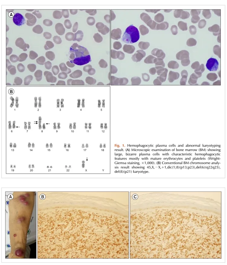
Numerous binucleated or multinucleated plasma cells were observed, of which 5.7% displayed prominent phagocytosis, primarily of erythrocytes and platelets (Fig. 1A). Flow cyto- metric analysis revealed that the plasma cells lacked CD56 expression, which is frequently found in PCL, and no other aberrant expression was observed. In addition, im- munohistochemical analysis showed that the plasma cells were positive for CD138 and negative for CD20. Karyotypic analysis revealed an abnormal 45,X,−X,+1,dic(1;8)(p13;
p23),del(6)(q22q25),del(8)(p21) karyotype (Fig. 1B). The pa- tient was diagnosed with primary PCL, fulfilling the diag- nostic criteria of the International Myeloma Working Group, which suggested more than 20% circulating plasma cells and an absolute plasma cell count of greater than 2×10⁹/L [9]. Bortezomib, melphalan, and prednisolone (VMP) therapy was initiated, which reverted her serum lambda light-chain level to normal after one treatment cycle.
A second treatment cycle with VMP was planned, but the patient refused further chemotherapy. After 5 months from the termination of the first cycle of VMP therapy, multiple subcutaneous nodules developed on her left upper limb, trunk, back, and lower extremities (Fig. 2A), and her serum lambda light-chain level increased again to 127.65 mg/L.
bloodresearch.or.kr Blood Res 2017;52:316-39.
Letters to the Editor 325
Fig. 2. Multiple skin nodules and their biopsy showing infiltration of plasma cells. Multiple skin nodules in the patient’s extremities (A). Biopsy of the skin-infiltrating plasma cells showing positive immunohistochemical staining for CD138 (B) and lambda light-chain (C) (×200).
Skin biopsy showed the nodules as plasmacytoma, and im- munohistochemistry showed CD138 and lambda light-chain positivity (Fig. 2B, C). The patient was treated with lenalido- mide and dexamethasone chemotherapy, but this salvage therapy was effective for preventing nodule dissemination
only for 3 months. The cutaneous plasmacytoma aggravated again, and she is now on palliative radiotherapy.
Cutaneous plasmacytoma is an extremely rare condition with poor prognosis, and the median overall survival is only 8.5 months [10, 11]. This condition is not associated
Fig. 1. Hemophagocytic plasma cells and abnormal karyotyping result. (A) Microscopic examination of bone marrow (BM) showing large, bizarre plasma cells with characteristic hemophagocytic features mostly with mature erythrocytes and platelets (Wright- Giemsa staining, ×1,000). (B) Conventional BM chromosome analy- sis result showing 45,X,−X,+1,dic(1;8)(p13;p23),del(6)(q22q25), del(8)(p21) karyotype.
Blood Res2017;52:316-39. bloodresearch.or.kr
326 Letters to the Editor
with a specific myeloma immunoglobulin type, although a more aggressive course is observed in light-chain-only subtypes [10]. Requena et al. [11] analyzed 8 cases of cuta- neous plasmacytoma and revealed that malignant plasma- cytes are homogeneous in their immunophenotype with strong expression of CD79a, CD138, and epithelial mem- brane antigen. In addition, RB1 gene deletion in skin-in- filtrated plasmacytes was reported to be associated with poor prognosis [11]. A recent retrospective study of 53 cuta- neous plasmacytoma cases showed no correlation between CD56 negativity or cytogenetic abnormality with skin in- filtration of malignant plasma cells and that the plasmablastic morphology in the skin lesion indicated a worse overall survival [10]. In the present case, the malignant plasma cells were positive for CD138 and negative for CD56, CD19, CD20, and CD22, which correlated to the immuno- phenotypic characterization of malignant plasma cells. The RB1 gene deletion could not be analyzed, and no significant plasmablastic appearance of plasma cell infiltration was ob- served in the patient’s skin lesion.
Although hemophagocytosis by neoplastic plasma cells has rarely been described in the literature, this rare condition does not appear to be associated with a specific immuno- phenotype, immunoglobulin or light-chain subtype, or kar- yotype [6]. One hypothesis is that hemophagocytic plasma cell formation may be attributed to the expansion of rare B-cell clones with innate phagocytic potential, although this proposition remains to be confirmed [5, 7]. Similar to our case, the hemophagocytic feature of plasma cells is more frequently found in female patients, and it appears to be dominant in mature erythrocytes and platelets [4, 7]. Some reports also suggest that phagocytosis by neoplastic plasma cells resulted in peripheral blood cytopenia; how- ever, whether this complication is a direct consequence of hemophagocytosis by plasma cells remains to be de- termined [2, 4].
Narae Hwang, Ji Yeon Ham, Jang Soo Suh Department of Clinical Pathology, Kyungpook National
University School of Medicine, Daegu, Korea Correspondence to: Jang Soo Suh Department of Clinical Pathology, Kyungpook National
University School of Medicine, 680 Gukchebosang-ro, Chung-gu, Daegu 41944, Korea
E-mail: [email protected]
Received on May 22, 2017; Revised on Jun. 1, 2017; Accepted on Jul. 13, 2017 https://doi.org/10.5045/br.2017.52.4.324
AuthorsÊ Disclosures of Potential Conflicts of Interest No potential conflicts of interest relevant to this article were reported.
REFERENCES
1. Deng S, Xu Y, An G, et al. Features of extramedullary disease of multiple myeloma: high frequency of p53 deletion and poor sur- vival: a retrospective single-center study of 834 cases. Clin Lymphoma Myeloma Leuk 2015;15:286-91.
2. Swerdlow SH, Campo E, Harris NL, et al, eds. WHO classification of tumours of haematopoietic and lymphoid tissues. 4th ed. Lyon, France: IARC Press, 2008.
3. Invernizzi R, Pecci A. A case of phagocytic multiple myeloma.
Haematologica 2000;85:318.
4. Kanoh T, Saigo K. Phagocytic myeloma cells in asymptomatic multiple myeloma. Tohoku J Exp Med 1987;153:207-10.
5. Kucukkaya RD, Hacihanefioglu A, Yenerel MN, et al. CD15-ex- pressing phagocytic plasma cells in a patient with multiple myeloma. Blood 2001;97:581-3.
6. Ludwig H, Pavelka M. Phagocytic plasma cells in a patient with multiple myeloma. Blood 1980;56:173-6.
7. Ramos J, Lorsbach R. Hemophagocytosis by neoplastic plasma cells in multiple myeloma. Blood 2014;123:1634.
8. Savage DG, Zipin D, Bhagat G, Alobeid B. Hemophagocytic, non-secretory multiple myeloma. Leuk Lymphoma 2004;45:
1061-4.
9. Fernández de Larrea C, Kyle RA, Durie BG, et al. Plasma cell leu- kemia: consensus statement on diagnostic requirements, re- sponse criteria and treatment recommendations by the International Myeloma Working Group. Leukemia 2013;27:
780-91.
10. Jurczyszyn A, Olszewska-Szopa M, Hungria V, et al. Cutaneous involvement in multiple myeloma: a multi-institutional retro- spective study of 53 patients. Leuk Lymphoma 2016;57:2071-6.
11. Requena L, Kutzner H, Palmedo G, et al. Cutaneous involvement in multiple myeloma: a clinicopathologic, immunohistochem- ical, and cytogenetic study of 8 cases. Arch Dermatol 2003;139:475-86.
Differential diagnosis of primary cutaneous CD4+ small/medium T-cell lymphoproliferative lesions:
A report of three cases
TO THE EDITOR: Primary cutaneous CD4+ small/medium T-cell lymphoproliferative disorder (CD4+ PCSM-TLPD) is characterized by the proliferation of small-to-medium-sized T-helper lymphocytes within the dermis. According to the 2016 World Health Organization (WHO) classification, the prognosis of CSMTLPD is considered excellent, and it should not be diagnosed as lymphoma [1]. We studied three cases of CD4+ PCSM-TLPD, focusing on its clinicopathological characteristics and differential diagnoses.