Brief Report
640 Ann Dermatol
Received June 9, 2016, Revised August 30, 2016, Accepted for publication September 9, 2016
Corresponding author: Eun Jung Kim, Department of Dermatology, Wonkwang University Hospital, Wonkwang University School of Medicine, 895 Muwang-ro, Iksan 54538, Korea. Tel: 82-63-859-1590, Fax: 82-63-842-1895, E-mail: [email protected]
This is an Open Access article distributed under the terms of the Creative Commons Attribution Non-Commercial License (http://creativecommons.org/
licenses/by-nc/4.0) which permits unrestricted non-commercial use, distribution, and reproduction in any medium, provided the original work is properly cited.
Copyright © The Korean Dermatological Association and The Korean Society for Investigative Dermatology
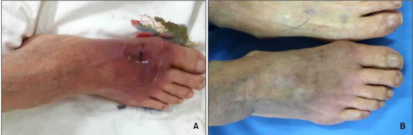
Fig. 1. (A) Painful erythematous purpuric swelling with scales on the dorsal surface of the right foot. (B) Improved lesions with residual hyperpigmentation after 3 months of therapy.
https://doi.org/10.5021/ad.2017.29.5.640
A Case of Localized Cutaneous Infection Caused by Scedosporium apiospermum Presenting as Cellulitis
Soo Hyeon Noh, Ga Hye Na, Kun Park, Eun Jung Kim
Department of Dermatology, Wonkwang University School of Medicine, Iksan, Korea
Dear Editor:
Scedosporium apiospermum is a rare, opportunistic fungal pathogen. There are two types of cutaneous infections:
mycetomas and localized skin infections without grain production. Localized skin infections are much rarer than mycetomas1.
An 80-year-old woman presented with painful, purpuric swelling on the dorsum of her foot (Fig. 1A). She did not have any systemic disease other than hypertension, and she was not on any medication impairing her immunity.
She had undergone needle aspiration therapy, for a 0.5 cm pustule on her right first toe in July 2015. After 1 week, she developed painful swelling over the dorsum of her foot and visited Wonkwang University Hospital.
Laboratory tests showed elevated leukocyte counts.
Histopathological analysis of a skin biopsy specimen re- vealed diffuse inflammation indicated by the presence of neutrophils in the dermis and subcutaneous fat (Fig. 2A).
However, Periodic acid Schiff staining did not reveal fun- gal hyphae. Accordingly, she was admitted under the sus- picion of cellulitis. We initiated empirical first-generation cephalosporin, but the lesion did not improve; instead, pustules started developing. Fungal culture of the pustules revealed velvety, grayish-white colonies on the surface of a Sabouraud agar plate (Fig. 2B). Upon lactophenol cotton blue staining, the fungi showed single ovoid conidia, borne laterally or terminally on conidiophores (Fig. 2C).
The isolate was identified as S. apiospermum; the nucleo-
Brief Report
Vol. 29, No. 5, 2017 641 Fig. 2. (A) Diffuse inflammation indicated by the presence of neutrophils in the dermis and the subcutaneous fat (H&E; left: ×40, right: ×400). (B) Velvety, grayish-white colonies on the surface of a Sabouraud agar plate as revealed by a fungal culture. (C) Branched conidiophores with single ovoid conidia, borne laterally or terminally on conidiophores (lactophenol cotton blue stain, ×400).
tide sequence of an internal transcribed spacer for the clinical sample was identical to that of S. apiospermum strain CBS 101.22 (GenBank accession number U43913).
She was diagnosed with a localized cutaneous infection due to S. apiospermum. She started receiving intravenous voriconazole, 400 mg/d, for 1 month. Depending on symptom improvement, the drug regimen was changed from an intravenous to an oral form. However, new pus- tules developed; hence, itraconazole (400 mg/d) was orally administered instead of voriconazole. After itraconazole treatment for 2 months, the lesions improved with residual post-inflammatory hyperpigmentation (Fig. 1B).
Ishii et al.2 reported the frequent occurrence of skin mani- festations with localized Scedosporium infections, the most common being a subcutaneous node-type infection, followed by subcutaneous abscesses, ulcers, folliculitis, and lymphocutaneous-type sporotrichosis-like nodes. Thir- teen cases have been reported in Korean literature3. Most cases involve the cutaneous nodule, followed by plaques, ulcers, abscesses, and pustules, with itraconazole being the treatment of choice. However, this case demonstrates that Scedosporium infections can present as cellulitis. In a recent study with an in vitro susceptibility test, vorico- nazole was considered the most effective agent against all Scedosporium strains4. In this case, the patient could not achieve therapeutic levels with oral voriconazole regimens.
Oral regimens have the risk of subtherapeutic exposure compared with intravenous regimens, as food influences the bioavailability of voriconazole by reducing drug ab- sorption5.
Herein, we report a rare case of a cutaneous infection sim- ilar to cellulitis caused by S. apiospermum. This case high- lights that fungal infections should be considered in the differential diagnosis of cutaneous erythematous swelling presenting as cellulitis.
ACKNOWLEDGMENT
This paper was supported by Wonkwang University in 2017.
CONFLICTS OF INTEREST
The authors have nothing to disclose.
REFERENCES
1. Kim HU, Kim SC, Lee HS. Localized skin infection due to Scedosporium apiospermum: report of two cases. Br J Dermatol 1999;141:605-606.
2. Ishii S, Hiruma M, Hayakawa Y, Sugita T, Makimura K, Hiruma M, et al. Cutaneous Pseudallescheria boydii/Scedo-
Brief Report
642 Ann Dermatol
Received June 21, 2016, Accepted for publication September 9, 2016
Corresponding author: Nobuo Kanazawa, Department of Dermatology, Wakayama Medical University, Kimiidera 811-1, Wakayama 641-0012, Japan. Tel:
81-73-441-0661, Fax: 81-73-448-1908, E-mail: nkanazaw@ wakayama-med.ac.jp
This is an Open Access article distributed under the terms of the Creative Commons Attribution Non-Commercial License (http://creativecommons.org/
licenses/by-nc/4.0) which permits unrestricted non-commercial use, distribution, and reproduction in any medium, provided the original work is properly cited.
Copyright © The Korean Dermatological Association and The Korean Society for Investigative Dermatology sporium apiospermum Complex (Molecular type: Scedo-
sporium apiospermum [Clade 4]) infection: a case report and literature review of cases from Japan. Med Mycol J 2015;
56:E25-E30.
3. Yoo JY, Song YB, Suh MK, Ha GY, Lee JI, Sohng SH. A case of localized skin infection due to Scedosporium apiospermum diagnosed by DNA sequencing of the internal transcribed spacer region. Korean J Med Mycol 2014;19:45-51.
4. Wang H, Wan Z, Li R, Lu Q, Yu J. Molecular identification and susceptibility of clinically relevant Scedosporium spp. in China. Biomed Res Int 2015;2015:109656.
5. Purkins L, Wood N, Kleinermans D, Greenhalgh K, Nichols D. Effect of food on the pharmacokinetics of multiple-dose oral voriconazole. Br J Clin Pharmacol 2003;56 Suppl 1:
17-23.
https://doi.org/10.5021/ad.2017.29.5.642
A Case of Hailey-Hailey Disease with a Novel Nonsense Mutation in the ATP2C1 Gene
Hazuki Yasuda, Nobuo Kanazawa, Mitsuhiro Matsuda
1, Takahiro Hamada
1, Minao Furumura
1, Takashi Hashimoto
1,2, Takekuni Nakama
1, Fukumi Furukawa
Department of Dermatology, Wakayama Medical University, Wakayama, 1Department of Dermatology, Kurume University School of Medicine, 2Kurume University Institute of Cutaneous Cell Biology, Kurume, Japan
Dear Editor:
Hailey-Hailey disease (HHD) is an autosomal dominant hereditary skin disease typically presenting with vesicles, erosions and crusts on the intertriginous areas such as the neck, axillae, groins, and perineum after the middle age.
The responsible gene for HHD is ATP2C1, which encodes human secretory pathway Ca2+/Mn2+-ATPase protein 1 (SPCA1), a Ca2+ pump expressed in the Golgi apparatus1. Although over 150 pathological mutations have been identi- fied throughout ATP2C1, no clear genotype-phenotype correlation has been revealed2.
A 50-year-old Japanese male had a 2-year history of re- current erythemas with vesicles and erosions on the poste- rior neck, axillae and popliteal fossae. Despite application of topical steroids, he had difficulty in walking due to painful inguinal erosions and was admitted. Skin lesions of the patient included erosions with pustular discharge
on the inguinal area and erythemas with vesicles and crusts on the back, abdomen, axillae and thighs (Fig. 1A, B). The serum level of C-reactive protein was elevated (2.9 mg/dl), and Streptococcus agalactiae was cultured from in- guinal discharge. Histopathological examination of vesicu- lar erythema on the back revealed a separation of kerati- nocytes (acantholysis) at the suprabasal layers of the epi- dermis, which gave the appearance of “dilapidated brick wall” (Fig. 1C). Direct immunofluorescence was negative and generalized HHD with secondary bacterial infection was suspected. The lesions were improved by staying calm with oral clarithromycin and olopatadine hydro- chloride, as well as topical zinc oxide for inguinal lesions and difluprednate for other lesions. After discharge, skin lesions occasionally flared and temporal administration of oral steroid was required.
By genetic analysis of the patient’s peripheral blood, a