CASE REPORT
J Korean Surg Soc 2012;82:195-199
http://dx.doi.org/10.4174/jkss.2012.82.3.195
JKSS
Journal of the Korean Surgical Society pISSN 2233-7903ㆍeISSN 2093-0488
Received July 18, 2011, Revised September 16, 2011, Accepted October 5, 2011 Correspondence to: Dong-Ik Kim
Division of Vascular Surgery, Department of Surgery, Samsung Medical Center, 81 Irwon-ro, Gangnam-gu, Seoul 135-710, Korea Tel: +82-2-3410-3467, Fax: +82-2-3410-0040, E-mail: [email protected]
cc Journal of the Korean Surgical Society is an Open Access Journal. All articles are distributed under the terms of the Creative Commons Attribution Non-Commercial License (http://creativecommons.org/licenses/by-nc/3.0/) which permits unrestricted non-commercial use, distribution, and reproduction in any medium, provided the original work is properly cited.
A hybrid operation in a patient with complex right subclavian artery aneurysm
Young-Nam Roh, Kwang-Bo Park
1, Young-Soo Do
1, Wook-Sung Kim
2, Young-Wook Kim, Dong-Ik Kim
Division of Vascular Surgery, Departments of Surgery, 1Radiology, and 2Thoracic & Cardiovascular Surgery, Samsung Medical Center, Sungkyunkwan University School of Medicine, Seoul, Korea
We report a hybrid surgery including endovascular aneurysm repair and debranching procedures to treat a patient with a complex right subclavian artery aneurysm. The patient was a 70-year-old woman who presented with dry cough and hoarseness. The aneurysm was characterized by the absence of a proximal neck, and involvement of the origin of the right vertebral artery. She underwent carotid-vertebral artery bypass, stent graft from the innomiate artery to the common carotid artery and carotid-axillary artery bypass. Great saphenous vein was used for the carotid-vertebral artery bypass and 7 mm reinforced polytetrafluoroethylene graft was used for the carotid-axillary artery bypass. The postoperative course was uneventful.
Key Words: Subclavian artery, Aneurysm, Hybrid operation
INTRODUCTION
Subclavian artery aneurysms are rare. Open surgery can be performed through a median sternotomy, a left thoracotomy or a supraclavicular approach, depending on the location of the aneurysms [1,2]. A less invasive endo- vascular treatment using stent-grafts has been recently re- ported, which requires acceptable proximal and distal landing zones [1,2]. And according to the anatomic rela- tion of the aneurysm with the carotid artery and vertebral artery, complex extra-anatomic bypass is needed.
We present here a case of hybrid surgery to treat a pa- tient with a complex right subclavian artery aneurysm.
Endovascular stent grafting of innominate to carotid ar- tery was combined with carotid-subclavian and car- otid-vertebral bypass.
CASE REPORT
A 70-year old woman with a history of hypertension presented with an aneurysm of the right subclavian artery.
She had suffered from dry cough and hoarseness for 1 year.
Computed tomography (CT) angiography revealed a 50
× 49 mm size, true aneurysm of the right subclavian artery.
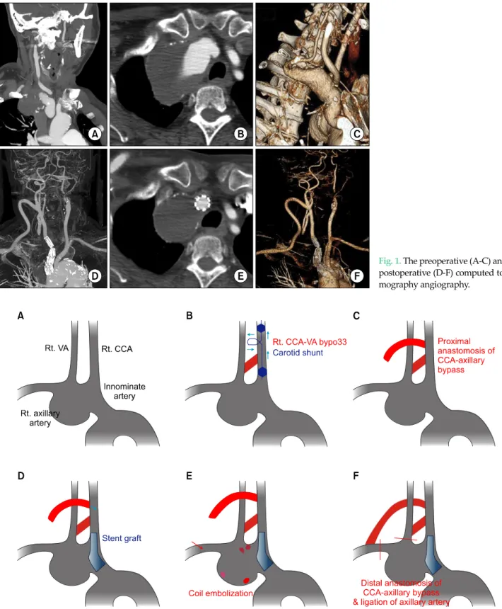
Fig. 1. The preoperative (A-C) and postoperative (D-F) computed to- mography angiography.
Fig. 2. Preoperative plan of the hybrid surgery. (A) The anatomy of the right subclavian artery aneurysm. (B) Right common carotid-vertebral bypass with the great saphenous vein graft. (C) Proximal anastomosis of the right common carotid-axillary bypass with a 7 mm ringed polytetrafluorethylene (PTFE) graft. (D) Stent graft from the innominate artery to the right common carotid artery via the PTFE graft. (E) Coil embolization of the branches. (F) Distal anastomosis of the right common carotid artery-right axillary artery and ligation of the axillary artery.
Rt., right; VA, vertebral artery; CCA, common carotid artery.
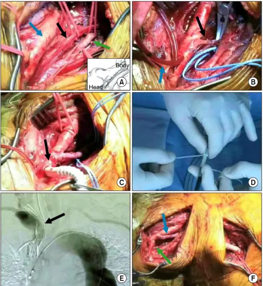
Fig. 3. Intra-operative pictures of the hybrid surgery. (A) Exposure of the right common carotid artery and right vertebral artery (blue arrow, carotid artery; green arrow, vertebral artery; black arrow, va- gus nerve). (B) Right common carotid-vertebral bypass with the great saphenous vein (GSV) graft (black arrow, GSV graft; blue arrow, carotid shunt). (C) Proximal anastomosis of the right common carotid-axillary bypass (black ar- row). (D) Reloading of the stent graft. (E) The stent graft from the innominate artery to the right common carotid artery (black arrow). (F) Distal anastomosis of the right common carotid artery- right axillary artery (blue arrow, right common carotid-vertebral bypass with the GSV graft; green arrow, right common carotid-axil- lary bypass with polytetrafluoroe- thylene graft).
The aneurysm developed from the innominate artery proximally and extended to the right subclavian artery distally. The right vertebral artery was originated the aneurysm, while the left vertebral artery was occluded (Fig. 1A-C). We planned to adopt a less invasive approach including a stent graft of innominate artery to carotid ar- tery and extra-anatomic bypass as shown in Fig. 2.
Under general anesthesia, after harvest of the great sa- phenous vein (GSV) for carotid-vertebral bypass, a sub- clavicular incision was made and the axillary artery was isolated. Then an oblique skin incision was made along the anterior border of the sternocleidomastoid muscle, and the common carotid and vertebral arteries were exposed.
An 1 cm-long vertical incision was made below the carotid bifurcation. A Pruitt-Inahara carotid shunt (LeMaitre
Vascular Inc., Burlington, MA, USA) was inserted through the arteriotomy to reduce the cerebral ischemic time. An anastomosis was made between the common carotid ar- tery below the arteriotomy and the vertebral artery using GSV (Fig. 3B).
The proximal anastomosis of the carotid-axillary by- pass was made with a 7-mm reinforced polytetrafluoro- ethylene (PTFE) graft (W.L. Gore and Associates Inc., Newark, DE, USA) in an end-to-side fashion at the site where the carotid shunt was inserted (Fig. 3C). The carotid shunt was maintained until the completion of the posteri- or wall suture and the beginning of the anterior wall su- ture of the anastomosis.
And then percutaneous access was obtained through the right groin and a 5-French pigtail catheter was ad-
vanced into the ascending thoracic aorta. An angiography confirmed the patency of the carotid-vertebral bypass.
A 6-French sheath was introduced through the side of the PTFE graft in the right common carotid artery. A Berenstein catheter (Terumo Co., Tokyo, Japan) with a guide wire was inserted, with the tip of the catheter ad- vanced into the ascending aorta. The size and position of the stent graft were assessed on the angiography. The Zenith stent graft with a diameter of 12 to 16 mm and a length of 72 mm (Cook Medical Inc., Bloomington, IN, USA) was reloaded in a reverse manner using umbilical tape (Fig. 3D). The stent graft was deployed from the in- nominate artery to the common carotid artery for exclud- ing the origin of the subclavian artery (Fig. 3E).
Since the thyrocervical trunk, costocervical trunk and internal mammary artery were already occluded on the angiography, further embolization was not required.
Finally, the graft was anastomosed to the axillary artery in end-to-side fashion with ligation of the axillary artery just distal to the aneurysm (Fig. 3F). Completion angiog- raphy of the aortic arch showed good flow through the stent-graft and bypasses with complete exclusion of the aneurysm.
There were not any perioperative events including is- chemic symptoms of the brain or arm. Postoperative course of the patient was uneventful during 1-year fol- low-up. And in the second month after operation, the hoarseness and dry cough disappeared completely. A CT angiography showed complete exclusion of the aneurysm without any endoleaks, and the maximal diameter of aneurysm decreased to 40 mm from 50 mm (Fig. 1D-F).
DISCUSSION
Subclavian artery aneurysm is the rarest peripheral ar- tery aneurysm. Up to one third of the patients with this aneurysm are asymptomatic, and intrathoracic aneur- ysms are more likely to be asymptomatic and they are very difficult to be found on a physical examination [1]. The symptoms of subclavian artery aneurysm are brachial plexipathy, Horner’s syndrome, dysphagia and hemop- tysis. Other symptoms can develop as the result of compli-
cations such as rupture, thrombosis or upper extremity embolism. Physicians dealing with these aneurysms have to be fully aware of the potential for multiple aneurysms that can be aortic, visceral or peripheral [2,3].
The natural history of subclavian artery aneurysms is not well known due to the limited number of cases and the high rate of surgical intervention. No reports have docu- mented any correlation between the absolute size of aneurysms and the potential risk for rupture. But elective surgical repair is mandatory for subclavian aneurysms even when they are asymptomatic, because they tend to increase in size with an increased risk of rupture, throm- bosis, embolization and compression of adjacent struc- tures [4]. Any complications such as thrombosis, embo- lism, rupture and rapid growth with increasing symptoms are absolute indications of surgery [1-3].
The treatment of large sized subclavian artery aneur- ysms has been surgical. Open surgical repair of the sub- clavian artery necessitates an invasive approach with ster- notomy or lateral thoracotomy. In a series of 13 patients who underwent open surgery for subclavian artery aneur- ysms, the postoperative complication rate was reported to be 46% [5]. Endovascular treatment of the subclavian ar- tery with a variety of devices and approaches has been re- ported in the literature [6-8].
There have been two case reports that are similar to ours [9,10]. But the operation in our case was different from these cases. First, the operations were done via a supra- clavicular incision in other two reports. But in our case, two separate incisions including a subclavicular incision and an oblique incision along the anterior border of the sternocleidomastoid muscle were made. We think that the separate oblique incision is better for the exposure of the carotid and vertebral arteries. Second, the left vertebral ar- tery was occluded in our case, so right vertebral artery by- pass was inevitable and we tried to reduce the cerebral is- chemic time during the anastomosis. We decided to use a carotid shunt with the method shown in Fig. 3B, and a car- otid-vertebral bypass with a GSV graft was performed with a minimal cerebral ischemic time. Third, in the other reports, the costocervical and thyrocervical trunks were li- gated, but we tried to embolize them with coils (Fig. 2E).
But on the angiography, the costocervical and thyrocer-
vical trunks were already occluded by the thrombus in the aneurysm, and so we didn’t perform coil embolization.
In conclusion, we successfully treated a complex true intrathoracic right subclavian artery aneurysm by a stent-graft combined with extra-anatomic bypasses. This hybrid operation can be a useful, less-invasive alternative to open surgical approaches via sternotomy or thoraco- tomy.
CONFLICTS OF INTEREST
No potential conflict of interest relevant to this article was reported.
REFERENCES
1. Halldorsson A, Ramsey J, Gallagher C, Meyerrose G.
Proximal left subclavian artery aneurysms: a case report and review of the literature. Angiology 2007;58:367-71.
2. Dougherty MJ, Calligaro KD, Savarese RP, DeLaurentis DA. Atherosclerotic aneurysm of the intrathoracic sub- clavian artery: a case report and review of the literature. J
Vasc Surg 1995;21:521-9.
3. McCollum CH, Da Gama AD, Noon GP, DeBakey ME.
Aneurysm of the subclavian artery. J Cardiovasc Surg (Torino) 1979;20:159-64.
4. Bin HG, Kim MS, Kim SC, Keun JB, Lee JH, Kim SS.
Intrathoracic aneurysm of the right subclavian artery pre- senting with hoarseness: a case report. J Korean Med Sci 2005;20:674-6.
5. Salo JA, Ala-Kulju K, Heikkinen L, Bondestam S, Ketonen P, Luosto R. Diagnosis and treatment of subclavian artery aneurysms. Eur J Vasc Surg 1990;4:271-4.
6. Becker GJ, Benenati JF, Zemel G, Sallee DS, Suarez CA, Roeren TK, et al. Percutaneous placement of a balloon-ex- pandable intraluminal graft for life-threatening subclavian arterial hemorrhage. J Vasc Interv Radiol 1991;2:225-9.
7. Hilfiker PR, Razavi MK, Kee ST, Sze DY, Semba CP, Dake MD. Stent-graft therapy for subclavian artery aneurysms and fistulas: single-center mid-term results. J Vasc Interv Radiol 2000;11:578-84.
8. Kasirajan K, Matteson B, Marek JM, Langsfeld M. Covered stents for true subclavian aneurysms in patients with de- generative connective tissue disorders. J Endovasc Ther 2003;10:647-52.
9. Resch TA, Lyden SP, Gavin TJ, Clair DG. Combined open and endovascular treatment of a right subclavian artery aneurysm: a case report. J Vasc Surg 2005;42:1206-9.
10. Van Leemput A, Maleux G, Heye S, Nevelsteen A.
Combined open and endovascular repair of a true right subclavian artery aneurysm without proximal neck.
Interact Cardiovasc Thorac Surg 2007;6:406-8.