ISSN 2234-3806 • eISSN 2234-3814
http://dx.doi.org/10.3343/alm.2015.35.6.657 www.annlabmed.org 657
Ann Lab Med 2015;35:657-659
http://dx.doi.org/10.3343/alm.2015.35.6.657
Letter to the Editor
Clinical Microbiology
Campylobacter hyointestinalis Isolated From a Human Stool Specimen
Do-kyun Kim, M.D.1, Sung Kuk Hong, M.D.1, Myungsook Kim, M.T.1, Jin Young Ahn, M.D.2, Dongeun Yong, M.D.1, and Kyungwon Lee, M.D.1
Department of Laboratory Medicine and Research Institute of Bacterial Resistance1; Department of Internal Medicine2, Yonsei University College of Medicine, Seoul, Korea
Dear Editor
Campylobacter are curved, gram-negative, non-spore-forming rods that usually grow in a microaerophilic environment. To date, 22 species have been assigned to Campylobacter. Although most Campylobacter species are zoonotic, C. jejuni, C. coli, and C. fetus are well-known human pathogens; C. upsaliensis and C.
lari are also considered enteropathogenic bacteria [1].
C. hyointestinalis was first isolated from pigs with proliferative enteritis in 1983 [2] and was initially considered a pathogen of pigs and rodents; however, there have been two previous re- ports of isolation from patients with proctitis and diarrhea [3, 4].
C. hyointestinalis is now thought to be a human pathogen, but reported cases are rare [1, 3, 4]. We report a case of gastroen- teritis possibly induced by C. hyointestinalis, which was identi- fied in stool specimens and confirmed with 16S ribosomal RNA (rRNA) gene sequencing, biochemical testing, and matrix-as- sisted laser desorption/ionization time-of-flight mass spectrome- try (MALDI-TOF MS).
The patient was an 88-yr-old male who visited the emergency room because of right-side weakness. He had a history of left middle cerebral artery infarction and rectal cancer and under- went low anterior resection with coloanal anastomosis and post- operative adjuvant radiotherapy five years earlier. He was diag-
nosed as having recurrent left middle cerebral artery infarction and admitted for antithrombotic therapy. On day three after ad- mission, he complained of diarrhea, which was unresponsive to antidiarrheal agents. Physical examination showed mild hypoes- thesia of the right extremities unrelated to the diarrhea. Labora- tory investigation showed hemoglobin level of 13.0 g/dL, leuko- cyte count of 5.58×10⁹/L, platelet count of 270×109/L, C-reac- tive protein level of 1.6 mg/L, and erythrocyte sedimentation rate of 9 mm/hr.
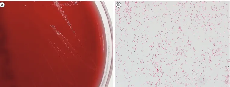
Stool culture was performed consecutively on days three and four. No remarkable growth of enteropathogenic bacteria was observed on MacConkey agar or salmonella-shigella agar. A large number of gray colonies positive for oxidase and catalase (Fig. 1A) were observed in a selective medium for Campylo- bacter species under microaerophilic conditions below 42°C [5].
Curved gram-negative rods were observed on the smear prepa- ration (Fig. 1B). The hippurate test was negative, and H2S pro- duction was positive in triple sugar iron (TSI) agar. Species iden- tification performed with a Bruker Microflex MALDI-TOF MS system (Bruker Daltonics, Bremen, Germany) showed C. hyoin- testinalis in both stool cultures (day 3 score: 2.199; day 4 score:
2.159). For confirmation, the 605 base pairs of the 16S rRNA gene were sequenced, and the ezTaxon database (http://www.
Received: February 12, 2015 Revision received: March 31, 2015 Accepted: July 9, 2015
Corresponding author: Kyungwon Lee
Department of Laboratory Medicine and Research Institute of Bacterial Resistance, Severance Hospital, Yonsei University College of Medicine, 50 Yonsei-ro, Seodaemun-gu, Seoul 03722, Korea
Tel: +82-2-2228-2446, Fax: +82-2-313-0956 E-mail: [email protected]
© The Korean Society for Laboratory Medicine.
This is an Open Access article distributed under the terms of the Creative Commons Attribution Non-Commercial License (http://creativecommons.org/licenses/by-nc/3.0) which permits unrestricted non-commercial use, distribution, and reproduction in any medium, provided the original work is properly cited.
Kim D-K, et al.
C. hyointestinalis isolated from human stool
658 www.annlabmed.org http://dx.doi.org/10.3343/alm.2015.35.6.657 ezbiocloud.net/eztaxon) showed 99.5% identity with GenBank
sequence JHQP01000019 (C. hyointestinalis) and 99.3% iden- tity with GenBank sequence AY621303 (C. fetus). These results confirmed the isolate as C. hyointestinalis (C. fetus: negative for H2S production in TSI agar). However, an examination for para- sites and ova and tests for Clostridium difficile were negative, in- cluding toxigenic culture and toxin PCR assay using the GeneX- pert system (Cepheid, Sunnyvale, CA, USA).
Antimicrobial susceptibility testing (AST) was performed via the disk diffusion method on blood agar incubated microaerobi- cally for 24 hr. Zone diameters of ciprofloxacin and erythromy- cin were 33 mm and 36 mm, respectively, which suggested susceptibility according to the interpretive criteria of the Clinical and Laboratory Standard Institute guidelines for C. jejuni and C.
coli. [6]. The patient was discharged without antibiotic treatment on day six. After two weeks, he returned to the infectious dis- eases outpatient clinic owing to persistent diarrhea with hema- tochezia. Oral ciprofloxacin (500 mg daily) for 14 days was pre- scribed without further examination. The patient did not return to the clinic; therefore, additional follow-up was not performed.
The clinical significance of C. hyointestinalis has not been clearly established. However, previous reports [3, 4] suggest that it is a human pathogen causing proctitis and enteritis. Al- though other pathogens for diarrhea including enterohemor- rhagic Escherichia coli, Yersinia, rotavirus, norovirus, and en- teric adenovirus were not considered, C. hyointestinalis was the only pathogen identified in this case. This species was isolated from two consecutive stool cultures, which indicate C. hyointes-
tinalis as a possible pathogen of diarrhea. Furthermore, this iso- late was initially identified as C. hyointestinalis with MALDI-TOF MS. Because identification with conventional biochemical meth- ods including the hippurate test, H2S production, and growth under specific conditions is cumbersome and time-consuming, MALDI-TOF MS may be a rapid and accurate method for identi- fying Campylobacter species [7].
In conclusion, we isolated C. hyointestinalis from a patient stool specimen for the first time in Korea and confirmed its iden- tification with MALDI-TOF MS and 16S rRNA gene sequencing.
This isolate appeared to be a pathogenic cause for enteritis.
Authors’ Disclosures of Potential Conflicts of Interest
No potential conflicts of interest relevant to this article were re- ported.
REFERENCES
1. Fitzgerald C and Nachamkin I. Camphylobacter and Arcobacter. In:
Manual of clinical microbiology. 10th ed. Washington DC: ASM press, 2011:885-99.
2. Gebart CJ, Ward GE, Chang K, Kurtz HJ. Campylobacter hyointestinalis (new species) isolated from swine with lesions of proliferative ileitis. Am J Vet Res 1983;44:361-7.
3. Fennell CL, Rompalo AM, Totten PA, Bruch KL, Flores BM, Stamm WE.
Isolation of “Campylobacter hyointestinalis” from a human. J Clin Mi- crobiol 1986;24:146-8.
4. Edmonds P, Patton CM, Griffin PM, Barrett TJ, Schmid GP, Baker CN,
A B
Fig. 1. Colony and microscopic characteristics of Campylobacter hyointestinalis. (A) Gray, flat, irregular and spreading colonies were ob- served on a blood agar plate after 48 hr of microaerobic incubation. (B) Curved gram-negative rods from a smear of colonies obtained from the blood agar plate (× 1,000).
Kim D-K, et al.
C. hyointestinalis isolated from human stool
http://dx.doi.org/10.3343/alm.2015.35.6.657 www.annlabmed.org 659
et al. Campylobacter hyointestinalis associated with human gastrointes- tinal disease in the United States. J Clin Microbiol 1987;25:685-91.
5. Chung Y, Lee K, et al. Diagnostic Microbiology. 5th ed. Seoul: Seoheung, 2014:374-85.
6. Clinical and Laboratory Standards Institute. Methods for antimicrobial dilution and disk susceptibility testing of infrequently isolated or fastidi- ous bacteria; approved guideline. M45-A2, 2nd ed. Wayne, PA: Clinical
and Laboratory Standards Institute, 2010.
7. Alispahic M, Hummel K, Jandreski-Cvetkovic D, Nöbauer K, Razzazi- Fazeli E, Hess M, et al. Species-specific identification and differentiation of Arcobacter, Helicobacter and Campylobacter by full-spectral matrix- associated laser desorption/ionization time of flight mass spectrometry analysis. J Med Microbiol 2010;59:295-301.