Mid-Term Outcomes in Patients Implanted with Cardiac Resynchronization Therapy
We applied cardiac resynchronization therapy (CRT) for desynchronized heart failure patients. We evaluated clinical outcomes including morbidity, mortality, and
echocardiographic parameters in 47 patients with implanted CRT in Korea from October 2005 to May 2013. The combined outcomes of hospitalization from heart failure, heart transplantation and death were the primary end point. Median follow-up period was 17.5 months. The primary outcomes listed above occurred in 10 (21.3%) patients. Two patients (4.3%) died after CRT and 8 (17%) patients were hospitalized for recurrent heart failure.
Among patients hospitalized for heart failure, 2 (4.3%) patients underwent heart transplantation. The overall free rate of heart failure requiring hospitalization was 90.1%
(95% CI, 0.81-0.99) over one year and 69.4% (95% CI, 0.47-0.91) over 3 yr. We observed improvement of the New York Heart Association classification (3.1 ± 0.5 to 1.7 ± 0.4), decreases in QRS duration (169.1 to 146.9 ms), decreases in left ventricular (LV) end- diastolic (255.0 to 220.1 mL) and end-systolic (194.4 to 159.4 mL) volume and increases in LV ejection fraction (22.5% to 31.1%) at 6 months after CRT. CRT improved symptoms and echocardiographic parameters in a relatively short period, resulting in low mortality and a decrease in hospitalization due to heart failure.
Keywords: Cardiac Resynchronization Therapy; Echocardiography; Heart Failure Sung Ho Lee,1 Seung-Jung Park,2
June Soo Kim,2 Dae-Hee Shin,3 Dae Kyoung Cho,4 and Young Keun On2
1Division of Cardiology, Kangbuk Samsung Medical Center, Sungkyunkwan University School of Medicine, Seoul; 2Division of Cardiology, Department of Medicine, Heart Vascular Stroke Institute, Samsung Medical Center, Sungkyunkwan University School of Medicine, Seoul; 3Division of Cardiology, Gangneung Asan Hospital, University of Ulsan College of Medicine, Gangneung; 4Division of Cardiology, S-Jungang Hospital, Jeju, Korea Received: 14 February 2014
Accepted: 12 August 2014 Address for Correspondence:
Young Keun On, MD
Division of Cardiology, Department of Medicine, Heart Vascular Stroke Institute, Samsung Medical Center, Sungkyunkwan University School of Medicine, 81 Irwon-ro, Gangnam-gu, Seoul 135-710, Korea
Tel: +82.2-3410-3419, Fax: +82.2-3410-3849 E-mail: [email protected]
http://dx.doi.org/10.3346/jkms.2014.29.12.1651 • J Korean Med Sci 2014; 29: 1651-1657
INTRODUCTION
Cardiac resynchronization therapy (CRT) is indicated for the treatment of New York Heart Association (NYHA) functional class III or IV heart failure, with a wide QRS duration (QRS du- ration ≥ 120 ms) and an ejection fraction ≤ 35% (1). CRT has been shown to improve symptoms, exercise capacity, and left ventricular (LV) function; in addition, it reduced mortality and hospitalization rates for heart failure in several large multicenter clinical trials (2-5). CRT devices have recently become a more common treatment for desynchronized heart failure patients in Korea. However, follow-up data regarding the effectiveness of CRT are sparse.
We analyzed the effectiveness of CRT by comparing clinical and echocardiographic parameters. We also evaluated mortali- ty and morbidities such as hospitalization from heart failure and heart transplantation in patients with an implanted CRT device in Korea.
MATERIALS AND METHODS Study population
We enrolled 47 patients who underwent CRT implantation at
Samsung Medical Center, Gangneung Asan Hospital, and Han- maeum General Hospital between October 2005 and May 2013.
The criteria for CRT include New York Heart Association (NYHA) function class III/IV symptoms despite optimal medical thera- py due to either ischemic or nonischemic cardiomyopathy with a left ventricular (LV) ejection fraction ≤ 35% and a QRS dura- tion ≥ 120 ms on electrocardiography.
The primary end point was a composite of death from any cause, hospitalization from heart failure, or need of heart trans- plantation. Hospitalization with heart failure was defined by symptoms such as dyspnea, chest discomfort, and increased edema resulting in the need for admission for treatment with intravenous diuretics or inotropics.
Study design
Patients meeting the criteria for enrollment were evaluated for NYHA class, QRS duration on 12-lead electrocardiogram (ECG), and two-dimensional Doppler echocardiography measures (LV ejection fraction, LV end-diastolic diameter, LV end-systolic di- ameter, LV end-diastolic volume, LV end-systolic volume) at baseline. After this initial evaluation, patients underwent im- plantation of CRT with a right atrial lead, right ventricular lead, and a left ventricular lead, which was inserted into the lateral or Cardiovascular Disorders
posterolateral cardiac vein by a transvenous approach. NYHA functional class, QRS duration, and echocardiographic param- eters were assessed at the 6-month follow-up visit. Combined outcomes of death, hospitalization from heart failure, and heart transplantation were assessed during the follow-up period.
CRT device implantation
The CRT device was implanted under local anesthesia with a transvenous approach via the left subclavian vein. The right ven- tricular lead was positioned in the RV apex or septum, and the right atrial lead was conventionally located in the right atrial appendage. The left ventricular lead was placed preferentially in a posterolateral or lateral vein after the coronary sinus veno- gram using an 8-Fr guiding catheter. The great cardiac vein or the middle cardiac vein were used only when other sites were not suitable or accessible. Only one patient required a left ven- tricular lead implanted via a thoracoscopic epicardial route; in this patient, the transvenous approach failed as there was no appropriate coronary venous branch.
Electrocardiograms
Standard 12-lead ECGs were obtained at baseline and at 6 months after CRT. Intraventricular conduction disturbances were de- fined according to criteria approved by the World Health Orga- nization (6). Left bundle branch block (LBBB) was defined as QRS duration ≥ 120 ms, QS or rS in lead V1, broad (frequently notched or slurred) R waves in leads I, aVL, V5 or V6, and absent q waves in lead V5 and V6. Right bundle branch block (RBBB) was defined as QRS duration ≥ 120 ms, rsr’, rsR’, Rsr’, or Qr in leads V1 or V2, and, occasionally, a wide and notched R wave and wide S waves in leads I, V5 and V6. Intraventricular con- duction delay (IVCD) was defined as QRS ≥ 110 ms without typical features of LBBB or RBBB.
Echocardiography
Baseline and subsequent echocardiograms were obtained at 6 months after CRT implantation. The LV end-diastolic diameter (LVEDD), LV end-systolic diameter (LVESD), LV end-diastolic volume (LVEDV), LV end-systolic volume (LVESV), LV ejection fraction (LVEF), left atrial (LA) width, and LA volume index (LAVI) were assessed according to the guidelines of the American So- ciety of Echocardiography (7). LV and LA volume were estimat- ed by Simpson’s equation in 2- and 4-chamber views.
Statistical analysis
Data were expressed as the median with interquartile range or mean ± standard deviation. Comparisons between groups were performed with the Student’s t-test for continuous variables and the chi-square test for categorical data. For comparison of para- metric variables between baseline and 6 months after CRT, the paired sample t-test was used. Cumulative clinical event-free
rate curves for heart failure, heart transplantation, and mortali- ty were determined according to the Kaplan-Meier method. Cox proportional hazards models were used to assess clinical fac- tors and primary outcomes. Values of P < 0.05 were considered significant. Statistical analysis was performed with SPSS 18.0 (SPSS Interactive Graphics, Version 18.0, SPSS Inc., Chicago, IL, USA).
Ethics statement
The study was approved by the institutional review board (IRB) of Samsung medical center (IRB No. 2014-03-104). In addition, the local IRB at each participating hospital approved this study and waived the requirement for informed consent.
RESULTS
Clinical characteristics of study subjects
Forty-seven patients (27 men and 20 women) with a mean age of 62.3 yr were enrolled in this study. The median duration of follow-up was 17.5 months (interquartile range, 6-26.5 months).
The baseline characteristics of the study population are described in Table 1. The cause of heart failure was ischemic in 6 (12.8%) patients and nonischemic in 41 (87.2%) patients. The mean NYHA class was 3.1 ± 0.5, despite the use of optimal treatment, including angiotensin-converting enzyme inhibitors or angio- tensin-receptor blockers in 36 (76.6%) patients, beta-blockers in 34 (72.3%) patients, and diuretics in 33 (70.2%) patients. Im- plantation of a CRT device alone was performed in 7 (14.9%) patients, while a CRT-D device was implanted in 39 (85.1%) pa- tients. Right ventricle lead placement was apical in 24 (51.1%) patients and septal in 22 (46.8%) patients. There were 32 (68.1%) patients with left bundle branch block, 4 (6.4%) patients with right bundle branch block, and 12 (25.5%) patients with non- specific intraventricular conduction delay. A history of atrial fi- brillation (AF) was observed in 10 patients (21.3%). Four patients underwent a CRT upgrade from a permanent pacemaker (PPM), initially implanted because of complete atrioventricular (AV) block. Although these patients had an atrial flutter fibrillation with slow ventricular rhythm in device interrogation after PPM implantation, the CRT upgrade was performed due to a high percentage of ventricular pacing, persistent severe HF symp- toms and depressed EF. Two patients were in sinus rhythm after direct current cardioversion or catheter ablation. Three patients had a paroxysmal AF. Only 1 patient was implanted with CRT after AV junction ablation for biventricular pacing because of persistent AF.
Clinical, electrocardiographic, and echocardiographic parameters 6 months after CRT
Clinical, electrocardiographic, and echocardiographic changes during the first 6 months after CRT are shown in Table 2 and
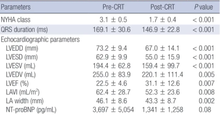
Table 2. Comparison of changes in clinical, electrocardiographic and echocardio- graphic parameters before and after CRT
Parameters Pre-CRT Post-CRT P value
NYHA class 3.1 ± 0.5 1.7 ± 0.4 < 0.001
QRS duration (ms) 169.1 ± 30.6 146.9 ± 22.8 < 0.001 Echocardiographic parameters
LVEDD (mm) LVESD (mm) LVESV (mL) LVEDV (mL) LVEF (%) LAVI (mL/m2) LA width (mm) NT-proBNP (pg/mL)
73.2 ± 9.4 62.9 ± 9.9 194.4 ± 62.8 255.0 ± 83.9 22.5 ± 4.6 62.4 ± 28.7 46.1 ± 8.6 3,697 ± 5,054
67.0 ± 14.1 55.0 ± 15.9 159.4 ± 99.7 220.1 ± 111.4
31.1 ± 12.6 52.3 ± 23.6 43.3 ± 8.7 1,341 ± 1,258
< 0.001
< 0.001
< 0.001 0.005 0.007 0.008 0.002 0.08 CRT, Cardiac resynchronization therapy; LA, left atrium; LAVI, left atrial volume index;
LVEDD, left ventricular end-diastolic diameter; LVEDV, left ventricular end-diastolic volume; LVEF, left ventricular ejection fraction; LVESD, LV end-systolic diameter; LVESV, left ventricular end-systolic volume; NT-proBNP, N-terminal pro-brain natriuretic pep- tide; NYHA, New York Heart Association.
Table 1. Baseline clinical characteristics of the study population (n = 47)
Characteristics Value
Age (yr) 62.3 ± 11.5
Male, No. (%) 27 (57.4)
Etiology, No. (%) Ischemic heart disease
Nonischemic heart disease 6 (12.8)
41 (87.2)
NYHA functional class 3.1 ± 0.5
Type of CRT device, No. (%) CRT
CRT-D 7 (14.9)
40 (85.1) Implantation technique, No. (%)
Transvenous
Epicardiac 46 (97.8)
1 (0.2) RV lead position, No. (%)
Apex Septum
24 (51.1) 22 (46.8) Comorbidities, No. (%)
Hypertension Diabetes mellitus Valvular heart disease Dyslipidemia Atrial fibrillation history Chronic renal failure
27 (57.4) 15 (31.9) 13 (27.7) 12 (25.5) 10 (21.3) 9 (19.1) Electrocardiographic parameters
QRS duration, ms LBBB, No. (%) RBBB, No. (%) IVCD, No. (%)
169.1 ± 30.6 32 (68.1)
3 (6.4) 12 (25.6) Echocardiographic parameters
LVEF (%) LVESV (mL) LFEDV (mL) LAVI (mL/m2)
22.9 ± 4.7 202.0 ± 65.5 263.2 ± 85.7 60.5 ± 26.0 Medication
Diuretics, No. (%) ACE inhibitor or ARB, No. (%) β-blocker, No. (%) Amiodarone, No. (%)
33 (70.2) 36 (76.6) 34 (72.3) 34 (72.3)
Data are presented as percentage or mean ± SD. ACE, angiotensin- converting en- zyme; ARB, angiotensin-receptor blocker; CRT, cardiac resynchronization therapy;
CRT-D, cardiac resynchronization therapy defibrillators; IVCD, interventricular conduc- tion delay; LAVI, left atrial volume index; LBBB, left bundle branch block; LVEDV, left ventricular end-diastolic volume; LVEF, left ventricular ejection fraction; LVESV, left ventricular end-systolic volume; RBBB, right bundle branch block.
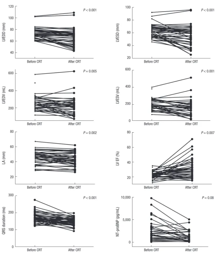
Fig. 1. NYHA functional class significantly improved from 3.1 ± 0.5 to 1.7 ± 0.4 (P < 0.001). The duration of the QRS interval de- creased from 169.1 ± 30.6 ms to 146.9 ± 22.8 ms (P < 0.001). LV- EDD decreased from 73.2 ± 9.4 mm to 67.0 ± 14.1 mm (P < 0.001), LVESD decreased from 62.9 ± 9.9 mm to 55.0 ± 15.9 mm (P <
0.001), LVESV decreased from 194.4 ± 62.8 mL to 159.4 ± 99.7 mL (P < 0.001), LVEDV decreased from 255.0 ± 83.9 mL to 220.1
± 111.4 mL (P = 0.005), and LVEF improved from 22.5% ± 4.6%
to 31.1% ± 12.6% (P = 0.007). LAVI decreased from 62.4 ± 28.7 mL/m2 to 52.3 ± 23.6 mL/m2 (P = 0.008), LA width decreased from 46.1 ± 8.6 mm to 43.3 ± 8.7 mm (P = 0.002), and N-termi- nal pro-brain natriuretic peptide (NT-proBNP) levels decreased from 3,697 ± 5,054 pg/ mL to 1,341 ± 1,258 pg/mL (P = 0.08).
The me dian decrease in the LVESV observed during the first six months after CRT was 21.9% (interquartile range 7.5%-47.7%),
the median decrease in the LVEDV was 10.4% (interquartile range 1.9%-38.4%), the median increase in the EF was 24.5%
(interquartile range 2.7%-57.7%), and the median decrease in LAVI was 15% (interquartile range 4.9%-34.3%).
In the patients with primary outcomes, baseline NYHA func- tional class was higher (3.5 ± 0.5 vs. 3.1 ± 0.5, P = 0.03), baseline QRS duration was prolonged (174.3 ± 36.3 vs. 167.8 ± 29.4 ms, P = 0.12), and baseline LV cavity dilation was more severe (LV- EDD, 78.2 ± 11.5 vs. 71.4 ± 7.9 mm, P = 0.03; LVESD, 67.9 ± 12.6 vs. 60.9 ± 8.0 mm, P = 0.03). Blunted improvement of echocar- diographic parameters after CRT were observed in patients who reached primary outcomes; we observed a smaller decrease in LVEDD (73.4 ± 16.4 vs. 65.0 ± 12.9 mL, P = 0.1), LVESD (60.5 ± 18.7 vs. 53.2 ± 14.8 mL, P = 0.21). LVEDV (7.3% ± 29.8% vs. 15.9%
± 29.0%, P = 0.49), LVESV (10.7% ± 39.9% vs. 22.1% ± 34.0%, P = 0.45), and EF (19.7% ± 61.5% vs. 49.4% ± 62.1%, P = 0.19) (Table 3) also chan ged less in patients with primary outcomes.
Primary end point
The Kaplan-Meier curves of free rate of hospitalization from heart failure, any death, and heart transplantation are shown in Fig. 2. Of the 47 patients, 2 (4.3%) patients died; one patient died suddenly 2 months after CRT, while the other patient died from heart failure after orthopedic surgery. Eight (17.0%) patients were hospitalized for worsening heart failure; 2 (4.3%) of these patients underwent heart transplantation during the follow-up period. The overall free rate of heart failure requiring hospital- ization was 90.1% (95% CI, 0.81-0.99) in one year, 69.4% (95%
CI, 0.47-0.91) in 3 yr, and 46.2% (95% CI, 0.06-0.85) in 5 yr (Fig.
1). The heart transplantation free rate of patients was 97% (95%
CI, 0.91-1.02) in 3 yr and 64% (95% CI, 0.12-1.17) in 6 yr (Fig. 2).
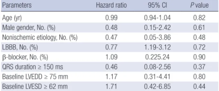
The primary end point was not influenced by the use of beta- blockers, the cause of heart failure (ischemic or nonischemic), the configuration of the QRS complex (left or non-left bundle branch block), the base-line duration of the QRS interval (QRS
Fig. 1. Comparison of changes in echocardiographic, electrocardiographic, and NT-pro BNP parameters before and after CRT.
LVEDD (mm)
Before CRT After CRT
120 100 80 60 40
LVESD (mm)
Before CRT After CRT
100 80 60 40 20
P < 0.001 P < 0.001
LVEDV (mL)
Before CRT After CRT
600
400
200
LVESV (mL)
Before CRT After CRT
600
400
200
0
P = 0.005 P < 0.001
LA (mm)
Before CRT After CRT
80
60
40
20
LV EF (%)
Before CRT After CRT
80
60
40
20
P = 0.002 P = 0.007
QRS duration (ms)
Before CRT After CRT
300
200
100
0
NT-proBNP (pg/mL)
Before CRT After CRT
10,000
5,000
0
P < 0.001 P = 0.08
duration < 150 ms or ≥ 150 ms), or the baseline echocardio- graphic parameters (LVEDD ≥ 75 mm or < 75 mm, LVESD ≥ 62 mm or < 62 mm) (Table 4).
DISCUSSION
We found that CRT substantially improved symptoms, reduced
QRS duration and improved echocardiographic parameters within the relatively short period of six months in Korean de- synchronized heart failure patients. The clinical, electrocardio- graphic, and echocardiographic parameters in this study are similar to those of previous clinical trials (8, 9). The data are con- sistent with a CRT reduction in the degree of ventricular dys- synchrony (as evidenced by a shortened QRS interval), and this
effect was accompanied by both an increase in the left ventric- ular ejection fraction and a decrease in the left ventricular end- diastolic and end-systolic dimension (5, 10). As a result, Korean patients with CRT experienced significant clinical improvements.
Ventricular remodeling consisting of LV dilation, cavity dis- tortion, and deterioration in pump function was a sign of poor prognosis in moderate to severe heart failure (11). Reversed re- modeling induced by CRT resulted in improved NYHA func- tional class, exercise capacity, and quality of life (4, 12). In our study, patients reaching primary outcomes had more baseline LV cavity dilatation and less reverse LV remodeling after CRT
Heart failure requiring hospitalization free rate
Month
0 12 24 36 48 60 72 84 100
80 60 40 20 0
Heart transplantation free rate
Month
0 12 24 36 48 60 72 84 100
80 60 40 20 0
Any death free rate
Month
0 12 24 36 48 60 72 84 96 100
90
80
70
Composite outcomes of HF, HT, Death free rate
Month
0 12 24 36 48 60 72 84 100
80 60 40 20 0
Fig. 2. Kaplan-Meier curve of the free rate of hospitalization from heart failure, heart transplantation and death, along with composite primary outcomes.
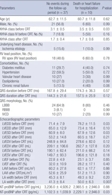
Table 3. Clinical and echocardiographic parameters in patients without and with pri- mary end points
Parameters
No events during the follow-up period (n = 37)
Death or heart failure for hospitalization
(n = 10) P value
Age (yr) 62.7 ± 11.5 60.7 ± 11.8 0.62
Male, No. (%) 21 (56.8) 6 (60) 0.99
NYHA class before CRT 3.1 ± 0.5 3.5 ± 0.5 0.03
NYHA class IV before CRT, No. (%) 7 (18.9) 5 (50) 0.16
NYHA class after CRT 1.7 ± 0.4 1.7 ± 0.6 0.85
Underlying heart disease, No. (%)
Ischemia etiology 5 (15.6) 1 (10.0) 0.99
RV lead position, No. (%)
RV apex (RV lead position) 18 (48.6) 6 (60.0) 0.78 Comorbidities, No. (%)
Diabetes mellitus Hypertension Valvular heart disease Dyslipidemia Chronic renal failure
11 (29.7) 22 (59.5) 10 (27) 10 (27.0)
5 (13.5)
4 (40.0) 5 (50.0) 3 (30) 2 (20.0) 4 (40)
0.74 0.72 0.99 0.99 0.08 QRS duration before CRT (ms) 167.8 ± 29.4 174.3 ± 36.3 0.57 QRS duration after CRT (ms) 144.3 ± 21.6 157.5 ± 25.9 0.12 QRS morphology, No. (%)
LBBB RBBB IVCD
24 (64.9) 3 (8.1) 10 (27)
8 (80) 0 2 (20)
0.46 0.99 0.99 Echocardiographic parameters
LVEDD before CRT (mm) LVEDD after CRT (mm) LVESD before CRT (mm) LVESD after CRT (mm) LVEDV before CRT (mL) LVEDV after CRT (mL) LVESV before CRT (mL) LVESV after CRT (mL) LVEF before CRT (%) LVEF after CRT (%) LAVI before CRT (mL/m2) LAVI after CRT(mL/m2) LA width before CRT (mm) LA width after CRT (mm)
71.4 ± 7.9 65.0 ± 12.9 60.9 ± 8.0 53.2 ± 14.8 248.9 ± 84.8 209.1 ± 106.6 190.1 ± 62.4 149.7 ± 93.3 22.8 ± 4.9 32.0 ± 10.9 60.2 ± 27.8 52.6 ± 25.9 45.3 ± 8.1 41.3 ± 8.4
78.2 ± 11.5 73.4 ± 16.4 67.9 ± 12.6 60.5 ± 18.7 278.6 ± 82.0 262.7 ± 127.8 211.0 ± 66.2 196.6 ± 121.8
23.1 ± 3.7 28.2 ± 17.1 62.1 ± 17.7 51.2 ± 11.3 49.3 ± 9.2 50.0 ± 6.3
0.03 0.10 0.03 0.21 0.12 0.20 0.14 0.18 0.85 0.40 0.86 0.90 0.20 0.007 NT-proBNP before CRT (pg/mL) 3,236.0 ± 4,926.2 2,965.5 ± 2,946.0 0.88 NT-proBNP after CRT (pg/mL) 1,102.3 ± 1,039.9 2,229.1 ± 2,946.0 0.03 ACE-I, ACE inhibitors; ARB, angiotensin-receptor blockers; LAVI, left atrial volume in- dex; LBBB, left bundle branch block; LVEDD, left ventricular end-diastolic diameter;
LVEDV, left ventricular end-diastolic volume; LVEF, left ventricular ejection fraction;
LVESD, LV end-systolic diameter; LVESV, left ventricular end-systolic volume; RBBB, right bundle branch block; NT-proBNP, N-terminal pro-brain natriuretic peptide; NYHA, New York Heart Association.
Table 4. Univariate analysis of associations between clinical factors and primary out- comes
Parameters Hazard ratio 95% CI P value
Age (yr) 0.99 0.94-1.04 0.82
Male gender, No. (%) 0.48 0.15-2.42 0.61
Nonischemic etiology, No. (%) 0.47 0.05-3.86 0.48
LBBB, No. (%) 0.77 1.19-3.12 0.72
β-blocker, No. (%) 1.09 0.225.24 0.90
QRS duration ≥ 150 ms 0.46 0.08-2.56 0.37
Baseline LVEDD ≥ 75 mm 1.17 0.31-4.41 0.80
Baseline LVESD ≥ 62 mm 1.71 0.42-6.85 0.44
CI, confidence interval; LBBB, left bundle branch block; LVEDD, left ventricular end- diastolic diameter; LVESD, LV end-systolic diameter; LVESV, left ventricular end-sys- tolic volume.
compared to those without primary outcomes; therefore, early implantation of a CRT device should be considered in these pa- tients before progressive LV dilation.
To our knowledge, this was the first study that investigated mortality and morbidity after CRT in Korea. The free rate of hos- pitalization from heart failure was as good as 90% after 1 yr, and 69% after 3 yr; however, it decreased to 46% in 5 yr after CRT.
Clinical outcomes after CRT confer greater benefits in a rela- tively short period (3 yr); however, that benefit was decreased over longer periods (5 yr). There were only two cases of death (4.3%) during the follow up period; this low mortality shows relatively good results in Korean patients who underwent CRT during a mid-term follow up period. Previous studies showed that the mortality rate was 10%/yr during 3 yr of follow up after CRT (9). Idiopathic dilated cardiomyopathy has a better respon- se to treatment and a better prognosis than heart failure from ischemic heart disease in a number of studies (13, 14). This dif- ference by etiology was explained by the rate of cardiac death due to progressive cardiac failure in ischemic heart disease (9).
First and subsequent heart failure episodes after CRT were as- sociated with 7- and nearly 19-fold respective increases in the risk of subsequent all-cause mortality in the MADIT-CRT study (15). In our study, the majority of the study population had id- iopathic dilated cardiomyopathy. Heart transplantation was performed at the appropriate time in patients with progressive heart failure. This selection bias and appropriate treatment such as heart transplantation might underestimate the mortality and hospitalization from heart failure. However, our data showed that the benefit of CRT therapy for the prevention of heart fail- ure was pronounced for 3 yr after CRT. CRT appears to be the optimal treatment in certain patients; our study showed a prom- inent decrease in mortality along with a decrease in hospital- ization from heart failure.
This study has several limitations. First, comparisons to this analysis are limited by retrospective design, as confounders that were not evaluated might have influenced outcomes. Second, although each center recruited patients consecutively and fol- low-up loss is rarely observed, the inclusion of two tertiary re-
ferral centers may have led to selection bias because more pa- tients with advanced heart failure were included. Third, because the main etiology of CRT indication was non-ischemic heart disease, it is possible that the prognosis for primary outcomes such as heart failure or death was fairly good. Lastly, our study population was small.
In conclusion, CRT improves clinical symptoms and electro- cardiographic/echocardiographic parameters over a relatively short period, resulting in a decrease in mortality and hospital- ization due to heart failure.
DISCLOSURE
The authors have no conflicts to disclose.
ORCID
Young Keun On http://orcid.org/0000-0003-1025-7283
REFERENCES
1. Epstein AE, DiMarco JP, Ellenbogen KA, Estes NA 3rd, Freedman RA, Gettes LS, Gillinov AM, Gregoratos G, Hammill SC, Hayes DL, et al. ACC/
AHA/HRS 2008 Guidelines for Device-Based Therapy of Cardiac Rhythm Abnormalities: a report of the American College of Cardiology/Ameri- can Heart Association Task Force on Practice Guidelines (Writing Com- mittee to Revise the ACC/AHA/NASPE 2002 Guideline Update for Im- plantation of Cardiac Pacemakers and Antiarrhythmia Devices): devel- oped in collaboration with the American Association for Thoracic Sur- gery and Society of Thoracic Surgeons. Circulation 2008; 117: e350-408.
2. Abraham WT, Fisher WG, Smith AL, Delurgio DB, Leon AR, Loh E, Ko- covic DZ, Packer M, Clavell AL, Hayes DL, et al. Cardiac resynchroniza- tion in chronic heart failure. N Engl J Med 2002; 346: 1845-53.
3. Bristow MR, Saxon LA, Boehmer J, Krueger S, Kass DA, De Marco T, Carson P, DiCarlo L, DeMets D, White BG, et al. Cardiac-resynchroniza- tion therapy with or without an implantable defibrillator in advanced chronic heart failure. N Engl J Med 2004; 350: 2140-50.
4. Cazeau S, Leclercq C, Lavergne T, Walker S, Varma C, Linde C, Garrigue S, Kappenberger L, Haywood GA, Santini M, et al. Effects of multisite bi- ventricular pacing in patients with heart failure and intraventricular conduction delay. N Engl J Med 2001; 344: 873-80.
5. Cleland JG, Daubert JC, Erdmann E, Freemantle N, Gras D, Kappen- berger L, Tavazzi L, Cardiac Resynchronization-Heart Failure (CARE- HF) Study Investigators. The effect of cardiac resynchronization on mor- bidity and mortality in heart failure. N Engl J Med 2005; 352: 1539-49.
6. Surawicz B, Childers R, Deal BJ, Gettes LS, Bailey JJ, Gorgels A, Han- cock EW, Josephson M, Kligfield P, Kors JA, et al. AHA/ACCF/HRS rec- ommendations for the standardization and interpretation of the electro- cardiogram: part III: intraventricular conduction disturbances: a scien- tific statement from the American Heart Association Electrocardiogra- phy and Arrhythmias Committee, Council on Clinical Cardiology; the American College of Cardiology Foundation; and the Heart Rhythm So- ciety. Endorsed by the International Society for Computerized Electro-
cardiology. J Am Coll Cardiol 2009; 53: 976-81.
7. Lang RM, Bierig M, Devereux RB, Flachskampf FA, Foster E, Pellikka PA, Picard MH, Roman MJ, Seward J, Shanewise JS, et al. Recommenda- tions for chamber quantification: a report from the American Society of Echocardiography’s Guidelines and Standards Committee and the Cham- ber Quantification Writing Group, developed in conjunction with the European Association of Echocardiography, a branch of the European Society of Cardiology. J Am Soc Echocardiogr 2005; 18: 1440-63.
8. Abreu CD, Xavier RM, Nascimento JS, Ribeiro AL. Long-term outcome after Cardiac Resynchronization Therapy: a nationwide database. Int J Cardiol 2012; 155: 492-3.
9. Gasparini M, Lunati M, Santini M, Tritto M, Curnis A, Bocchiardo M, Vincenti A, Pistis G, Valsecchi S, Denaro A, et al. Long-term survival in patients treated with cardiac resynchronization therapy: a 3-year follow- up study from the InSync/InSync ICD Italian Registry. Pacing Clin Elec- trophysiol 2006; 29: S2-10.
10. Knappe D, Pouleur AC, Shah AM, Cheng S, Uno H, Hall WJ, Bourgoun M, Foster E, Zareba W, Goldenberg I, et al. Dyssynchrony, contractile function, and response to cardiac resynchronization therapy. Circ Heart Fail 2011; 4: 433-40.
11. Pitt B, Zannad F, Remme WJ, Cody R, Castaigne A, Perez A, Palensky J, Wittes J. The effect of spironolactone on morbidity and mortality in pa- tients with severe heart failure. Randomized Aldactone Evaluation Study Investigators. N Engl J Med 1999; 341: 709-17.
12. Gras D, Leclercq C, Tang AS, Bucknall C, Luttikhuis HO, Kirstein-Ped- ersen A. Cardiac resynchronization therapy in advanced heart failure the multicenter InSync clinical study. Eur J Heart Fail 2002; 4: 311-20.
13. Likoff MJ, Chandler SL, Kay HR. Clinical determinants of mortality in chronic congestive heart failure secondary to idiopathic dilated or to isch- emic cardiomyopathy. Am J Cardiol 1987; 59: 634-8.
14. Adams KF Jr, Dunlap SH, Sueta CA, Clarke SW, Patterson JH, Blauwet MB, Jensen LR, Tomasko L, Koch G. Relation between gender, etiology and survival in patients with symptomatic heart failure. J Am Coll Car- diol 1996; 28: 1781-8.
15. Goldenberg I, Hall WJ, Beck CA, Moss AJ, Barsheshet A, McNitt S, Po- lonsky S, Brown MW, Zareba W. Reduction of the risk of recurring heart failure events with cardiac resynchronization therapy: MADIT-CRT (Mul- ticenter Automatic Defibrillator Implantation Trial With Cardiac Resyn- chronization Therapy). J Am Coll Cardiol 2011; 58: 729-37.