Midgut volvulus is a very uncommon cause of acute abdomenal distress in adults (1). Midgut volvulus is mostly related to intestinal malrotation, and it is rarely associated with postoperative adhesions and a bulky di- et after fasting (2). Although acquired jejunoileal diver- ticula are uncommon and asymptomatic in the majority of patients, the association of midgut volvulus and a large small-bowel diverticulum has been mentioned on- ly in a radiologic report (3). To the best of our knowl- edge, this condition has never been reported in the Korean literature. Therefore, we present our imaging findings of midgut volvulus associated with a large,
small-bowel diverticulum in an adult, together with a re- view of the literature.
Case Report
A 77-year-old woman presented with acute, cramping epigastric pain and she had bilious vomiting for 1 day.
She had a history of severe abdominal pain persisting for several hours, and these symptoms occurred two or three times for a year during the past 30 years. The physical examination revealed epigastric tenderness without any peritoneal signs. The laboratory findings were all within normal limits.
A contrast-enhanced abdomen CT showed the typical whirl sign of the mesenteric root resulting from loops of bowel and mesenteric vessels twisted several times around the superior mesenteric artery (Fig. 1A). The di- agnosis of midgut volvulus was suggested, but the se- vere abdominal pain of a cramping nature subsided sev-
J Korean Radiol Soc 2004;50:365-368
─ 365 ─
Imaging Findings of Midgut Volvulus Associated with a Large Small-Bowel Diverticulum in an
Adult Patient: Case Report1
Jee Young Kim, M.D., Sung Eun Rha, M.D., Soon Nam Oh, M.D., Seal Hwang-bo, M.D., Jae Young Byun, M.D.
Although most patients with jejunoileal diverticulum are asymptomatic, a large, small-bowel diverticulum can be associated with midgut volvulus in an adult. We pre- sent a rare case of midgut volvulus that was associated with a large, small-bowel diver- ticulum in a 77-year-old woman presenting with chronic recurrent abdominal pain.
The CT showed the characteristic whirl sign of twisted mesentery, the small bowel loops along the superior mesenteric artery and a large sac-like small-bowel diverticu- lum. A small bowel series also demonstrated a corkscrew appearance of proximal je- junum, a finding suggestive of midgut volvulus, and a large jejunal diverticulum.
During the laparotomy, the small bowel was seen twisted counterclockwise 270°. The mesenteric root was very shortened. A 4 cm sized diverticulum was seen on the mesenteric border of jejunum, on the portion about 40 cm distal from the Treitz liga- ment.
Index words :Intestines, CT Intestines, diverticula
Intestines, stenosis or obstruction
1Department of Radiology, Kangnam St. Mary’s Hospital, College of Medicine, The Catholic University of Korea
Received December 24, 2003 ; Accepted February 27, 2004
Address reprint requests to : Sung Eun Rha, M.D., Department of Radiology, Kangnam St. Mary’s Hospital, College of Medicine, The Catholic University of Korea, 505 Banpo-dong, Seocho-gu, Seoul 137-040, South Korea.
Tel. 82-2-590-2468 Fax. 82-2-599-6771 E-mail: [email protected]
Jee Young Kim, et al: Imaging Findings of Midgut Volvulus Associated with a Large Small-Bowel Diverticulum in an Adult Patient
─ 366 ─
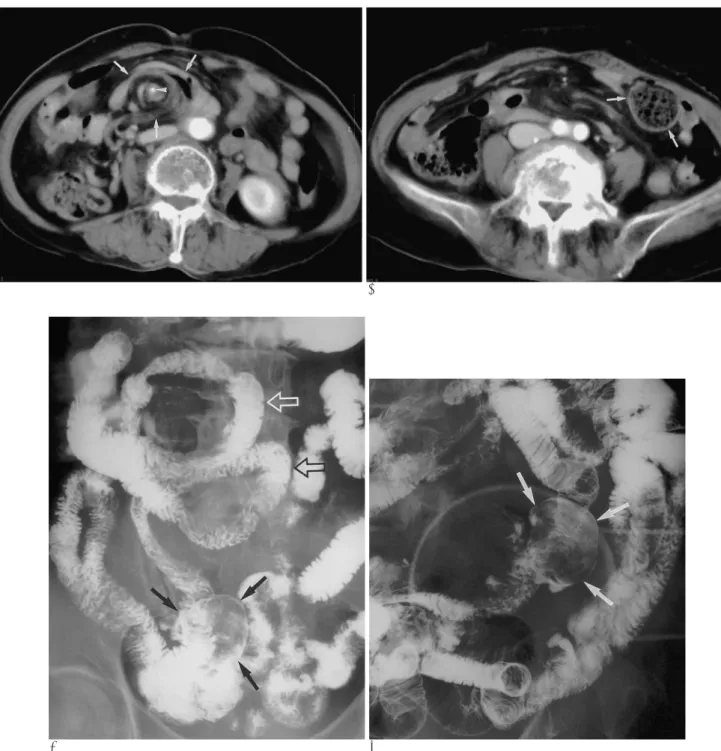
A B
C D
Fig. 1. Midgut volvulus associated a large small-bowel diverticulum in a 77-year-old woman.
A. Contrast-enhanced CT scan shows the typical whirl sign (arrows) of the mesenteric root. This is resulting from twisted loops of bowel and mesenteric vessels several times around the superior mesenteric artery (arrowhead).
B. Contrast-enhanced CT scan, 5 cm below A, reveals a large sized sac-like structure (arrows) filled with a stool-like mass of digest- ed food in the left side of lower abdomen. This is a finding of a large, small-bowel diverticulum.
C. Small bowel series shows typical corkscrew appearance (open arrows) of the proximal small bowel loops, a characteristic find- ing of midgut volvulus. The duodenojejunal junction is located just to the left of midline and to the right of its expected position, a finding suggestive of intestinal malrotation. About a 5.5×4 cm sized divertuculum (arrows) is also showing in the mesenteric bor- der of distal jejunum.
D. Spot radiograph of a small bowel series showing a large diverticulum (arrows) filled with a large well-defined mass lesion, which might be a bezoar or impacted fecal material.
eral hours after CT, and only the mild intermittent peri- umbilical pain persisted.
A small bowel series was performed during the next 6 days and these tests demonstrated the typical corkscrew appearance of the proximal small bowel loops, a charac- teristic finding of midgut volvulus. The duodenojejunal junction was located to the right of its expected position, a finding that is very suggestive of intestinal malrota- tion. In addition, about a 5.5×4 cm sized diverticulum was shown in the mesenteric border of distal jejunum (Fig. 1C). The lumen of the diverticulum was filled with a large well-defined mass-like lesion, which might have been a bezoar, or possibly impacted fecal material (Fig.
1D). The retrospective analysis of the CT revealed a large sized sac-like lesion filled with stool-like mass of digested food in the lower abdomen (Fig. 1B).
Surgical exploration confirmed midgut volvulus and a large jejunal diverticulum. There was no bowel necrosis present. The adhesions from recurrent episode of volvu- lus were lysed, the small bowel was untwisted and exci- sion of the diverticulum was performed. The patient did well after the operation and she has had no return of symptoms upon follow-up for 1 year.
Discussion
Midgut volvulus is the torsion of a segment or of all of small bowel and its mesentery, leading to a closed-loop obstruction and the vascular compromise to the bowel.
It is a potentially fatal surgical emergency. Midgut volvulus is mostly related to intestinal malrotation, and it’s rarely associated with pregnancy, postoperative ad- hesion, bulky diet after fasting and mass lesions, such as mesenteric lipoma or large pedunculated subserosal my- oma that may act as a leading mass. Any disturbance in the normal 270°counterclockwise return of the intes- tine into the abdominal cavity could produce a range of rotational and attachment abnormalities. The lack of normal peritoneal attachment predisposes the bowel to- wards development of a volvulus, with the twisting oc- curring around its attachment point and fulcrum, the su- perior mesenteric artery (4). In infancy, intestinal malro- tation almost always presents with a high intestinal ob- struction as a result of duodenal compression, obstruc- tion and often volvulus. However, intestinal malrotation in adults is usually an incidental finding, and it presents as chronic nonspecific gastrointestinal tract symptoms and chronic intermittent midgut volvulus, or less com- monly, as acute abdominal pain (5, 6). The nonspecific
clinical manifestations of midgut volvulus make diagno- sis difficult in adults. In this regard, imaging studies play an important role in the diagnosis.
Diagnosis of midgut volvulus can be made by means of ultrasound, CT and small bowel sevies. A gray-scale ultrasound finding of midgut volvulus is a whirl-like mass in the right upper abdomen and color Doppler ul- trasound shows a whirl sign with a clockwise rotation of the SMV around the SMA (7). CT is also very useful in diagnosis of midgut volvulus, as well as its complication of ischemia. CT reveals the characteristic whirl sign;
twisted loops of bowel and the branching mesenteric vessels create swirling strands of soft-tissue attenuation within a background of mesenteric fat attenuation, giv- ing the appearance of a hurricane on a weather map (8, 9). The volvulus causes the mesenteric veins and lym- phatics to become congested. Thickening of bowel wall and intraperitoneal fluid or gas in the bowel wall can al- so suggest the associated bowel infarction. A small bow- el series may demonstrate a spiral or corkscrew appear- ance resulting from a small bowel’s wrapping around the superior mesenteric artery, which is diagnostic of midgut volvulus.
The incidence of jejunoileal diverticula is 1.1 to 2.3%, and it is found upon enteroclysis, at postmortem by in- sufflating the intestine with air or during major abdomi- nal surgical procedures (10). Jejunoileal diverticula are asymptomatic in the majority of patients, but they may be the underlying cause of vague, chronic symptoms and such acute complications that include obstruction, hemorrhage and perforation. Mechanial intestinal ob- struction occurs in 2.3% to 4.6% of the cases of je- junoileal diverticulosis (11). This may be the result of pressure on the intestinal wall from distended diverticu- la, inflammatory mass associated with diverticulitis, stricture or adhesions from diverticulitis, intussuscep- tion at the site of the diverticulum, enteroliths devel- oped within the diverticula, or volvulus of the diverticu- la-containing segment (10).
In 1998, Chou et al. reported CT findings of a large small-bowel diverticulum in five cases (3). It was inter- esting that in all five cases, the CT demonstrated that midgut volvulus coexisted with a large, small bowel di- verticulum, as was seen in our case. All the diverticula were larger than 3 cm and located in the jejunum. The authors mentioned that it was not clear whether these two conditions have any direct relationship or not.
However, a large, small-bowel diverticulum might play a predisposing role in the occurrence of a midgut volvu-
J Korean Radiol Soc 2004;50:365-368
─ 367 ─
lus for the following reasons. (1) It may act as a leading mass if it were filled with fluid, and (2), it may interfere with the returning of an abnormally moved small-bowel loop and make the small-bowel rotate still further until a volvulus occurs.
In our case, both CT and a small bowel series showed the typical imaging findings of midgut volvulus. In addi- tion, a small bowel series showed the underlying intesti- nal malrotation and a large jejunal diverticulum (more than 3 cm). A retrospective CT analysis can also detect a large jejunal diverticulum, because of its large size (> 3 cm), its different intraluminal contents as compared with the surrounding small-bowel loops and the ab- sence of valvular conniventes (3). Indeed, the size of the diverticulum may play an important role in inducing small-bowel volvulus.
Considering the patient’s past history of recurrent cramping abdominal pain, the chronic abdominal symp- toms are thought to be from a chronic intermittent midgut volvulus precipitated by intestinal malrotation and a large jejunal diverticulum.
In summary, a midgut volvulus associated with in- testinal malrotation is uncommonly seen in adults, espe- cially with a large small-bowel diverticulum. Awareness of the imaging findings and the clinical significance of a large small-bowel diverticulum associated with midgut
volvulus can be helpful for the exact diagnosis and the management of this rare condition when it presents in the adult with recurrent abdominal pain.
References
1. Pelucio M, Haywood Y. Midgut volvulus: an unusual cause of adolescent abdominal pain. Am J Emerg Med 1994;12:167-171 2. Rowsom JT, Sullivan SN, Girvan DP. Midgut volvulus in the adult.
A complication of intestinal malrotation. J Clin Gastroenterol 1987;
9:212-216
3. Chou CK, Mak CW, Hou CC, Chang JM. CT of large small-bowel diverticulum. Abdom Imaging 1998;23:132-134
4. Fisher JK. Computer tomographic diagnosis of volvulus in intesti- nal malrotation. Radiology 1981;140:145-146
5. Bernstein SM, Russ PD. Midgut volvulus: a rare cause of acute ab- domen in an adult patient. AJR Am J Roentgenol 1998;171:639-641 6. Izes BA, Scholz FJ, Munson JL. Midgut volvulus in an elderly pa-
tient. Gastrointest Radiol 1992;17:102-104
7. Yeh WC, Wang HP, Chen C, Wang HH, Wu MS, Lin JT.
Preoperative sonographic diagnosis of midgut malrotation with volvulus in adults: the “whirlpool” sign. J Clin Ultrasound 1999;27:
279-283
8. Puvaneswary M, Rajaratham S. Midgut volvulus in adult.
Australas Radiol 2003;47:83-84
9. Khurana B. The whirl sign. Radiology 2003;226:69-70
10. de Bree E, Grammatikakis J, Christodoulakis M, Tsiftsis D. The clinical significance of acquired jejunoileal diverticula. Am J Gastroenterol 1998;93:2523-2538
11. Palder SB, Frey CB. Jejunal diverticulosis. Arch Surg 1988;123:889- 894
Jee Young Kim, et al: Imaging Findings of Midgut Volvulus Associated with a Large Small-Bowel Diverticulum in an Adult Patient
─ 368 ─
대한영상의학회지 2004;50:365-368
성인에서 발생한 소장 게실과 동반된 중장 염전: 증례 보고1
1가톨릭대학교 강남성모병원 진단방사선과
김지영・나성은・오순남・황보설・변재영
대부분의 소장 게실은 증상이 없지만 성인에서 발견되는 커다란 소장 게실은 중장 염전과 동반될 수 있다. 저자들은 만성 재발성 복통을 호소하는 77세 여자에서 발생한 커다란 소장 게실과 동반된 중장 염전의 드문 증례를 보고하고자 한다. 전산화단층촬영에서 상장간막 동맥을 따라 꼬인 장간막과 소장의 특징적인 와류증후(whirl sign)와 커다란 낭 모 양의 소장 게실이 보였다. 소장조영술(small bowel series)에서도 중장 염전을 시사하는 소견인 근위부 공장의 코르크 따개 모양(corkscrew appearance)과 커다란 공장 게실이 보였다.
수술시 소장은 반시계방향으로 270°돌아있었고 장간막의 기저부는 매우 짧았다. 약 4 cm 크기의 게실이 트라이츠 인대(Treitz ligament) 40 cm 하방의 공장에서 발견되었다.