Long-Term Survival Analysis of Korean Breast Cancer Patients at a Single Center: Improving
Outcome Over Time
Sung Gwe Ahn,
1Hak Min Lee,
1Seung Ah Lee,
2Joon Jeong,
1and Hy-De Lee
11Department of Surgery, Gangnam Severance Hospital, Yonsei University College of Medicine, Seoul;
2Department of Surgery, Eulji General Hospital, Eulji University College of Medicine, Seoul, Korea.
Received: October 31, 2013 Revised: January 9, 2014 Accepted: January 9, 2014
Corresponding author: Dr. Joon Jeong, Department of Surgery,
Gangnam Severance Hospital, Yonsei University College of Medicine, 211 Eonju-ro, Gangnam-gu, Seoul 135-720, Korea.
Tel: 82-2-2019-3379, Fax: 82-2-3462-5994 E-mail: [email protected]
∙ The authors have no financial conflicts of interest.
© Copyright:
Yonsei University College of Medicine 2014 This is an Open Access article distributed under the terms of the Creative Commons Attribution Non- Commercial License (http://creativecommons.org/
licenses/by-nc/3.0) which permits unrestricted non- commercial use, distribution, and reproduction in any medium, provided the original work is properly cited.
Purpose: The prognosis of breast cancer has been consistently improving. We an- alyzed our cohort of breast cancer patients with a long-term follow up at a single center over time. Materials and Methods: A total of 1889 patients with known cancer stages were recruited and analyzed between January 1991 and December 2005. Patients were classified according to the time periods (1991--1995; 1996-- 2000; 2001--2005). To determine intrinsic subtypes, 858 patients whose human epidermal growth receptor-2 status and Ki67 were reported between April 2004 and December 2008 were also analyzed. Results: At a median follow up of 9.1 years, the 10-year overall survival (OS) rate was 80.5% for the entire cohort. On multivariate analysis for OS and recurrence-free survival (RFS), the time period was demonstrated to be a significant factor independent of conventional prognos- tic markers. In the survival analysis performed for each stage (I to III), OS and RFS significantly improved according to the time periods. Adoption of new agents in adjuvant chemotherapy and endocrine therapy was increased according to the elapsed time. In the patients with known subtypes, OS and RFS significantly dif- fered among the subtypes, and the triple-negative subtype showed the worst out- come in stages II and III. Conclusion: In the Korean breast cancer cohort with a long-term follow up, our data show an improved prognosis over the past decades, and harbor the contribution of advances in adjuvant treatment. Moreover, we pro- vided new insight regarding comparison of the prognostic impact between the tu- mor burden and subtypes.
Key Words: Breast cancer, survival, subtype, long-term
INTRODUCTION
Breast cancer is the most common cancer in women, with about 1.5 million new cases being diagnosed annually worldwide, a lifetime risk of up to 12%, and a risk of death of up to 5% in Western countries.1 The incidence of breast cancer in Ko- rea has been increasing constantly, although it remains low compared with its inci- dence in Western countries.2-5 According to the hospital-based cancer registry
MATERIALS AND METHODS
Patient population
A prospectively maintained database of breast cancer pa- tients treated at Gangnam Severance Hospital, Seoul, Re- public of Korea, was used to identify patients who under- went operation with a diagnosis of breast cancer between January 1991 and December 2005. The period was divided into three corresponding periods: 1991--1995, 1996--2000, and 2001--2005. The follow-up protocol included planned regular visits every 6 months and requests for missed ap- pointments with a telephone call were made to minimize patient loss and raise the accuracy of survival data. The last update of the clinical database was in September 2012.
Among the patients receiving an operation, six male pa- tients with breast cancer, one patient with occult cancer, and 18 patients with breast cancer of non-epithelial origin (such as a phyllodes tumor, sarcoma, or lymphoma) were exclud- ed. For survival analysis based on stages, patients with an unknown tumor size and nodal status were also excluded.
Bilateral breast cancer was counted as a single patient. Fi- nally, 1889 patients were included in the primary analysis.
The institutional review board of Gangnam Severance Hos- pital, Yonsei University, Seoul, Korea, approved the study to be in accordance with good clinical practice guidelines and the Declaration of Helsinki (Local IRB number: #3- 2013-0152). The need for informed consent was waived due to the retrospective design.
For analysis using intrinsic subtypes, we required infor- mation regarding HER-2 status. However, we reported reli- able HER-2 data from the institute for April 2002. There- fore, to overcome a small sample size of analysis based on subtypes, with a restriction to this analysis, we extended the study period to December 2008. As a result, 858 patients treated between April 2004 and December 2008 were in- cluded in the present analysis.
The staging system was based on the American Joint Committee on Cancer (AJCC), 6th edition.14 We revised the final stage according to the criteria. In synchronous bilateral breast cancer, the higher stage between two tumors was se- lected. In metachronous bilateral breast cancer, the stage of the first cancer was chosen. In patients receiving neoadju- vant chemotherapy, the clinical stage before neoadjuvant chemotherapy was applied.
During the process of collecting information on adjuvant treatment, we investigated the change in drug regimen ac- called the Korea Central Cancer Registry, the age-standard-
ized breast cancer incidence rate per 100000 was 20.9 in 1999, and it was exponentially elevated to 39.8 in 2010, providing an annual percent change of 6.3%.6 Based on this national-wide database, the breast was the second most common cancer site following the thyroid in year 2010 and comprised 14.3% of all female cancers.6
Although there is an increasing incidence of breast can- cer in Korea, the survival outcome of breast cancer patients has also markedly improved. The 5-year survival rate be- tween 1993 and 1995 was 78.0%, and that rate jumped up to 89.5% between 2003 and 2007.2 Many reports have pro- vided relevant explanations for the recent survival improve- ment in breast cancer patients. In part, the incline in surviv- al rate can be attributed to nationwide screening programs, with expansion of a proportion of patients with ductal carci- noma in situ and early breast cancer.7 Other reasons for these improvements are the advances in adjuvant treatment, which includes the increasing use of adjuvant anthracycline-based regimens or taxane-based regimens8,9 and clinical adoption of new agents: for instance, aromatase inhibitors for hor- mone receptor-positive tumors and the monoclonal anti- body trastuzumab for human epidermal growth factor re- ceptor-2 (HER-2)-positive tumors.10,11 However, almost all these studies have been reported in Western countries, in which Asian races had seldom participated. In contrast to these countries, very few investigations have been reported to explain the underlying cause of the survival improve- ment of Korean breast cancer patients over time. Thus, it would be clinically relevant to discriminate the influences between the incremental changes in early-stage cancer and time periods that suggest the advancement in cancer man- agement.
Recently, molecular subtyping of breast cancer was logi- cally accepted in clinical practice12,13 hence, the prognostic influence of the subtypes has become increasingly impor- tant. Therefore, we explored survival analysis according to subtype in the present investigation.
To discriminate the impact on survival between tumor stage and time periods, we analyzed survival outcome ac- cording to the time trend at a single institution. We sought to delineate the improving trend of survival outcome according to time periods at each stage and uncover factors of survival prolongation using our database of well over 1000 patients.
To evaluate a prognostic influence of the intrinsic subtypes, we compared survival among subtypes defined by immu- nohistochemistry (IHC) markers.
Kaplan-Meier method were compared using the log-rank test. Factors, which were significantly demonstrated in the univariate analyses, were used in the multivariate analyses.
A Cox proportional hazards regression model was used to assess the effect of each potential prognostic variable on survival. A p value <0.05 was considered significant. The software used to perform these analyses was the SPSS ver- sion 18 (SPSS Inc., Chicago, IL, USA).
RESULTS
Demographic characteristics
In 1889 patients, we compared baseline characteristics ac- cording to the corresponding time period (Table 1). Accord- ing to the elapsed time, patients aged 40--49 years and 50--59 years showed a peak breast cancer incidence (41.0% and 23.1%, respectively), followed by patients aged 30--39 years (20.2%). These peak ages are concordant with breast cancer in Korea and are different from those observed in Western countries. In our cohort, pure in situ carcinoma was diag- nosed in 9.3% of the patients, and invasive carcinoma oc- curred in 91.7% of the patients. The composition of T stage was not significantly changed, but the proportion of N stage showed a significant change according to the time period. As a result, the proportion of stage 0 and I increased from 37.3%
from 1991 to 1995 to 42.7% during 15 years. The composi- tion of high histologic grade was not associated with elapsed time (Table 1). The rate of positive ER among the patients with known ER status was not significantly different (58.9%
in 1991--1995, 59.3% in 1996--2000, and 61.7% in 2001-- 2005; p=0.528). The results were similar for PR.
Survival outcomes
During the follow-up periods, 303 breast cancer-specific mortalities and 49 non-cancer related deaths occurred. At a median follow up of 9.1 years, the 10-year OS rate was 80.5% [95% confidence interval (CI), 79.5--81.5%] for the entire cohort. Tumor stages according to AJCC classification of the 1889 patients were as follows: stage 0 in 176 (9.3%), stage I in 540 (28.6%), stage II in 804 (42.6%), stage III in 871 (17.4%), and stage IV in 40 (2.1%) patients. The 10-year OS rates by each stage were 97.3% (95% CI, 96.0--98.6%) for stage 0, 91.4% (95% CI, 90.0--92.8%) for stage I, 83.5%
(95% CI, 82.0--84.0%) for stage II, 53.1% (95% CI, 50.0-- 56.2%) for stage III, and 12.0% (95% CI, 6.7--17.3%) for stage IV.
cording to the time period. We obtained information regard- ing changing regimens with endocrine therapy. Radiothera- py data were not included.
Subtyping
With regard to biomarker assays, before February 1999, es- trogen receptor (ER) status was determined using the li- gand binding assay, and tumors were considered ER posi- tive with a score greater than 10 fmol/mg.15 After February 1999, the IHC method for ER staining was introduced and replaced the biochemical method. Likely, progesterone re- ceptor (PR) expression was measured based on the ligand binding assay before 1999 and the IHC method after that time. As mentioned earlier, refined IHC evaluation for HER- 2 was established from April 2002 at our institute. HER-2 positivity was assessed by three positive results on IHC ex- amination or fluorescence in situ hybridization amplifica- tion. Since March 2002, Ki67 labelling index using MIB-1 monoclonal antibodies was clinically applied in our patho- logic laboratory. Ki67 expression was measured by an ex- perienced pathologist and was presented as a percentage score (from 0 to 100). Ki67 staining was stratified as a high or low score with a cut-off value of 14%.
For the intrinsic sub-classification, we analyzed the 858 patients with information regarding ER, PR, HER-2, and Ki67 status. According to the criteria suggested by the St.
Gallen panelists,16 we classified four subtypes as follows:
luminal A (ER-positive and/or PR-positive, HER-2-nega- tive and Ki-67 <14%); luminal B (ER-positive and/or PR- positive, HER-2-negative, and Ki67 ≥14% or ER-positive and/or PR-positive and HER-2-positive, and any Ki67);
HER-2 (ER-negative, PR-negative, and HER-2-positive);
and triple-negative breast cancer (ER-negative, PR-nega- tive, and HER-2-negative).
We reviewed the medical records for any discrepancies in the information and pathologic data of the patients and summarized the clinicopathologic characteristics of the pa- tients and details of adjuvant treatment.
Statistical analysis
Discrete variables were compared by χ2 test. Overall sur- vival (OS) was measured from the date of the first curative surgery to the date of the last follow up or death from any cause during follow up. Recurrence-free survival (RFS) was measured from the date of the first curative surgery to the date of the first locoregional or distant metastasis, or death without any type of relapse. Survival curves based on the
surgery were not completely registered in the database;
thus, our data could not represent survival of stage IV dis- ease. Therefore, to evaluate the effect of the time periods, we performed survival analysis in patients with stage I to Survival improvement according to the time period for
patients with stage I--III disease
We noted the increased rate of early breast cancer over time. Additionally, the patients in stage IV without breast Table 1. Baseline Characteristics Based on the Time Periods
Time cohort All patients 1991--1995 1996--2000 2001--2005 p value
n 1889 391 726 772
Age in yrs 0.552
<30 52 12 23 17
30--39 383 81 168 185
40--49 775 162 297 316
50--59 437 84 168 185
60--69 184 40 63 81
70--79 51 10 17 24
≥80 7 2 0 5
Histologic type <0.001
In situ, carcinoma 176 41 57 78
IDC 1528 327 604 607
ILC 41 3 18 20
Medullary 50 11 23 16
Mucinous 40 6 13 21
Tubular 14 2 7 5
Others 26 0 3 23
T stage 0.259
Tis 174 43 54 77
T1 746 152 278 316
T2 823 163 337 323
T3 146 33 57 56
N stage <0.001
N0 1129 233 406 490
N1 439 71 204 164
N2 162 40 52 70
N3 159 47 64 48
Stage <0.001
In situ 176 43 56 77
I 540 103 184 253
II 804 150 350 304
III 329 87 120 122
IV 40 8 16 16
Histologic grade 0.150
I, II 1052 222 400 430
III 347 76 147 124
Unknown 490 93 179 218
Estrogen receptor 0.528
Positive 993 175 366 452
Negative 651 122 251 278
Unknown 245 94 109 42
Progesterone receptor 0.623
Positive 1014 180 389 445
Negative 628 117 226 285
Unknown 247 94 111 42
IDC, invasive ductal carcinoma; ILC, invasive lobular carcinoma.
2005, whereas it was 33.0% during 1991--1995. At our in- stitute, taxane-related regimens were first prescribed during 1996--2000. Since then, they have become widely used in clinical practice, with 37.1% of patients receiving adjuvant chemotherapy during 2001--2005.
Survival based on molecular subtype
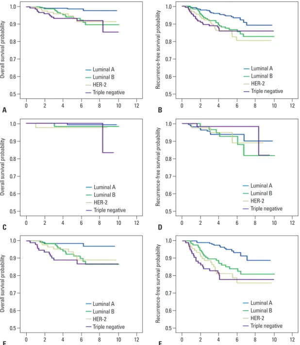
To evaluate the prognostic value of breast cancer subtypes, we performed survival analysis with subtypes defined by IHC markers. Stages of all patients were also I--III. Among the 858 patients with known molecular subtypes, luminal A, luminal B, HER-2, and triple-negative subtypes were 375 (43.8%), 190 (22.1%), 134 (15.6%), and 159 (18.5%), respectively. At a median follow up of 5.5 years, OS and RFS significantly differed among the groups classified by subtypes on the log-rank test (Fig. 3A and B). In univariate analysis for OS, luminal A showed the best survival, where- as the triple-negative type showed the worst outcome (Fig.
3A). The 5-year RFS rate was 94.2% in luminal A, 87.5%
in luminal B, 85.4% in HER-2, and 85.9% in triple-nega- tive subtypes (Fig. 3B).
To compare the influence on survival of the subtypes with tumor burden, we conducted survival analysis using the subtypes according to each stage. Among 326 patients with stage I disease, a significant difference in RFS and OS was not found among the subtypes (p=0.517 and p=0.747, respectively) (Fig. 3C and D). Of the 532 patients with stage II and III disease, RFS and OS were significantly dif- ferent according to subtype (p<0.001 and p=0.003, respec- tively) (Fig. 3E and F). For these patients in stage II and III, the triple-negative subtype showed a worse OS than the best results observed in the luminal A subtype (Fig. 3E).
These findings suggest that the influence of subtype has III disease according to the time period. In 1673 patients
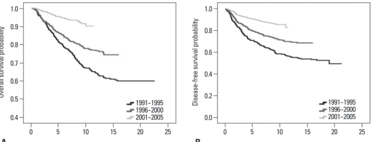
with stage I--III disease, the 7-year OS rates were 76.2% for 1991--1995, 82.1% for 1996--2000, and 93.7% for 2001-- 2005 (p<0.001) (Fig. 1A). The 7-year RFS rates were 65.6%
for 1991--1995, 75.4% for 1996--2000, and 88.2% for 2001-- 2005 (p<0.001) (Fig. 1B).
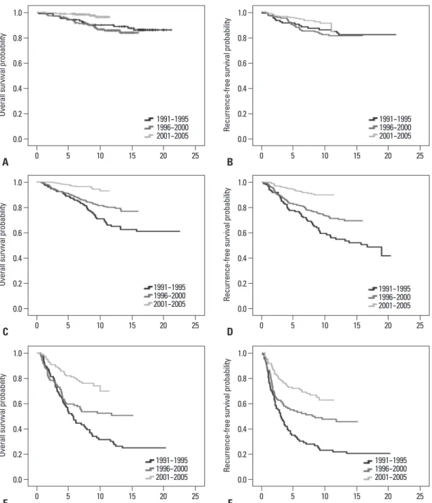
To discriminate between the impact of tumor stage and the time periods on survival, we performed survival analy- sis by each stage (Fig. 2). In patients with stage I disease, survival significantly differed according to the time period.
For these patients, however, OS and RFS did not seem to be much different between the 1991--1995 and 1996--2000 time periods (Fig. 1). In contrast to stage I, the difference between OS and RFS became larger in stage II or III by ev- ery time period (Fig. 2C-F). We found that mortality reduc- tion and recurrence reduction according to the time period were mainly achieved in stages II--III.
In the final step, we performed multivariate survival anal- ysis (Table 2). In this analysis, the time factor was demon- strated to be a significant prognostic factor for OS and RFS independent of age, tumor size, nodal status, and ER status.
Change in adjuvant chemotherapy and endocrine therapy
We investigated the regimens of chemotherapy and endo- crine therapy, which reflected the advancement of cancer management (Table 3). For endocrine therapy, selective ER modulators were the only option during 1991--1995. How- ever, aromatase inhibitors were introduced clinically during 1996--2000, and their use was expanded during 2001--2005.
For chemotherapy, the use of an anthracycline-based regi- men was constantly expanded during 15 years. The rate of anthracycline-based regimen use was 57.6% during 2001--
Fig. 1. Kaplan-Meier plots for overall survival (OS) and recurrence-free survival (RFS) according to the time period among patients with stage I--III disease. All p values are measured by the log-rank test. (A) OS (p<0.001). (B) RFS (p<0.001).
0.4 0.0
0.5 0.2
0.6 0.4
0.7
0.6 0.9
0.8
0.8
1.0 1.0
Overall survival probability Disease-free survival probability
0 5 10 15 20 25 0 5 10 15 20 25
A B
1991--1995 1996--2000 2001--2005
1991--1995 1996--2000 2001--2005
role in the survival improvement of Korean breast cancer patients. Until now, this advance in survival outcome by time trend has been mainly explained by the increasing pro- portions of early-stage cancer. It has been suggested that the wide application of newly developed agents in chemothera- py or endocrine therapy may be an underlying cause. In our results, an incremental trend in the proportion of stage 0 and I disease was similarly noted (37.3% during 1991-- 1995 to 42.7% during 2001--2005).
To discriminate the influences on survival improvement been attenuated in survival of early breast cancer.
DISCUSSION
In the first part of the present study, we evaluated the fac- tors associated with survival improvement in breast cancer using the database of a single institution, which is prospec- tively maintained and less affected by an interdisciplinary variability. We found that the time period played a major
Fig. 2. Kaplan-Meier plots for overall survival (OS) and recurrence-free survival (RFS) according to the time period in each stage (I--III). All p values are measured by the log-rank test. (A) OS in stage I (p=0.002). (B) RFS in stage I (p=0.039). (C) OS in stage II (p<0.001). (D) RFS in stage II (p<0.001). (E) OS in stage III (p<0.001). (F) RFS in stage III (p<0.001).
0.0 0.0 0.0
0.0 0.0 0.0
0.2 0.2 0.2
0.2 0.2 0.2
0.4 0.4 0.4
0.4 0.4 0.4
0.6 0.6 0.6
0.6 0.6 0.6
0.8 0.8 0.8
0.8 0.8 0.8
1.0 1.0 1.0
1.0 1.0 1.0
Overall survival probabilityOverall survival probabilityOverall survival probability Recurrence-free survival probabilityRecurrence-free survival probabilityRecurrence-free survival probability
0 0 0
0 0 0
15 15 15
15 15 15
5 5 5
5 5 5
20 20 20
20 20 20
10 10 10
10 10 10
25 25 25
25 25 25
E C A
F D B
1991--1995 1996--2000 2001--2005 1991--1995 1996--2000 2001--2005 1991--1995 1996--2000 2001--2005
1991--1995 1996--2000 2001--2005 1991--1995 1996--2000 2001--2005 1991--1995 1996--2000 2001--2005
pansion of early-stage breast cancer to an important reason, and comparisons of survival in the same stage were not per- formed. Our results highlighted that survival improvement by time trend was accomplished in each stage (stage I--III), as well as in the overall population.
In the second part of our study analyzing survival out- come with breast cancer intrinsic subtypes, our data from a cohort with known intrinsic subtypes provided results simi- lar to those of previous studies in which the triple-negative subtype showed the worst outcome.17-19 Furthermore, to isolate the prognostic influence of the subtype from the ef- fect of tumor burden, we conducted survival analysis using the subtypes in stage I or stages II and III. In stages II and III, survival outcome significantly varied according to sub- type. By contrast, in stage I, a significant difference was not observed in survival outcome among the subtypes, imply- ing that the intrinsic subtypes less affect prognosis in early breast cancer. However, conflicting data have been reported that the subtypes play an important role in prognosis in ear- ly breast cancer,20,21 even in node-negative T1ab breast can- cer.22 This discrepancy might be explained by the expanded use of chemotherapy in ER-negative patients because we between increases in early breast cancer and advances of
adjuvant therapy, we performed multivariate survival anal- ysis, including the time factor and survival analysis in each stage among the patients with AJCC stages I--III. The con- tributions of the time factor to survival improvement were observed independent of other important factors such as age, tumor size, nodal status, and ER status (Table 2).
Moreover, survival gains brought by elapsed time have been achieved in each stage (Fig. 2). Obviously, this phe- nomenon was remarkable in stages II and III. To indirectly evaluate the influence of changing regimens in adjuvant therapy, we compared the types of regimens used in endo- crine therapy and chemotherapy among the time periods, and our analysis showed that the proportion of new agents of chemotherapy and endocrine therapy incrementally changed over time. Advancement in adjuvant therapy may be asso- ciated with survival improvement of breast cancer patients during the investigated period, particularly in the patients with locally advanced breast cancer.
In the previous study based on a large cohort of Korean breast cancer patients,5 an enhanced survival outcome by time trend was reported. However, they suggested the ex-
Table 3. Change in Adjuvant Therapy Regimen for Patients with Stage I--III Disease Based on the Time Period
Time cohort All patients 1991--1995 1996--2000 2001--2005 p value
Endocrine therapy <0.001
SERM 758 135 283 340
AI 101 0 39 62
Chemotherapy <0.001
CMF 545 128 387 30
Anthracycline-based regimen 538 63 148 327
Taxane-based regimena* 248 0 37 211
SERM, selective estrogen receptor modulator; AI, aromatase inhibitor; CMF, cyclophosphamide-methotrexate-5-fluorouracil.
*Taxane-based regimen included the sequential regimen such as anthracycline followed by taxane.
Table 2. Multivariate Models for Survival Outcomes during 15 Years
Characteristics Overall Recurrence-free
Hazard ratio 95% CI p value Hazard ratio 95% CI p value
Age in yrs
≤35 vs. >35 0.60 0.44--0.81 0.001 0.49 0.38--0.64 <0.001
Tumor size
≤2 cm vs. >2 cm 1.64 1.26--2.14 <0.001 1.55 1.24--1.95 <0.001
Nodal status
Positive vs. Negative 2.98 2.30--0.86 <0.001 2.83 2.27--3.54 <0.001
Estrogen receptor status
Positive vs. Negative 0.68 0.54--0.86 0.001 0.80 0.65--0.99 0.039
Time period
1991--1995 vs. 1996--2000 0.58 0.45--0.76 <0.001 0.61 0.48--0.77 <0.001
1991--1995 vs. 2001--2005 0.23 0.17--0.33 <0.001 0.29 0.22--0.39 <0.001
CI, confidence interval.
clinical trials, showed heterogeneity in adjuvant therapy.
The reasons contributing to survival improvement in each stage are not fully elucidated and the influence of advance- ment in adjuvant therapy could not be directly evaluated.
However, our findings have advantages over these limita- tions regarding the well-outlined survival improvement by time trend, and the unique analysis with the intrinsic sub- types based on a single-center database interfered less with heterogeneous treatment policies. The strengths of the study actively used chemotherapy for ER-negative tumors, even
for small tumors (data not shown). To solve contradictory results between our data and other studies, longer follow-up duration is required to delineate a survival pattern according to subtype because survival outcome in stage I is very favor- able (the estimated 10-year OS rate is 90.9% in stage I).
The present retrospective study possesses several limita- tions. The retrospective design is associated with inherent limitations. The patients in our study, which is not based on
Fig. 3. Kaplan-Meier plots for overall survival (OS) and recurrence-free survival (RFS) according to subtype. All p values are measured by the log-rank test. (A) OS by subtype in stages I--III (p=0.002). (B) RFS by subtype in stages I--III (p=0.039). (C) OS by subtype in stage I (p=0.517). (D) RFS in stage I (p=0.747). (E) OS by subtype in stages II--III (p=0.003). (F) RFS by subtype in stages II--III (p<0.001). HER-2, hu- man epidermal growth factor receptor-2.
0.5
0.5
0.5
0.5
0.5
0.5 0.6
0.6
0.6
0.6
0.6
0.6 0.7
0.7
0.7
0.7
0.7
0.7 0.8
0.8
0.8
0.8
0.8
0.8 0.9
0.9
0.9
0.9
0.9
0.9 1.0
1.0
1.0
1.0
1.0
1.0
Overall survival probabilityOverall survival probabilityOverall survival probability Recurrence-free survival probabilityRecurrence-free survival probabilityRecurrence-free survival probability
0
0
0
0
0
0 8
8
8
8
8
8 6
6
6
6
6
6 2
2
2
2
2
2 10
10
10
10
10
10 4
4
4
4
4
4 12
12
12
12
12
12
A
C
E
B
D
F
Luminal A Luminal B HER-2 Triple negative
Luminal A Luminal B HER-2 Triple negative
Luminal A Luminal B HER-2 Triple negative
Luminal A Luminal B HER-2 Triple negative
Luminal A Luminal B HER-2 Triple negative
Luminal A Luminal B HER-2 Triple negative
IV breast cancer. J Clin Oncol 2008;26:4891-8.
8. Early Breast Cancer Trialists’ Collaborative Group (EBCTCG).
Effects of chemotherapy and hormonal therapy for early breast cancer on recurrence and 15-year survival: an overview of the ran- domised trials. Lancet 2005;365:1687-717.
9. Trudeau M, Charbonneau F, Gelmon K, Laing K, Latreille J, Mackey J, et al. Selection of adjuvant chemotherapy for treatment of node-positive breast cancer. Lancet Oncol 2005;6:886-98.
10. Piccart-Gebhart MJ, Procter M, Leyland-Jones B, Goldhirsch A, Untch M, Smith I, et al. Trastuzumab after adjuvant chemotherapy in HER2-positive breast cancer. N Engl J Med 2005;353:1659-72.
11. Romond EH, Perez EA, Bryant J, Suman VJ, Geyer CE Jr, David- son NE, et al. Trastuzumab plus adjuvant chemotherapy for opera- ble HER2-positive breast cancer. N Engl J Med 2005;353:1673- 12. Cancer Genome Atlas Network. Comprehensive molecular por-84.
traits of human breast tumours. Nature 2012;490:61-70.
13. Perou CM, Sørlie T, Eisen MB, van de Rijn M, Jeffrey SS, Rees CA, et al. Molecular portraits of human breast tumours. Nature 2000;406:747-52.
14. Singletary SE, Allred C, Ashley P, Bassett LW, Berry D, Bland KI, et al. Staging system for breast cancer: revisions for the 6th edition of the AJCC Cancer Staging Manual. Surg Clin North Am 2003;
83:803-19.
15. Hammond ME, Hayes DF, Dowsett M, Allred DC, Hagerty KL, Badve S, et al. American Society of Clinical Oncology/College of American Pathologists guideline recommendations for immuno- histochemical testing of estrogen and progesterone receptors in breast cancer (unabridged version). Arch Pathol Lab Med 2010;
134:e48-72.
16. Goldhirsch A, Wood WC, Coates AS, Gelber RD, Thürlimann B, Senn HJ, et al. Strategies for subtypes--dealing with the diversity of breast cancer: highlights of the St. Gallen International Expert Consensus on the Primary Therapy of Early Breast Cancer 2011.
Ann Oncol 2011;22:1736-47.
17. Carey LA, Perou CM, Livasy CA, Dressler LG, Cowan D, Con- way K, et al. Race, breast cancer subtypes, and survival in the Carolina Breast Cancer Study. JAMA 2006;295:2492-502.
18. Lee JA, Kim KI, Bae JW, Jung YH, An H, Lee ES, et al. Triple negative breast cancer in Korea-distinct biology with different im- pact of prognostic factors on survival. Breast Cancer Res Treat 2010;123:177-87.
19. O’Brien KM, Cole SR, Tse CK, Perou CM, Carey LA, Foulkes WD, et al. Intrinsic breast tumor subtypes, race, and long-term survival in the Carolina Breast Cancer Study. Clin Cancer Res 2010;16:6100-10.
20. Park YH, Lee SJ, Cho EY, Choi YL, Lee JE, Nam SJ, et al. Clini- cal relevance of TNM staging system according to breast cancer subtypes. Ann Oncol 2011;22:1554-60.
21. Sanpaolo P, Barbieri V, Genovesi D. Prognostic value of breast cancer subtypes on breast cancer specific survival, distant metasta- ses and local relapse rates in conservatively managed early stage breast cancer: a retrospective clinical study. Eur J Surg Oncol 2011;37:876-82.
22. Theriault RL, Litton JK, Mittendorf EA, Chen H, Meric-Bernstam F, Chavez-Macgregor M, et al. Age and survival estimates in pa- tients who have node-negative T1ab breast cancer by breast can- cer subtype. Clin Breast Cancer 2011;11:325-31.
include a large patient population, the long-term follow up duration, and the uniform initial staging and follow-up sur- veillance protocol. Our attempt to ameliorate survival in Ko- rean breast cancer patients will be facilitated by the findings that we were successful in improving outcome over time.
In conclusion, the present study provides significant evi- dence of improvement in the prognosis of Korean breast cancer patients with AJCC stage I--III during the recent 15 years, while considering the beneficial effect of significant prognostic factors. Our data imply that advancement of ad- juvant treatment plays an integral part in producing a sur- vival benefit and in expanding the therapeutic options for breast cancer patients. Moreover, we concordantly showed a clinical significance of the intrinsic subtypes and provided a novel insight regarding comparison of the prognostic im- pact between tumor burden and subtypes.
ACKNOWLEDGEMENTS
We dedicated this study to Prof. Hy-De Lee, who largely contributed to establish the Koran Breast Cancer Society and improve in the management for Korean breast cancer patients. The authors also thank nurse Keum Son Jeung for help in the construction and management of database.
REFERENCES
1. Parkin DM, Bray F, Ferlay J, Pisani P. Global cancer statistics, 2002. CA Cancer J Clin 2005;55:74-108.
2. Jung KW, Park S, Kong HJ, Won YJ, Boo YK, Shin HR, et al.
Cancer statistics in Korea: incidence, mortality and survival in 2006-2007. J Korean Med Sci 2010;25:1113-21.
3. Lee JH, Yim SH, Won YJ, Jung KW, Son BH, Lee HD, et al. Pop- ulation-based breast cancer statistics in Korea during 1993-2002:
incidence, mortality, and survival. J Korean Med Sci 2007;22 Suppl:S11-6.
4. The Korean Breast Cancer Society. Nationwide Korean Breast Cancer Data of 2004 Using Breast Cancer Registration Program. J Breast Cancer 2006;9:151-61.
5. Son BH, Kwak BS, Kim JK, Kim HJ, Hong SJ, Lee JS, et al.
Changing patterns in the clinical characteristics of Korean patients with breast cancer during the last 15 years. Arch Surg 2006;141:
155-60.
6. Jung KW, Won YJ, Kong HJ, Oh CM, Seo HG, Lee JS. Cancer statistics in Korea: incidence, mortality, survival and prevalence in 2010. Cancer Res Treat 2013;45:1-14.
7. Dawood S, Broglio K, Gonzalez-Angulo AM, Buzdar AU, Horto- bagyi GN, Giordano SH. Trends in survival over the past two de- cades among white and black patients with newly diagnosed stage