Surgical outcomes and prognostic factors influencing long-term survival in patients with gallbladder cancer
Sung Ha Lee, Jae Do Yang, Hong Pil Hwang, Hee Chul Yu, and Baik Hwan Cho
Department of Surgery, Chonbuk National University Medical School and Hospital, Jeonju, Korea
Backgrounds/Aims: The aim of this study is to analyze surgical outcomes and prognostic factors affecting survival after surgical resection in patients with gallbladder cancer. Methods: We retrospectively reviewed 86 patients treated surgically for gallbladder cancer from January 2000 to December 2009 at Chonbuk National University Hospital.
Clinicopathologic factors, surgical treatment and outcome data were analyzed. Results: Among the 86 patients (44 male, 42 female) with gallbladder cancer, the mean age was 62.9 years (range: 32-80) and the median survival was 42.4±3.5 month. The overall cumulative survival rates of 86 patients were for 1 year, 83.7%; 3 year, 67.4%; 5 year survival, 61.7%. Univariate analysis revealed that preoperative serum alanine aminotransferase, alkaline phosphatase, total bilirubin, carcinoembryonic antigen (CEA), T staging, N staging were statistically significantly associated with survival. CEA (p=0.004) and T staging (p=0.005) were associated with survival in multivariate analysis. Two-year surviv- al rates were analyzed according to the methods of surgical resection, with simple cholecystectomy showing 100%, whereas extended cholecystectomy showed about 83% in T1b. We could not find out any adverse effect of the simple cholecystectomy for survival. Conclusions: CEA and T stage are independent significant prognostic factor associated with patient survival in our study. Simple cholecystectomy can be regarded as curative resection in stage T1b. Longer observation periods and more cases will be needed to confirm these conclusions. (Korean J Hepatobiliary Pancreat Surg 2012;16:59-64)
Key Words: Gallbladder cancer; Prognostic factors; Survival rate
Received: February 8, 2012; Revised: April 9, 2012; Accepted: April 20, 2012 Corresponding author: Hee Chul Yu
Department of Surgery, Chonbuk National University Medical School and Hospital, 634-18, Keumam-dong, Dukjin-gu, Jeonju 561-712, Korea Tel: +82-63-250-1576, Fax: +82-63-271-6197, E-mail: [email protected]
Copyright Ⓒ 2012 by The Korean Association of Hepato-Biliary-Pancreatic Surgery Korean Journal of Hepato-Biliary-Pancreatic Surgery ∙ pISSN: 1738-6349
INTRODUCTION
Gallbladder cancer is the most common cancer of the biliary tract. According to the Korea Central Cancer Registry report in 2008, gallbladder cancer is the fifth most frequent cancer in the gastrointestinal tract.1 Early detection of gallbladder cancer is difficult due to asympto- matic growth. Many patients have infiltration of surround- ing structures such as the portal vein and hepatic artery at time of diagnosis. Therefore gallbladder cancer has the shortest median survival duration in biliary cancers.2,3
Five-year survival rate has been reported as 17.3%.1 However, recent studies show improved survival rates, at- tributable to early detection through the development of pre-operative radiologic diagnostic tools, increased aware- ness of personal health and spread of routine health checkups, as well as improved surgical methods and post operative care.
The aim of this study is to analyze surgical outcomes and prognostic factors affecting survival after surgical re- section in patients with gallbladder cancer at our institu- tion.
METHODS
We retrospectively reviewed age, gender, clinical fac- tors, stage distribution, and surgical method of 133 pa- tients operated with primary gallbladder cancer at Chonbuk National University Hospital from January 2000 to December 2009. This study design was approved by the institutional review board of our institution. Of these 133 patients, 86 patients were followed for post-operative survival. Survival status and cause of death were con- firmed through phone calling and reviewing medical records. Age, gender, pre-operative clinical factors within one month, T stage, N stage and surgical method were
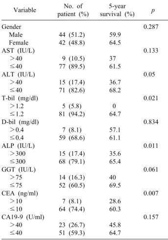
Table 1. Survival according to clinical factors of patients Variable No. of
patient (%)
5-year
survival (%) p Gender
Male Female AST (IU/L)
>40
≤40 ALT (IU/L)
>40
≤40 T-bil (mg/dl)
>1.2
≤1.2 D-bil (mg/dl)
>0.4
≤0.4 ALP (IU/L)
>300
≤300 GGT (IU/L)
>75
≤75 CEA (ng/ml)
>10
≤10 CA19-9 (U/ml)
>40
≤40
44 (51.2) 42 (48.8) 9 (10.5) 77 (89.5) 15 (17.4) 71 (82.6) 5 (5.8) 81 (94.2) 7 (8.1) 59 (68.6) 15 (17.4) 68 (79.1) 14 (16.3) 52 (60.5) 7 (8.1) 64 (74.4) 23 (26.7) 51 (59.3)
59.9 64.5 37 61.5 36.7 68.2 0 64.7 57.1 61.1 35.6 65.4 40 69.5 28.6 60.3 45.8 64.7
0.287
0.133
0.05
0.021
0.834
0.011
0.061
0.007
0.157
AST, aspartate aminotransferase; ALT, alanine aminotransfe- rase; T-bil, total bilirubin; D-bil, direct bilirubin; ALP, alka- line phosphatase; GGT, Gamma-glutamyl transferase; CEA, carcinoembryonic antigen; CA19-9, carbohydrate antigen 19-9
Fig. 1. Cumulative survival rates related to T stage (AJCC 7th edition).
analyzed. Stages of the cancer were classified according to the American Joint Committee on Cancer (AJCC) 7th edition. Curative resection defined as simple chol- ecystectomy with no remnant cancer on permanent patho- logic report in Tis and T1a, and radical cholecystectomy combined hepatectomy or not with no remnant cancer on permanent pathologic report in T1b, T2 and T3.
Univariate analysis was performed using the Kaplan- Meier method and compared with the log-rank test.
Multivariate analysis was performed using the Cox re- gression hazards model to identify independent prognostic factors. All statistical analyses used SPSS 18.0 for Windows (SPSS Inc. Chicago, III). A p-value less than 0.05 was considered statistically significant.
RESULTS
Age and gender distribution
Of the 86 patients, gender distribution is similar, 44 male and 42 female. Mean age was about 62.9 years, with a range of 32 to 80. Highest prevalence was seen in the 5th decade, 29 cases (33.7%).
Survival rates according to clinical factors Preoperative serum aspartate aminotransferase (AST), alanine aminotransferase (ALT), total bilirubin (T-bil), di- rect bilirubin (D-bil), alkaline phosphatase (ALP), Gam- ma-glutamyl transferase (GGT), carcinoembryonic antigen (CEA), carbohydrate antigen 19-9 (CA 19-9) were ana- lyzed, and among these factors, ALT, ALP, T-bil, and CEA showed a statistically significant association with 5-year survival in univariate analysis (Table 1).
Survival rates according to T stage and N stage T stage and N stage were classified by the 7th AJCC classification system, with 6 cases of Tis (7.0%), 5 cases of T1a (5.8%), 13 cases of T1b (15.1%), 32 cases of T2 (37.2%), 26 cases of T3 (30.2%) and 4 cases of T4 (4.7%). According to T stage, the 5-year survival rate de- creased as stages increased. Tis and T1a showed 100%
5-year survival rate, and T1b showed 84.6%, T2 61.4%, T3 40.9%, T4 0% survival rate. All 11 patients of the Tis and T1a stages showed more than 10 year survival (p=0.000) (Fig. 1).
Excluding 30 patients (34.9%) who did not undergo
Fig. 2. Cumulative survival rates related to N stage (AJCC 7th edition).
Table 2. Multivariate analysis of variables as prognostic fac- tors for survival rate
Variables Univariate p-value
Multivariate p-value ALT (IU/L)
T-bil (mg/dl) ALP (IU/L) CEA (ng/ml) T stage N stage
0.05 0.021 0.011 0.007 0.000 0.003
0.889 0.986 0.469 0.004 0.005 0.055 ALT, alanine aminotransferase; T-bil, total bilirubin; ALP,
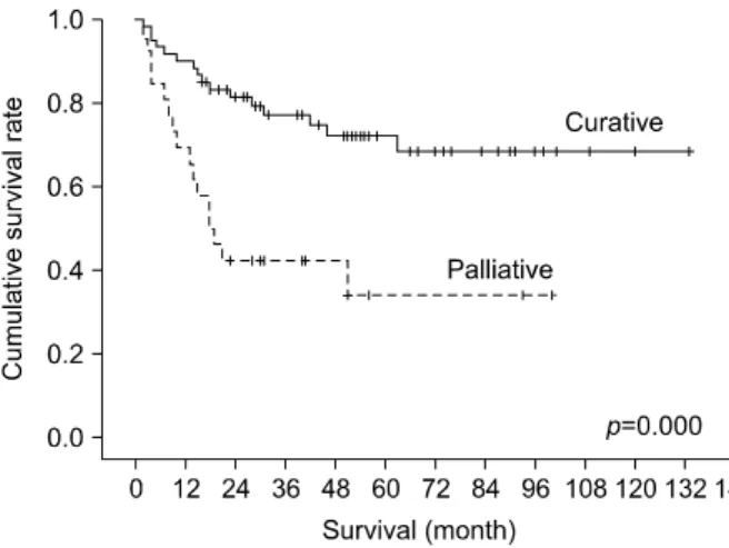
alkaline phosphatase; CEA, carcinoembryonic antigen Fig. 3. Cumulative survival rates related to types of surgical resection.
lymph node dissection, 56 patients were classified accord- ing to the N stage of the 7th AJCC classification. Results were 32 cases (37.2%) of N0, 17 cases (19.8%) of N1, and 7 cases (8.1%) of N2 stages. The 5-year survival rates according to N stage were 80.6% in N0, 27.6% in N1, and 19.0% in N2 (p=0.003) (Fig. 2).
Prognostic factors
ALT, ALP, T-bil and CEA, T and N stage were sig- nificant factors on univariate analysis.
Of these factors, CEA and T stage were statistically significant independent prognostic factors on multivariate analysis (Table 2).
Survival rates according to surgical method in T stage
Of the 86 cases, 60 cases underwent curative resection, while 26 cases were operated palliatively. In the curative resection groups, the survival rates were 1-year (90.0%),
3-year (77.0%), and 5-year (72.1%). Survival in the pal- liative resection group was 1-year 69.2%; 3-year 42.3%;
and 5-year 33.8%. These suggested a higher survival rate in curative resection (p=0.000) (Fig. 3).
Comparison between curative and palliative resection was carried out in the T1b, T2, and T3 groups. Tis and T1a groups, in which only curative resection were per- formed, were not comparable and therefore excluded, along with the T4 groups, in which no case was treated with curative resection at all.
Survival in patients with T1b tumor: In group T1b, excluding one case where second-look operation was rejected despite a positive margin of cancer on the cystic duct, 6 cases of simple cholecystectomy and 6 cases of extended cholecystectomy were compared. Medial surviv- al was 54.7±33.8 months in the simple cholecystectomy subgroup, and 32.0±19.7 months in the extended chol- ecystectomy subgroup, with no statistically significant difference. During the observation period, one patient ex- pired due to liver metastases 15 months after extended cholecystectomy. 2-year survival rates were compared ac- cording to surgical method. All of the simple chol- ecystectomy subgroup survived, while in the extended cholecystectomy subgroup 83.3% survived.
Survival in patients with T2 tumor: Twenty-four cases of curative resection and 8 cases of palliative re- section were performed in the T2 group (3 cases in which second-look operations were not performed; 3 cases in which lymph node dissection was not performed; and 2 cases in which only simple cholecystectomy was per-
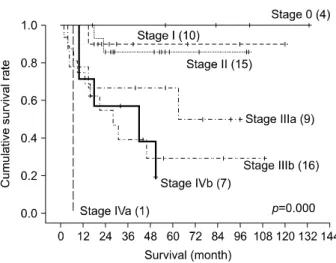
Fig. 4. Cumulative survival rates related to stage system (AJCC 7th edition).
formed due to diagnosis of distant metastasis during oper- ation). Cases treated with curative resection showed 74%
5-year survival rates, whereas cases operated palliatively resulted in 0% 5-year survival rates, a statistically sig- nificant difference (p=0.003).
Survival in patients with T3 tumor: Nineteen cases of curative resection and 7 cases of palliative resection were performed in the T3 group. The 5-year survival rates were 51.5% when treated with curative and 0% for pallia- tive resection not a significant difference (p=0.103).
Overall survival rates and survival rates according to stage
Medial survival of the 86 cases was 42.4±32.5 months, with the shortest of 2 months and the longest of 133 months. Overall survival rates were 83.7% at 1 year, 67.4% at 3 years, and 61.7% at 5 years.
Except for the 24 cases in which lymph node dissection was not performed, final pathologic stages of the remain- ing 62 cases were as followed: 4 cases of stage 0 (4.7%), 10 cases of stage I (11.6%), 15 cases of stage II (17.4%), 9 cases of stage IIIa (10.5%), 16 cases of stage IIIb (18.6%), 1 case of stage IVa (1.2%), and 7 cases of stage IVb (8.1%).
Five-year survival rates according to pathologic stage were stage 0 (100%), in stage I (90%), II (85.7%), IIIa (66.7%), IIIb (29.3%), IVa (0%), IVb (0%), showing de- creased survival as stage increased (p=0.000) (Fig. 4).
DISCUSSION
Despite increased early detection through the develop- ment of preoperative diagnostic tools, greater acceptance of routine health checkups, and increased survival rate through advanced operative procedures and post-operative care, gallbladder cancer still has a poor prognosis, espe- cially since many patients are inoperable at diagnosis.3,4 In a study of 724 cases carried out by the French Surgical Association in 1994, the median survival period was 3 months, and 5 year survival rates were 5%.5 Konstantinidis et al.6 reported a 40-year follow-up study which showed medial survival of 3.5 months between 1962 and 1979, 6.5 months between 1980 and 1997, and 12 months between 1998 and 2008. Liang et al.7 reported a 25 year follow-up study which showed medial survival of 12.3 months and survival rates at 1 year (50.5%), 3 years (29.5%), and 5 years (26.2%). In our study, com- parable survival rates are 1 year (83.7%), 3 years (67.4%), and 5 years (61.7%).
Various clinical prognostic factors in gallbladder cancer have been reported. Generally, incidence is 2 to 6 times higher in women, which is probably due to the fact that cholelithiasis is more frequent in women. Despite the low- er incidence rate, male gender is a poor prognostic factor, showing shorter medial survival periods.2,8 There was no survival difference according to gender in our study.
There are no characteristic symptoms in early gall- bladder cancer, but as disease progresses various symp- toms arise, generally showing poorer prognosis with acute/chronic cholecystitis or focal biliary complications.9 In our study, poorer prognosis was observed when there were abnormal results in the liver function tests, such as ALT, bilirubin, and ALP, in univariate analysis.
CEA is a typical tumor marker elevated not only in gallbladder cancer but also in colon cancer and other types of cancer. Chakravarty et al.10 reported that serum CEA levels were independent prognostic factors that af- fect long-term survival regardless of T stage. Likewise, CEA was an independent prognostic factor in our study.
CA19-9 is a tumor marker frequently elevated in gall- bladder cancer, especially associated with intra-epithelial dysplasia and adenocarcinoma. It has been reported to show significant association with other important prog- nostic factors such as a history of jaundice and lymph
node metastasis.11 Study to distinguish CA19-9 as a prog- nostic factor in gallbladder cancer has been insufficient, and in our study there was no association in univariate analysis.
The major principle in treatment of gallbladder cancer is surgical resection, with radiation therapy, chemo- therapy, immunotherapy as optional choices, although ef- fectiveness is meager. Recently, there has been a tendency towards determining surgical methods in gallbladder can- cer according to T stage.2,12
Tis and T1a have shown complete remission with sim- ple cholecystectomy alone.13 In our study, all cases of Tis and T1a cancers were treated with simple chol- ecystectomy, and although comparison with radical chol- ecystectomy was unavailable, both groups showed 100%
survival rates at 5 and 10 years. In the case of stage T1b, the extent of resection is controversial. Lee et al.14 re- ported that radical cholecystectomy had no benefit over simple cholecystectomy, whereas studies by Pilgrim et al.15 and Abramson et al.13 showed increased survival with radical resection, due to extraction of lymph node meta- stases and recurrence. There was no statistically sig- nificant difference according to surgical method in this study. There was no disadvantage to simple cholecystec- tomy, when compared with extended cholecystectomy ac- cording to median survival and 2-year survival rates in this study. As there were no deaths in the simple chol- ecystectomy subgroup, simple cholecystectomy in stage T1b could be regarded as curative resection.
There have also been disagreements on the extent of resection in stage T2. Ambramson et al.13 calculated mean 5-year cancer - specific survival of 61.3% in simple chol- ecystectomy alone group, but 87.5% in radical chol- ecystectomy group. Pilgrim et al.15 reported much higher 5 year survival rates in radical cholecystectomy (61% to 100%) when compared with simple cholecystectomy (19% to 50%). Zhu et al.2 and Kang et al.12 supported rad- ical resection over simple resection, which showed higher 5-year survival rates in the T2 groups. On the contrary, Konstantinidis et al.6 reported that there was no significant difference between radical and simple cholecystectomy. In addition, Kohya et al.16 subdivided stage T2 patients into whether or not there was hepatic, biliary, lymphatic, ve- nous, peri-neural or lymph node invasion, and proposed that radical resection was unnecessary in negative sub-
groups, whereas liver and/or biliary resection was needed when positive findings were present. Five year survival rates were confirmed to be higher in stage T2 radical re- section subgroups in his study.
Stages T3 and T4 lead to poor prognosis, even after radical cholecystectomy. According to the French Surgical Association survey, 90% of patients with completed radi- cal cholecystectomy expired within 12 months.3 Likewise, this study also had no significant difference in surgical method in the T3 subgroups, with 5 year survival rates at 40.9% in the T3 and 0% in the T4 subgroups. On the other hand, Kondo et al.17 reported T3, T4 with N1 pa- tients were improved survival rate by lymph node dis- section and with no detectable difference in 5-year surviv- al rates between N0 (66%) and N1 (53%) patients.
In conclusion, CEA and T stage were independent prognostic factors significantly associated with patient survival in multivariate analysis. Although observation pe- riods were short and number of cases was small, both simple and extended cholecystectomy showed similar sur- vival rates in the T1b subgroups. Longer observation peri- ods and more cases will be needed to confirm these conclusions.
REFERENCES
1. Jung KW, Park S, Kong HJ, et al. Cancer statistics in Korea:
incidence, mortality, survival, and prevalence in 2008. Cancer Res Treat 2011;43:1-11.
2. Zhu AX, Hong TS, Hezel AF, et al. Current management of gall- bladder carcinoma. Oncologist 2010;15:168-181.
3. Cubertafond P, Gainant A, Cucchiaro G. Surgical treatment of 724 carcinomas of the gallbladder. Results of the French Surgical Association Survey. Ann Surg 1994;219:275-280.
4. Smith GC, Parks RW, Madhavan KK, et al. A 10-year experience in the management of gallbladder cancer. HPB (Oxford) 2003;5:
159-166.
5. Moon HH, Yoon M. The survival rate of gallbladder carcinoma based on the presence of lymph node metastasis and the depth of the primary tumor invasion. Korean J Hepatobiliary Pancreat Surg 2008;12:128-133.
6. Konstantinidis IT, Deshpande V, Genevay M, et al. Trends in presentation and survival for gallbladder cancer during a period of more than 4 decades: a single-institution experience. Arch Surg 2009;144:441-447.
7. Liang JW, Dong SX, Zhou ZX, et al. Surgical management for carcinoma of the gallbladder: a single-institution experience in 25 years. Chin Med J (Engl) 2008;121:1900-1905.
8. Chan SY, Poon RT, Lo CM, et al. Management of carcinoma of the gallbladder: a single-institution experience in 16 years. J Surg Oncol 2008;97:156-164.
9. Misra S, Chaturvedi A, Misra NC, et al. Carcinoma of the gall- bladder. Lancet Oncol 2003;4:167-176.
10. Chakravarty KD, Yeh CN, Jan YY, et al. Factors influencing long-term survival in patients with T3 gallbladder adeno- carcinoma. Digestion 2009;79:151-157.
11. Park JI, Kim JS, Kim KH, et al. Analysis of prognostic factors affecting survival in patients with gallbladder cancer. Korean J Hepatobiliary Pancreat Surg 2010;14:173-183.
12. Kang SY, Lee SK, Kim JY, et al. Clinical features and prognostic factors influencing long-term survival in pT2 gallbladder carcino- ma patients. Korean J Hepatobiliary Pancreat Surg 2008;12:
173-179.
13. Abramson MA, Pandharipande P, Ruan D, et al. Radical resection for T1b gallbladder cancer: a decision analysis. HPB (Oxford) 2009;11:656-663.
14. Lee SE, Jang JY, Lim CS, et al. Systematic review on the surgi-
cal treatment for T1 gallbladder cancer. World J Gastroenterol 2011;17:174-180.
15. Pilgrim C, Usatoff V, Evans PM. A review of the surgical strat- egies for the management of gallbladder carcinoma based on T stage and growth type of the tumour. Eur J Surg Oncol 2009;35:
903-907.
16. Kohya N, Kitahara K, Miyazaki K. Rational therapeutic strategy for T2 gallbladder carcinoma based on tumor spread. World J Gastroenterol 2010;16:3567-3572.
17. Kondo S, Takada T, Miyazaki M, et al; Japanese Association of Biliary Surgery; Japanese Society of Hepato-Biliary-Pancreatic Surgery; Japan Society of Clinical Oncology. Guidelines for the management of biliary tract and ampullary carcinomas: surgical treatment. J Hepatobiliary Pancreat Surg 2008;15:41-54.