Korean Circulation Journal
Introduction
The remodeling process after acute myocardial infarction (AMI) is
Print ISSN 1738-5520 • On-line ISSN 1738-5555
Progressive Dilation of the Left Atrium and Ventricle after Acute Myocardial Infarction Is Associated with High Mortality
Hyun Ju Yoon, MD, Myung Ho Jeong, MD, Yuna Jeong, MD, Kye Hun Kim, MD, Ji Eun Song, MD, Jae Yeong Cho, MD, Su Young Jang, MD, Hae Chang Jeong, MD, Ki Hong Lee, MD, Keun Ho Park, MD, Doo Sun Sim, MD, Nam Sik Yoon, MD, Young Joon Hong, MD, Hyung Wook Park, MD, Ju Han Kim, MD, Youngkeun Ahn, MD, Jeong Gwan Cho, MD, Jong Chun Park, MD, and Jung Chaee Kang, MD
The Heart Center of Chonnam National University Hospital, Korea Cardiovascular Stent Research Institute of Chonnam National University, Gwangju, Korea
Background and Objectives: The purpose of this study is to identify the prevalence of progressive dilation in patients with acute myo- cardial infarction (AMI) combined with heart failure (HF) and determine the prognostic significance and associated factors with a geometric change of an infarcted heart.
Subjects and Methods: A total of 1310 AMI patients with HF (63.9±12.5 years, 70% male) between November 2005 and April 2011 un- derwent echocardiography at admission and one year later. Left ventricular (LV) remodeling is defined as 20% progression, and left atria (LA) remodeling is 10% compared with the initial volume index.
Results: The prevalence of both LA and LV remodeling was 13.9%; LV only was 9.3%, LA only 22.8% and non-remodeling was 55.1%, re- spectively. In the non-remodeling group, Killip class II was more frequent (83.9%, p<0.001) whereas in other remodeling groups, Killip class III was more frequent. Initial wall motion score index, ejection fraction, maximal cardiac enzyme, high sensitive C-reactive protein, B type natriuretic peptide, and triglyceride serum levels were significantly associated with heart remodeling. All causes of death occurred in 168 cases (12.8%) during the follow-up period. Mortality was the highest in the LV and LA remodeling group (20.9%) and the lowest in the non- remodeling group (11.4%). During the period of follow-up, the cumulative survival rate was significantly lower in the groups of LA and LV remodeling than in others (log rank p=0.006).
Conclusion: Total mortality was significantly increased in patients AMI with geometrically progressive LA and LV dilatation. (Korean Circ J 2013;43:731-738)
KEY WORDS: Myocardial infarction; Ventricular remodeling; Heart failure; Prognosis.
Received: July 10, 2013
Revision Received: September 3, 2013 Accepted: September 16, 2013
Correspondence: Myung Ho Jeong, MD, Heart Research Center Nominat- ed by Korea Ministry of Health and Welfare, Chonnam National University Hospital, 42 Jebong-ro, Dong-gu, Gwangju 501-757, Korea
Tel: 82-62-220-6243, Fax: 82-62-228-7174 E-mail: [email protected]
• The authors have no financial conflicts of interest.
This is an Open Access article distributed under the terms of the Creative Commons Attribution Non-Commercial License (http://creativecommons.
org/licenses/by-nc/3.0) which permits unrestricted non-commercial use, distribution, and reproduction in any medium, provided the original work is properly cited.
clinically characterized by progressive cavity dilatation. Left ventric- ular (LV) remodeling is a globally heterogeneous process, involving both infarcted and non-infarcted myocardium, which results from infarction expansion in the acute phase, whereas late cavity dilation is the result of the eccentric hypertrophy process. LV remodeling after AMI is stimulated by the interaction of a number of factors, playing a key role in the pathology of post-infarction ventricular dysfunction.
1-3)When reacting to aggression, the genetic, structur- al, and biochemical changes arising from that process will result in the deterioration of the functional ability of the heart in the long run. It was reported that patients who develop LV dilatation follow- ing AMI have significantly reduced survival rates.
4)5)Left atrial (LA) volume is considered to be a presentation of the di-
astolic burden, and increased LA volume usually reflects elevated
ventricular filling pressure. During ventricular diastole, the left atri-
um is directly exposed to LV pressure through the open mitral valve.
As an adaptation to the decreased ventricular compliance following myocardial infarction (MI), LA pressure rises, increasing LA wall ten- sion and stretching the atrial myocardium. Therefore, LA remodeling tends to be a result of adaptation to allow for the preservation of LV function and reflect increased LA pressure.
6)7)A previous report sh- owed that LA volume is a independent predictor of mortality and smaller LA volumes are associated with a good prognosis after MI.
8)However, the effect of both LV and LA remodeling in the post-MI period was not clear. The purpose of the present study was, there- fore, to assess the prevalence of LV and LA remodeling in patients with AMI combined with heart failure (HF) during the first one year after MI, and to define the long-term clinical prognostic significance and associated factors with a geometric change of an infarcted heart.
Subjects and Methods
Study population and grouping
A total of 1310 AMI patients with HF (63.9±12.5 years, 70% male) enrolled retrospectively from Chonnam University Hospital between November 2005 and April 2011 underwent echocardiography at admission and one year later. We excluded Killip class IV, due to in- appropriate image quality on admission period.
The subjects were divided into four groups according to the re- modeling of LV and/or LA during one year. LV remodeling is defined as 20% progression and LA remodeling 10%, compared with initial volume index. Group I was with progressive LA and LV dilatation (n=182, 64.2±12.3 years, 128 males), group II with only LV remodel- ing without LA (n=120, 65.7±11.9 years, 78 males), group III with LA remodeling without LV (n=286, 63.3±12.3 years, 195 males), and group IV without LA and LV dilatation (n=722, 62.9±12.5 years, 519 males). Hospital records collected on admission were registered.
Information on events and mortality were obtained from hospital records and phone calls. This study protocol was approved by the Institutional Review Board or ethics committee at Chonnam Nation- al University Hospital (No=2010-05-092).
Definition of hypertension, diabetes, dyslipidemia, myocardial infarction, and heart failure
Subjects were considered as having hypertension if their blood pressure was ≥140/≥90 mm Hg as Joint National Committee VII
9)or if they were undergoing treatment for hypertension. The American Diabetes Association criteria
10)were used to define diabetes (DM).
We considered a subject as having DM when the fasting plasma glucose levels were ≥126 mg/dL in 2 consecutive assessments or if they were on treatment for DM. Dyslipidemia was diagnosed ac- cording to the 2004 update of the National Cholesterol Education
Program guidelines.
11)According to these guidelines, high levels of low density lipoprotein-cholesterol ≥160 mg/dL, low high density lipoprotein-cholesterol ≤40 mg/dL, and high triglycerides ≥150 mg/
dL were included.
12)The presence of ST-segment elevation MI was determined by >30 minutes of continuous chest pain, a new ST-segment elevation ≥2 mm on at least two contiguous electrocardiographic leads, creatine kinase (CK)-MB or troponin (Tn) >3 times normal.
13)Infarct-related arteries were identified using a combination of electrocardiographic findings, LV wall motion abnormalities on two-dimensional echo- cardiography, and coronary angiography.
Acute HF is defined as the rapid onset of symptoms and signs se- condary to abnormal cardiac function. The symptoms include short- ness of breath, decreased exercise ability, orthopnea, profound fati- gue, and dizziness. The frequent signs were edema (no site specifi- city), ankle edema, palpitation, an irregular pulse, and abdominal ede- ma. The Killip classification is based on clinical signs and chest X-ray findings, and has been validated in HF after AMI. Killip stage I is no HF. There were no clinical signs of cardiac decompensation. Killip classification stage II is defined as HF. Diagnostic criteria include rales, S3 gallop and pulmonary venous hypertension, pulmonary congestion with wet rales in the lower half of the lung field. Stage III is severe HF with pulmonary edema and rales throughout the lung field fields. Stage IV is cardiogenic shock. Signs include hypo- tension and evidence of peripheral vasoconstriction, such as oliguria, cyanosis and diaphoresis.
14)According to this classification, we ch- ose patients with MI conditioned with Killip stage II and III. Alth- ough Killip IV is advanced HF, we excluded patients with Killip IV due to insufficient imaging quality. A family history of premature coro- nary artery disease (CAD) is defined as CAD before the age of 55 in a first-degree male relative, or before the age of 60 in a first-degree female relative. Hospital records of patients were reviewed to obtain information on clinical demographics.
Laboratory tests
A routine laboratory study was performed upon admission. Cardi-
ac enzyme CK-MB and Tn-I were followed up serially, and we ch-
oose the maximal values for data analysis. Blood samples to assess
the serum lipid profile and glucose were obtained the morning fol-
lowing admission. High sensitive C-reactive protein (hs-CRP) was
measured by immunoturbidimetric CRP-Latex (II) high sensitive as-
say using an Olympus 5431 auto analyzer (Olympus America Inc.,
Melville, NY, USA). Serum N-terminal-pro B-type-natriuretic pep-
tide (NT-pro BNP) was measured using an eletrochemiluminescence
sandwich immunoassay method with an Elecsys 2010 analyzer
(Roche Diagnostics, Mannheim, Germany). The analytic range of the
NT-pro BNP assay extends from 5 to 35000 pg/mL.
Measurement of left ventricular and left atrial dilatation Two-dimensional, M-mode echocardiography and Doppler ultra- sound examination were performed with an echocardiography (VIV- ID7, GE Healthcare, Milwaukee, WI, USA). Image-Point at the time of initial admission on day 1 or 2 and at one year after MI was as- sessed. LV volume and ejection fraction (EF) were measured using Simpson’s formula.
15)The LV and LA volume indices were obtained by dividing the volume of the body surface area. The mean values of three measurements of the technically best cardiac cycles were taken from each examination performed by two independent inter- observers. Intra-observer and inter-observer variabilities of Simp- son’s method were 4±5% and 5±4% (absolute difference divided by the mean value of measurement). In each patient, the wall mo- tion score index was derived. The LV was divided according to a 17-segment model.
16)For each segment, wall motion was scored from 1 (normal) to 4 (dyskinetic). The LA maximum volume was also measured by the biplane area-length method indexed to body sur- face area. An increase in LV end-diastolic volume index >20% be- tween the initial and one-year follow-up was considered as an LV remodeling pattern, whereas
17)an increase in LA volume index >10%
between the initial and one-year follow-up was defined as an LA remodeling pattern.
18)Statistical analysis
The Statistical Package for the Social Sciences (SPSS) for Win- dows, version 15.0 (SPSS Inc., Chicago, IL, USA) was used for all anal- yses. For each parameter, mean, median, and standard deviation were calculated. The statistical significance between means for different groups was calculated by analysis of variance. Statistical signifi- cance between frequencies was calculated using chi square tests.
A p of less than 0.05 was required to reject the null hypothesis. The event-free survival rates were calculates using the Kaplan-Meier analysis, and the event rates were compared using the log-rank test. We used the multivariate regression analysis method to seek the independent predictor. The variables that were significant in the univariate analysis were entered into multivariate methods.
Results
Baseline clinical characteristics
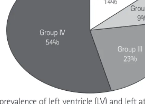
The prevalence of remodeling was presented in Fig. 1. Group I with progressive LA and LV dilatation was 13.9% (n=182, 64.2±
12.3 years, 128 males), group II with LV remodeling without LA dil- atation was 9.3% (n=120, 65.7±11.9 years, 78 males), group III with LA remodeling without LV dilatation was 22.8% (n=286, 63.3±12.3 years, 195 males), and group IV without LA and LV dilatation was 55.1% (n=722, 62.9±12.5 years, 519 males), respectively.
The baseline characteristics are as summarized in Table 1. The age and gender were not significant differences among groups. Weight and body mass index were higher in the remodeling groups than the non-remodeling group. The prevalence of hypertension was higher in remodeling groups than group IV (p<0.05). The proportion of Killip class III was more frequent in groups I, II, and III, whereas Killip class II was frequent in group IV (p<0.05). There was no difference in the groups with respect to the initial vital sign or percentage of ST-segment elevation MI or prior MI history.
Comparison of biochemical parameters, echocardiographic parameters, angiographic parameters, and medication according to left ventricular and left atrial remodeling
The levels of triglyceride, maximal CK-MB, Tn-I, and hs-CRP were more significantly increased in groups I and II than in groups III and IV (p<0.05) (Table 2). NT-pro BNP was highly increased in groups I, II, and III, whereas it presented a mild increase in group IV. LV end- diastolic and systolic dimension and volume were increased in groups I and II over other groups at admission. LA dimension and volume were increased in groups I and III over other groups. EF was the lowest in group I and wall motion score was the highest in group I. There were no significant differences among groups in dia- stolic parameters, such as mitral inflow pattern or e/e’ velocity. In coronary angiography, the types of infarct-related artery, multi ves- sel involvement, and reperfusion time were not different among groups. However, no-reflow phenomenon after percutaneous coro- nary intervention (PCI) was frequently observed in group I (Table 3).
Most of the patients enrolled in this study used aspirin and clopi- dogrel. There were no significant differences among groups for us- ing beta-blockers, angiotensin converting enzyme inhibiter (ACEI), or aldosterone receptor blocker (ARB), known to affect remodeling. Al- though the species of statin were various, statin usage rates were similar among groups. However, diuretics were used less in group IV than in other remodeling groups, quite significantly (Table 4).
Group I 14% Group II
9%
Group III 23%
Group IV 54%
Fig. 1. The prevalence of left ventricle (LV) and left atrium (LA) remodeling.
Group I: LV and LA remodeling, Group II: LV remodeling, Group III: LA re-
modeling, Group IV: no remodeling.
Mortality and long-term prognosis
All causes of death occurred in 168 cases (12.8%) in follow-up pe- riods (mean 1240±680, maximum 2210 days). Mortality was highest in the LA and LV remodeling group (20.9%) and lowest in the non- remodeling group (11.4%). Although the development of adverse cardiac events included in cerebrovascular events, re-MI, and re-ad- mission rates for HF tends to frequent in remodeled groups, there was no statistical significance among the groups during follow-up
(mean 945±746 days) (Fig. 2).
During the period of follow-up, the cumulative survival rate was significantly lower in the group of LV and LA remodeling than in others, but cardiac events were not significant (Fig. 3).
Independent predictors of mortality
There were several factors affecting death during follow-up. We found the independent predictors of mortality from multivariate re- Table 1. Baseline clinical characteristics
Group I Group II Group III Group IV p
Age (years) 64.2±12.3 65.7±11.9 63.3±12.3 62.9±12.5 0.098
Sex (Male, %) 70.8 65.0 68.2 71.8 0.373
Weight (kg) 64.2±10.5 65.0±11.8 66.2±12.8 63.6±11.2 0.014
Height (cm) 163.2±9.1 163.9±8.6 163.2±9.6 162.5±9.2 0.370
BMI 24.0±3.1 24.1±3.1 24.7±3.4 24.0±3.2 0.008
Hypertension (%) 51.6 59.1 56.3 45.3 0.012
Diabetes (%) 29.6 33.4 32.5 27.1 0.225
Smoking (%) 18.7 14.2 19.9 20.2 0.351
FHx (%) 4.3 5.0 3.5 5.9 0.386
Prior MI (%) 0.5 3.4 4.2 3.3 0.159
SBP (mm Hg) 124.7±30.7 128.8±36.7 128.4±31.2 130.1±29.6 0.209
DBP (mm Hg) 82.3±60.3 79.4±23.3 80.0±19.4 81.9±30.4 0.743
HR (/min) 77.7±21.2 74.3±23.0 77.4±21.5 75.9±19.3 0.319
Killip class II (%) 73.6 73.3 73.0 83.9 <0.001
Killip class III (%) 26.3 26.7 26.9 16.0 <0.001
STEMI (%) 58.7 56.7 61.8 58.3 0.705
Group I: LV and LA remodeling, Group II: LV remodeling, Group III: LA remodeling, Group IV: no remodeling, BMI: body mass index, FHx: family history, MI:
myocardial infarction, SBP: systolic blood pressure, DBP: diastolic blood pressure, HR: heart rate, STEMI: ST-segment elevation myocardial infarction, LA:
left atrium, LV: left ventricle
Table 2. Comparison of laboratory findings
Group I Group II Group III Group IV p
WBC (mg/dL) 7269 7290 7457 7322 0.793
Hb (g/dL) 13.7±8.0 14.1±11.6 13.5±6.1 13.9±9.75 0.892
Glucose (mg/dL) 170.6±81 169.5±78.0 185.2±98.0 171.5±83.5 0.168
Creatinine (mg/dL) 1.05±1.1 1.07±0.6 1.02±0.6 1.09±0.9 0.709
TC (mg/dL) 186.1±45.5 183.2±43.4 178.9±39.4 182.8±40.7 0.323
TG (mg/dL) 133.5±104.1 137.8±112.1 121.4±89.3 118.9±69.2 0.041
LDL-C (mg/dL) 121.5±39.3 118.5±38.4 115.0±35.6 118.4±36.1 0.313
HDL-C (mg/dL) 43.3±12.1 45.5±14.8 45.1±12.5 46.7±23.5 0.180
CK-MB (ng/mL) 136.3±17.8 103.1±12.6 63.8±12.8 63.6±11.2 <0.001
Tn-I (ng/mL) 90.6±50.5 65.6±71.1 46.2±61.6 30.6±41.4 <0.001
NT-Pro BNP (pg/mL) 3830.3±7532.2 3640.9±6660.1 4031.7±7877.4 2458.7±5530.3 0.002
hs-CRP (mg/dL) 2.94±4.7 2.51±3.8 1.88±3.3 1.65±2.9 0.001
HbA1c (%) 6.52±1.2 6.57±1.6 6.49±1.3 6.61±1.5 0.710
Group I: LV and LA remodeling, Group II: LV remodeling, Group III: LA remodeling, Group IV: no remodeling, WBC: white blood cell, Hb: hemoglobin, TC: to- tal cholesterol, TG: triglyceride, LDL-C: low density lipoprotein-cholesterol, HDL-C: high density lipoprotein-cholesterol, CK-MB: creatinine kinase MB, Tn-I:
troponin I, NT-pro BNP: N-terminal pro B natriuretic peptide, hs-CRP: high sensitivity C-reactive protein, HbA1c: glycosylated hemoglobin, LA: left atrium,
LV: left ventricle
gression using positive factors from univariate analysis. Age, ele- vated NT-pro BNP, no reflow after PCI, and the presence of LV & LA remodeling were (odd ratio; 3.688, confidence interval; 1.309-10.387, p=0.014) significant independent predictors of long-term mortality (Table 5).
Discussion
The major finding of this study is that the progressive dilation of both LA and LV resulted in a poor prognosis, especially mortality in MI patients with HF. Conversely, the preservation of LA and LV size Table 3. Comparison of echocardiographic and coronary angiographic findings
Group I Group II Group III Group IV p
LVEDD (mm
2) 49.80±7.3 47.50±7.0 52.23±6.9 51.04±5.4 0.002
LVESD (mm
2) 36.61±8.2 33.82±7.2 36.60±7.9 34.98±7.0 <0.001
LVEDV (mL) 139.20±80.0 111.37±76.8 155.83±85.9 117.4±89.1 <0.001
LVESV (mL) 75.65±55.2 56.62±45.0 76.53±52.7 55.61±49.8 <0.001
LVVI (mL/m
2) 24.02±3.1 24.10±3.1 24.73±3.4 23.95±3.2 <0.001
LAD (mm) 38.90±7.5 37.36±6.5 37.20±1.1 39.79±5.9 <0.001
LAV (mL) 65.69±63.4 44.27±52.4 61.59±52.2 45.33±57.2 <0.001
LAVI (mL/m
2) 35.40±30.5 40.14±37.5 35.61±30.6 27.95±35.3 <0.001
EF (%) 48.09±12.5 50.88±11.6 53.87±12.4 56.64±12.2 <0.001
WMS 24.54±5.5 22.99±5.9 22.36±5.6 20.84±5.4 <0.001
E (cm/seconds) 0.72±0.3 0.67±0.3 0.65±0.2 0.73±0.3 0.067
A (cm/seconds) 0.76±0.3 0.79±0.0 0.86±0.9 0.79±0.3 0.608
E’ (cm/seconds) 0.059±0.02 0.056±0.02 0.072±0.13 0.064±0.02 0.598
E/E’ 13.98±7.8 12.85±6.3 12.69±7.4 12.37±7.3 0.860
IRA LAD (%) 53.3 40.0 41.3 40.4 0.064
RCA (%) 15.4 17.5 15.4 15.0 0.670
LCX (%) 24.2 32.5 34.3 31.7 0.133
Left main (%) 2.7 1.7 0 2.2 0.071
≥2 vessel disease 52.8 51.3 48.0 49.3 0.764
Door to reperfusion time (minutes) 41.5±1.9 44.2±2.7 44.7±2.6 63.0±13.9 0.906
No reflow (%) 46.7 34.2 39.8 32.4 0.002
Group I: LV and LA remodeling, Group II: LV remodeling, Group III: LA remodeling, Group IV: no remodeling, LVEDD: left ventricle end diastolic dimension, LVESD: left ventricle end systolic dimension, LVEDV: left ventricle end diastolic volume, LVESV: left ventricle end systolic volume, LVVI: left ventricular vol- ume index, LAD: left atrial dimension, LAV: left atrial volume, LAVI: left atrial volume index, EF: ejection fraction, WMS: wall motion score, E: mitral inflow velocity, A: atrial contraction velocity, E’: systolic velocity of mitral septal annulus, IRA: infarct related artery, LAD: left anterior descending artery, RCA: right coronary artery, LCX: left circumflex artery, min: minute, LA: left atrium, LV: left ventricle
Table 4. Comparison of medication for treatment
% Group I Group II Group III Group IV p
Aspirin 99.5 100 99.3 99.6 0.273
Clopidogrel 98.9 98.3 99.3 97.9 0.086
Cilostazol 51.1 55.8 50.0 54.2 0.236
Beta blocker 78.6 85.3 82.9 81.0 0.708
ACEI 48.9 50.0 53.1 59.6 0.056
ARB 34.0 38.3 33.6 27.3 0.104
CCB 5.0 5.0 8.4 0.9 0.164
Diuretics 34.6 38.3 34.6 20.5 0.002
Nitrate 64.8 66.7 18.5 69.3 0.691
Statin 74.7 78.3 86.4 86.3 0.749
Group I: LV and LA remodeling, Group II: LV remodeling, Group III: LA remodeling, Group IV: No remodeling, ACEI: angiotensin converting enzyme inhibiter,
ARB: aldosterone receptor blocker, CCB: calcium channel blocker, LA: left atrium, LV: left ventricle
after MI was the best prognosis among the remodeling groups.
Initial and follow-up noninvasive measurements of LA and LV size provides long-term prognostic information in patients with AMI. Th- erefore, it may be important to identify patients at risk of LV and LA remodeling to prevent remodeling after AMI.
The remodeling pattern after MI is not precisely known. It might possibly be a time dependent progression, but not a linear sequence.
In our MI population with HF, cardiac remodeling was frequently ob- served in patients with hypertension, elevated cardiac enzyme, and elevated serum inflammatory markers. These factors are already reported to increase LV filling pressure and, therefore, affect LV and LA function. Traditionally, ventricular remodeling was regarded to continue for several months until the distending forces were coun- terbalanced by collagen scarring.
19)20)The underlying cellular mecha-
nism includes progressive myocyte lengthening without a propor- tional increase in the myocyte cross-sectional area.
21)22)Failure to normalize increased wall stresses results in progressive dilatation, recruitment of border zone myocardium into the scar, and deterior- ation in contractile function. This balance is determined by infarct size, location, and trans-murality, the extent of myocardial stunning, the patency of the infarct-related artery, and local tropic factors.
23)The biomarkers of myocardial damage, such as cardiac Tn-I and T, CK, and CK-MB appear to be useful in predicting late ventricular dila-
Fig. 2. Mortality and major adverse cardiac events in four groups. Group I:
LV and LA remodeling, Group II: LV remodeling, Group III: LA remodeling, Group IV: no remodeling. LV: left ventricle, LA: left atrium, MACE: major adverse cardiac event.
50 40 30 20 10
0 Group I Group II Group III Group IV Death p=0.010 Total MACE p=0.073
Fig. 3. Comparison of all-cause mortality and cardiac events. A: survival curve according to left ventricle (LV) and left atrium (LA) remodeling. B: major ad- verse cardiac events according to LV and LA remodeling. Group I: LV and LA remodeling; open red circle; ○, Group II: LV remodeling; open blue triangle; △, Group III: LA remodeling; open reverse green triangle; ▽, Group IV: no remodeling; open purple diamond; ◊.
1.0 0.9 0.8 0.7 0.6 0.5 0.4
1.0 0.9 0.8 0.7 0.6 0.5 0.4
Days Days
Cumulative survival Cumulative survival
0 500 1000 1500 2000 0 500 1000 1500 2000
Log rank p=0.005 Log rank p=0.325
A B
Table 5. Predictors of mortality by multivariate analysis
RR CI p
Age 1.072 1.000-1.148 0.050
Diabetes 1.279 0.981-1.668 0.069
Hypertension 1.259 0.954-1.662 0.104
Multi vessel disease 1.459 0.819-6.156 0.303
Ejection fraction 0.946 0.874-1.025 0.175
Wall Motion Score 1.023 0.916-1.090 0.478
E/E’ 1.013 0.962-1.066 0.629
No reflow 0.316 0.152-0.654 0.020
CK-MB 1.003 1.000-1.006 0.070
Tn-I 0.996 0.986-1.006 0.425
hs-CRP 1.060 1.006-1.116 0.376
NT-pro BNP 1.000 1.000-1.000 0.001
LV remodeling 1.376 0.776-2.097 0.337
LA remodeling 1.296 0.852-1.972 0.385
LV & LA remodeling 3.688 1.309-10.387 0.014 RR: relation risk, CI: confidence interval, CK-MB: creatinine kinase MB, Tn-I:
troponine I, hs-CRP: high sensitivity C-reactive protein, NT-pro BNP: N type pro brain type natriuretic peptide, LV: left ventricle, LA: left atrium
(%)