J Lipid Atheroscler 2016 December;5(2):163-167 https://doi.org/10.12997/jla.2016.5.2.163
pISSN 2287-2892 • eISSN 2288-2561
JLA
Major Trauma induced Left Ventricular Thrombus after Acute Myocardial Infarction
Dong Wook Lee, Ju Hee Ha, Jun Ho Kim, Ki Beom Park, Jae Joon Lee, Han Il Choi, Jin Hee Kim
Department of Internal Medicine, Busan Medical Center, Busan, Korea
급성 심근경색 치료 후 장기적으로 안정된 환자에서 중증외상 이후 발생한 좌심실 혈전
이동욱, 하주희, 김준호, 박기범, 이재준, 최한일, 김진희 부산의료원 내과
Left Ventricular Thrombus (LVT) formation after acute myocardial infarction is a serious complication. And the most feared complication of LVT is the systemic thromboembolic events, especially to the brain. Nowadays patients with acute myocardial infarction are treated with primary PCI and more aggressive anticoagulation therapies, resulting in the lower incidence of LVT. Early detection of LVT is very important, and echocardiography is the definitive test for detecting intracardiac thrombus. However, the need for serial echocardiography remains controversial. In this case report, we describe a 55-year-old man with major trauma induced LVT after acute myocardial infarction who underwent successful therapy. (J Lipid Atheroscler 2016 December;5(2):163-167)
Key Words: Left Ventricular Thrombus, Echocardiography, Major Trauma
Received:
Revised:
Accepted:
May 30, 2016 August 23, 2016 August 25, 2016
Corresponding Author: Jin Hee Kim, Division of Cardiology, Department of Internal Medicine, Busan Medical Center, Geoje 2-dong, Yeonje-gu, Busan 611-072, Korea
Tel: +82-51-507-3000, Fax: +82-51-507-3001, E-mail: [email protected]
This is an Open Access article distributed under the terms of the Creative Commons Attribution Non-Commercial License (http://creativecommons.org/licenses/by-nc/3.0) which permits unrestricted non-commercial use, distribution, and reproduction in any medium, provided the original work is properly cited.
서 론
좌심실 벽의 국소 또는 전반에 걸친 운동장애는 혈류정체의 원인이 되며, 때때로 좌심실 혈전(Left Ventricular Thrombus) 을 유발하기도 한다. 좌심실 혈전은 급성 심근경색(Acute Myocardial Infarction)이 있는 환자와 스트레스 유발 심근증 (Stress Induced Cardiomyopathy)이 있는 환자에서 각각 5%
정도에서 발생된다고 알려져 있고,1 말기의 확장성 심근병증(End Stage Dilated Cardiomyopathy) 환자의 11-44% 정도에서도 발생하는 것으로 알려져 있다.2
임상에서는 좌심실 혈전 생성 여부의 선별검사로 심장초음파가
널리 사용되고 있다.3 하지만 급성 심근경색 이후 발생할 수 있는 중요한 합병증 중에 하나인, 좌심실 혈전 여부의 확인을 위한 연속적이고 반복적인 심장초음파 검사의 시기 및 필요성에 대해서 는 논란이 있다.4
본 증례에서는 급성 심근경색으로 관상동맥 중재술을 시행 받고, 이후 정기적인 심장초음파 검사에서 좌심실 혈전이 관찰되 지 않았던 장기간의 안정적인 환자에서 중증외상 이후 좌심실 혈전이 확인되었고, 약물 치료 통해 혈전의 호전을 경험하였기에 보고하는 바이다.
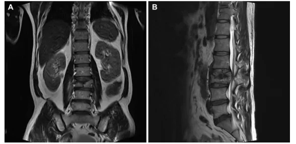
Fig. 1. Spinal magnetic resonanace imaging at coming to the orthopedics. Images show burst fracture of the 3rd lumbar.
(A) Coronal view, (B) Sagittal view.
증 례
환자: 우OO, 55세 남자 주소: 허리통증
현병력: 내원 이틀 전 1.5 m 가량의 높이에서 추락 이후 발생한 허리통증을 주소로 타병원 내원 이후 수술을 위해서 2014년 7월 2일 본원 정형외과로 전원되었고, 수술전 심장기능 평가를 위해서 심장내과로 협진의뢰 되었다.
과거력: ST 분절 상승 심근경색증(ST-Segment Elevation Myocardial Infarction)으로 좌전하행지 중간부 경피적 관상동 맥 중재술(2007년 4월) 시행하였고, 당시 시행한 경흉부 심장 초음파 검사에서 심첨부 전벽(Apical anterior), 전중격(Antero- septal), 전측부(Anterolateral), 하중격(Inferoseptal)에서 얇 아짐(Thinning)과 좌심실류(Left Ventricular Aneurysm)가 관 찰되었다. 좌심실 구혈률(Ejection Fraction)은 41%로 중등도의 좌심실 수축부전(LV Systolic dysfunction)이 관찰되었고, 하중 격, 하벽, 전벽에서의 무운동성(Akinesia)과 좌심방과 좌심실의 확장이 관찰되었다.
가족력: 특이사항 없음
사회력: 과거 흡연병력, 2007년부터 금연상태
진찰 소견: 두부 외상 소견은 없었고, 의식은 명료한 상황이었으 나 통증 및 자세 교정을 위해서 절대안정 및 자세고정을 유지하는 상황이었고, 생체징후는 혈압은 110/70 mmHg, 맥박 81회/분,
호흡수 20회/분, 체온 36.0℃로 관찰되었으며, 흉부청진과 신경 학적 검진 등 다른 이학적 검사에서 특이소견은 없었다.
검사실 소견: 말초혈액 검사에서 백혈구 11,800/mm3, 혈색소 12.5 g/dL, 혈소판 204,000/mm3였고, 생화학 검사에서 AST 23 IU/L, ALT 28 IU/L, BUN 12.4 mg/dL, Cr 0.7 mg/dL, 소변검사에서는 특이소견 관찰되지 않았다.
CRP 15.62 mg/L로 경한 상승소견 관찰되었고, CK-MB 1.8 ng/mL, Troponin-I 0.008 ng/mL로 정상소견을 보였다.
심전도 및 흉부방사선: 내원 시 심전도에서 분당 65회의 정상동 율동이었으며 ST 분절변화는 보이지 않았고, V4~V6 lead에서 T파 역전(inversion)이 관찰되었으나, 이전 심전도와의 차이는 없었다. 흉부 촬영에서 심비대 및 폐울혈 등의 이상소견은 관찰되 지 않았다.
척추 자기공명영상: 요추 손상 여부 및 정도를 확인하기 위해서 척추 자기공명영상(Spinal Magnetic Resonance Imaging)을 실시하였다. 그 결과 3번째 요추의 방출성 골절 소견 및 척추관 협착이 확인되었다(Fig. 1).
경흉부 심장 초음파 검사: 2007년 경흉부 심장 초음파 검사에서 중등도 이상의 좌심실 수축부전과 좌심실류가 관찰되어 이후 2년 간격으로 경흉부 심장 초음파 검사를 정기적으로 추적관찰 시행해 왔다. 외상이 있기 전, 마지막 추적검사였던 2013년의 경흉부 심장 초음파 검사에서 좌심실 구혈률과 국소 벽운동 장애 (Regional Wall Motion Abnormality)는 기존 추적검사와 큰
A B
Fig. 3. Echocardiographic findings at apical 4 chamber view. (A) Two days after trauma, large thrombus measuring 24 mm×15 mm in size were detected at the apex of the left ventricle, (B) Nine months treatment of thrombus, thrombus disappeared at the apex of the left ventricle.
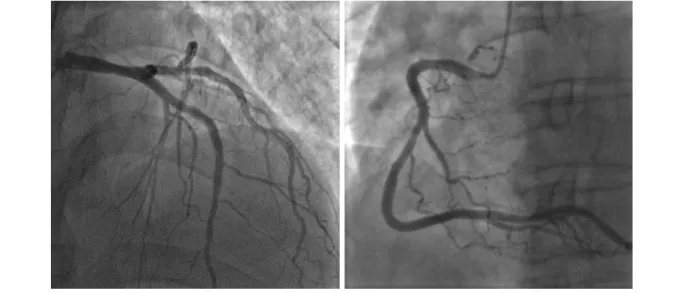
Fig. 2. Coronary angiography images show that there was no change compared with the previous results in Left Anterior Descending coronary artery middle part.
변화가 관찰되지 않았다. 2014년 7월, 외상이 있은지 2일이 경과 한 후에 시행한 경흉부 심장 초음파 검사에서 좌심실 구혈률 35%
로 오래된 심근 경색 후(Old Myocardial Infarction)에 전형적으 로 보이는 국소벽 운동 이상에 의한 좌심실 수축기 기능저하와 더불어, 2013년 경흉부 심장 초음파에서 관찰되지 않았던 심첨부 에 혈전이 새로이 관찰되었다.
관상 동맥 조영술: 좌심실 혈전의 확인 이후, 급성 심근경색의 합병증에 의한 좌심실 혈전의 발생 여부에 대해서 확인하기 위해서
관상 동맥 조영술을 시행하였다. 하지만 이전 마지막으로 시행한 2010년과 비교해서 4년만에 시행한 관상 동맥 조영술 검사에서는 의미있는 혈관의 변화는 관찰되지 않았다(Fig. 2).
치료 및 경과: 경흉부 심장 초음파 검사 결과를 바탕으로 환자의 수술 위험성 높다고 판단되었다. 하지만 환자의 주호소가 허리통 증이고, 척추 자기공명영상에서 제3요추부 방출성 골절 및 50%
이상의 척주관 침범으로 인한 하지 마비 가능성 높다고 판단되어 수술 위험성 등에 대해서 고지하고 좌심실 혈전을 치료하기 위해,
A B
헤파린(Heparin) 정맥 주사로 투여하는 항혈전요법과 더불어 척추 고정술 시행하였다(2014년 7월).
수술 이후 허리부위의 통증을 제외한 기타 이상소견은 관찰되지 않고, 안정적으로 유지 되어, 헤파린에서 와파린(Warfarin)으로 항응고제 약물의 단계적 교체를 시행하였고, 국제 정상화 비율 (International Normalized Ratio)을 2-3으로 유지 치료하였다.
3주의 입원치료 이후 통원치료로 전환하였으며, 연속적인 경흉 부 심장 초음파 검사를 시행하였다. 좌심실 혈전의 크기 감소를 확인할 수 있었으며, 수술 이후 대략 9개월 정도의 약물 치료 이후 좌심실 혈전은 관찰되지 않았다(Fig. 3).
본원 정형외과와 협진 통해서 통원치료 유지중이며, 현재까지 특이소견 및 자가 보행에 문제없는 상태로 외래 추적관찰중이다.
고 찰
현대 사회에서 심혈관 질환은 중요한 사망원인으로 알려져 있다. 최근 급성심근경색에서 항혈소판제, 항응고제, 경피적 관상 동맥 중재술 사용의 보편화로 인해서 사망률과 합병증의 빈도가 줄어들고 있는 실정이나, 그럼에도 불구하고 급성 심근경색증 이후 발생하는 합병증들은 여전히 많은 환자들에게 치명적인 결과 를 주고 있다.5 가장 심각한 합병증으로는 좌심실 혈전의 기동성으 로 인한 전신 혈전 색전증으로 뇌졸중 유발의 원인이 될 수 있다. 좌심실 혈전 발생의 기전은 급성 관상동맥 증후군(Acute Coronary Syndrome)으로 인한 응고항진(Hypercoagula- bility), 심내막하의 염증성 변화, 그리고 좌심실벽의 수축 기능 장애로 인한 혈액의 정체로 설명하고 있다.6 발생 위험인자로는 경색의 크기, 심실 수축 기능장애, 좌심실류, 전벽의 심근경색 등이 있다.7
2차원 경흉부 심장 초음파 검사는 좌심실 혈전 여부의 확인에 있어서 높은 특이도(85-90%)와 민감도(95%)를 나타낼 뿐만 아니라, 혈전의 크기 및 모양을 평가하는데 가장 유용하게 사용되 는 검사이다.8 또한 좌심실 내 혈전의 진단에서 심장 초음파 검사는 혈전의 유무뿐만 아니라 자연 경과 및 치료에 대한 반응을 평가하는 데 있어서 간편하고 매우 유용하게 사용된다.9
좌심실 혈전의 치료는 헤파린과 와파린을 사용하는 적절한 항혈전 및 항응고요법을 통하여 혈전 형성의 진행을 막고 색전증을 예방하는 데 있다. 특히 심장 초음파에서 색전의 위험성이 높은 혈전으로 판단되는 경우에는 조기에 항응고요법을 하고, 적어도
3개월에서 6개월은 와파린 사용이 권장되고 있으며,10 최근에는 저분자량 헤파린도 효과가 있고 안전하다고 보고되었다.11
급성심근경색의 중요한 합병증인 좌심실 혈전 발생은 급성 심근경색 발생이후 첫 3개월 이내에 발생하는 경우가 가장 많다고 알려져 있다.6 하지만 본 증례에서는 2007년 4월 좌전하행지 경피적 관상동맥 중재술 시행이후 정기적으로 추적 검사해오던 심장초음파 검사에서 7년 동안 좌심실 혈전이 관찰되지 않았던 환자였으나, 중증외상 이후 시행한 심장 초음파 검사에서 좌심실 혈전이 확인되었다.
혈전 발생의 위험성을 증가시킬 수 있는 응고항진은 암, 임신, 피임약 복용, 당뇨, 감염 등의 상황에서 발생할 수 있고, 두부외상이 나, 골절과 같은 중증 외상 환자에서도 혈전 발생의 위험성은 증가할 수 있다.12 한 문헌에서는 가슴 부위의 외상 이후 발생한 관상동맥 혈관 폐쇄에 따른 좌심실 혈전의 발생을 보고한 바 있 다.13 중증 외상은 트롬빈(Thrombin)의 생성을 증가시키고, 유지 하여 혈전 발생의 위험성을 증가시킨다고 알려져 있다.14 본 증례의 경우 급성 심근 경색 이후의 정기적 심장 초음파 검사에서 큰 변화 없이 유지되던 환자에서 중증외상 이후 좌심실 혈전이 확인되 었고, 약물 치료 통해 뇌졸중과 같은 중요한 전신 혈전 색전증 없이 혈전의 호전을 경험하였기에 보고하는 바이다.
급성 심근경색 이후, 좌심실 혈전 여부의 확인을 위한 연속적이 고 반복적인 심장초음파 검사의 시기 및 필요성에 대해서는 아직 정확하게 정해진 지침이 없는 실정이다. 본 증례의 환자에서는 2007년 급성 심근경색 이후에 2년 간격으로 시행하여 마지막 2013년까지 시행한 정기적인 추적 경흉부 심장 초음파 검사에서 7년 동안 장기간 안정적인 상태로 유지되었으나, 특별한 증상이나 검사상의 이상소견이 없었음에도 불구하고 중증외상 이후 치명적 인 합병증을 야기할 수 있는 좌심실 혈전이 발생한 경우이다.
따라서, 급성 심근경색이후 심장초음파의 주기적인 검사에 대한 필요성과 구체적인 가이드 라인 설정에 대한 추가적인 연구가 필요한 것으로 사료된다.
REFERENCES
1. Lee HJ, Kim HL, Hwang D, Park CS, Lim JS, Kang E, et al. Huge left ventricular thrombus and apical ballooning associated with recurrent massive strokes in a septic shock patient. Korean J Crit Care Med 2016;31:39-43.
2. de Gregorio C. Cardioembolic outcomes in stress-related cardiomyopathy complicated by ventricular thrombus: a systematic review of 26 clinical studies. Int J Cardiol 2010;141:11-17.
3. Cheitlin MD, Armstrong WF, Aurigemma GP, Beller GA, Bierman FZ, Davis JL, et al. ACC/AHA/ASE 2003 guide- line update for the clinical application of echocardio- graphy: summary article: a report of the American College of Cardiology/American Heart Association Task Force on Practice Guidelines (ACC/AHA/ASE Committee to Update the 1997 Guidelines for the Clinical Appli- cation of Echocardiography). Circulation 2003;108:
1146-1162.
4. Keren A, Goldberg S, Gottlieb S, Klein J, Schuger C, Medina A, et al. Natural history of left ventricular thrombi: their appearance and resolution in the post- hospitalization period of acute myocardial infarction. J Am Coll Cardiol 1990;15:790-800.
5. Porter A, Kandalker H, Iakobishvili Z, Sagie A, Imbar S, Battler A, et al. Left ventricular mural thrombus after anterior ST-segment-elevation acute myocardial infarction in the era of aggressive reperfusion therapy--still a frequent complication. Coron Artery Dis 2005;16:275- 279.
6. Delewi R, Zijlstra F, Piek JJ. Left ventricular thrombus formation after acute myocardial infarction. Heart 2012;
98:1743-1749.
7. Weinsaft JW, Kim HW, Crowley AL, Klem I, Shenoy C, Van Assche L, et al. LV thrombus detection by routine echocardiography: insights into performance characteri- stics using delayed enhancement CMR. JACC Cardiovasc
Imaging 2011;4:702-712.
8. Srichai MB, Junor C, Rodriguez LL, Stillman AE, Grimm RA, Lieber ML, et al. Clinical, imaging, and pathological characteristics of left ventricular thrombus: a comparison of contrast-enhanced magnetic resonance imaging, transthoracic echocardiography, and transesophageal echocardiography with surgical or pathological vali- dation. Am Heart J 2006;152:75-84.
9. Barbera S, Hillis LD. Echocardiographic recognition of left ventricular mural thrombus. Echocardiography 1999;16:
289-295.
10. Hirsh J, Fuster V, Ansell J, Halperin JL; American Heart Association; American College of Cardiology Foun- dation. American Heart Association/American College of Cardiology Foundation guide to warfarin therapy.
Circulation 2003;107:1692-1711.
11. Meurin P, Tabet JY, Renaud N, Weber H, Grosdemouge A, Bourmayan C, et al. Treatment of left ventricular thrombi with a low molecular weight heparin. Int J Cardiol 2005;98:319-323.
12. Gando S. Disseminated intravascular coagulation in trauma patients. Semin Thromb Hemost 2001;27:585- 592.
13. Kim JH, Kang TS. A case of distal embolization of left ventricular thrombus due to blunt chest trauma-induced coronary artery occlusion. Korean Circ J 2011;41:486- 489.
14. Selby R, Geerts W, Ofosu FA, Craven S, Dewar L, Phillips A, et al. Hypercoagulability after trauma: hemostatic changes and relationship to venous thromboembolism.
Thromb Res 2009;124:281-287.