Coronary Flow Reserve in the Remote Myocardium Predicts Left Ventricular Remodeling Following Acute Myocardial Infarction
Rongchao Cheng,
1Guoqian Wei,
1Longhao Yu,
1Zhendong Su,
1Li Wei,
1Xiuping Bai,
1Jiawei Tian,
2and Xueqi Li
11Department of Cardiology, The Fourth Affiliated Hospital of Harbin Medical University, Heilongjiang Province;
2Department of Echocardiology, The Second Affiliated Hospital of Harbin Medical University, Heilongjiang Province, China.
Received: September 23, 2013 Revised: December 2, 2013 Accepted: December 4, 2013 Corresponding author: Dr. Xueqi Li, Department of Cardiology,
The Fourth Affiliated Hospital of Harbin Medical University, No. 37, YiYuan Street, NanGang District, Harbin 150001, Heilongjiang, China.
Tel: 86-451-8593-9366, Fax: 86-451-8593-9366 E-mail: [email protected]
∙ The authors have no financial conflicts of interest.
© Copyright:
Yonsei University College of Medicine 2014 This is an Open Access article distributed under the terms of the Creative Commons Attribution Non- Commercial License (http://creativecommons.org/
licenses/by-nc/3.0) which permits unrestricted non- commercial use, distribution, and reproduction in any medium, provided the original work is properly cited.
Purpose: Coronary flow reserve (CFR) in the non-infarcted myocardium is often im- paired following acute myocardial infarction (AMI). However, the clinical signifi- cance of CFR in the non-infarcted myocardium is not fully understood. The objective of the present study was to assess whether a relationship exists between CFR and left ventricular remodeling following AMI. Materials and Methods: We enrolled 18 consecutive patients undergoing coronary intervention. Heart function was analyzed using real-time myocardial contrast echocardiography at one week and six months af- ter coronary angioplasty. Ten subjects were enrolled as the control group and were ex- amined using the same method at the same time to assess CFR. Cardiac troponin I (cTnI) levels were routinely analyzed to estimate peak concentration. Results: CFR was 1.55±0.11 in the infarcted zone and 2.05±0.31 in the remote zone (p<0.01) at one week following AMI. According to CFR values in the remote zone, all patients were divided into two groups: Group I (CFR <2.05) and Group II (CFR >2.05). The levels of cTnI were higher in Group I compared to Group II on admission (36.40 vs. 21.38, p<0.05). Furthermore, left ventricular end diastolic volume was higher in Group I compared to Group II at six months following coronary angioplasty. Conclusion: Mi- crovascular dysfunction is commonly observed in the remote myocardium. The CFR value accurately predicts adverse ventricular remodeling following AMI.
Key Words: Myocardial infarction, microvascular dysfunction, left ventricular re- modeling
INTRODUCTION
Restoration of adequate tissue perfusion is the goal of reperfusion therapy.1 Microvas- cular dysfunction in the infarcted myocardium is known to play a functional role in increasing infarct size, hindering left ventricular function and promoting left ventricu- lar remodeling (LVR) during the chronic phases of myocardial infarction.2 However, microvascular function has also been reported as impaired even in the remote regions supplied by normal coronary arteries.3 Nevertheless, the clinical significance of mi- crovascular dysfunction in the remote region has not yet been fully examined.4 Micro- vascular flow in humans is difficult to observe, and the methods of measuring micro-
revascularization with PCI within 12 hours following AMI.
Cardial TnI (cTnI) was analyzed on admission as a serum specific marker of myonecrosis, and the analysis was re- peated every six hours during the first 24 hours post-PCI in order to assess infarct size and severity of infarction.
All patients were subjected to two-dimensional echocar- diography (2-DE) and RT-MCE to assess left ventricular (LV) function and CFR one week following PCI. RT-MCE was performed both at rest and during dobutamine infusion in the infarcted region and remote regions to assess CFR. Af- ter six months, 2-DE was repeated to reassess LV function.
All patients received antiplatelet treatment with clopido- grel and aspirin. Angiotensin-converting enzyme inhibitor (ACE-I), angiotensin II receptor blocker (ARB) and β- blocker were routinely administered, if not contraindicated, and well tolerated.
Primary percutaneous coronary intervention
The infarct-related artery was treated with stent implanta- tion in all patients. Successful primary angioplasty was de- fined as a final TIMI flow grade 3 in the infarct-related ar- tery and residual stenosis of <20%. All patients received 300 mg aspirin and 300 mg clopidogrel prior to PCI. Coro- nary angiography was performed to select corresponding myocardial territory for each artery.
2-DE (Philips iE33, USA) was performed one week fol- lowing successful PCI and prior to RT-MCE. Left ventricu- lar end diastolic volume (LVEDV), left ventricular end sys- tolic volume (LVESV) and left ventricular ejection fraction (LVEF) were measured. LV volumes were calculated using the modified Simpson’s biplane method. p values <0.05 were considered statistically significant. LVEF was assessed as the percentage change in LV volume from end-diastole to end- systole. In order to assess the extent of post-infarct LVR, 2-DE was repeated at six months following PCI.
Real-time myocardial contrast echocardiography RT-MCE was performed in three apical views (apical 4-, 2-, and 3-chamber) one week following PCI using low- power continuous, power-modulation MCE in a mechani- cal index of 0.2. All subjects underwent infusion of Son- oVue (Bracco, Milan, Italy) at 50--70 mL/h with an infusion syringe-pump. The infusion rate was adjusted for each pa- tient to optimize myocardial opacification with minimal at- tenuation. Once optimized, the machine settings were main- tained throughout the study. To assess the replenishment kinetics, flash echocardiography at a high mechanical index vascular flow are also very complex. Coronary flow reserve
(CFR) is defined as the magnitude of increased coronary flow from the basal state to that achieved following maximal coro- nary vasodilation. Since flow resistance is primarily deter- mined by the microvasculature, CFR is therefore the primary method of measuring microvascular flow.
Previous studies utilizing CFR have consistently been performed with experimental animal models.5 Several stud- ies were performed using positron emission tomography, magnetic resonance imaging or Doppler-tipped flow wire to obtain measures of CFR. However, these methods are inva- sive or expensive, and difficult for wide utilization in the clinic. In contrast, real-time myocardial contrast echocar- diography (RT-MCE) is real-time, non-invasive, and a cost effective and simple method to measure CFR. To date, there are no data using RT-MCE to measure CFR in non-infarcted myocardium. In the present study, we performed RT-MCE with dobutamine to measure CFR in all subjects, because it is highly possible that CFR and LVR are related in the re- mote regions following acute myocardial infarction (AMI).
MATERIALS AND METHODS
Study population
Eighteen patients (13 males and 5 females, mean age of 58.8 years) suffering from AMI were enrolled. The inclu- sion criteria were as follows: 1) first AMI; 2) single-vessel disease; 3) successful recanalization by percutaneous coro- nary intervention (PCI) (defined as <20% of residual steno- sis and thrombolysis in myocardial infarction (TIMI) flow grade 3; and 4) within 12 hours following the onset of symp- toms. Myocardial infarction was diagnosed by chest pain lasting more than 30 minutes, ST-segment elevation of >0.2 mv in at least two contiguous electrocardiographic leads, and 2-fold increase in serum creatinine kinase or Troponin I (TnI) level. Patients with an unstable condition, malignant arrhythmias, severe extra-cardic disease, or severe valve disease were excluded. A control group included 10 sub- jects (6 males and 4 females, mean age of 53.4 years) with normal wall motion and coronary arteries. The study proto- col was approved by the Institutional Review Board of the Fourth Hospital of Harbin Medical University, and written informed consent was obtained from all patients.
Study design
This study included eighteen patients undergoing absolute
tomatically calculated mean acoustic intensity of each ROI and generated time-intensity curves that were subsequently fitted to a monoexponential function: y=A (1-e-βt) (q), where A represents the plateau acoustic intensity and β represents the rate of acoustic intensity increase and reflects microbub- ble velocity. MBF was calculated as the product of A×β.
The product of these parameters was correlated well with radiolabeled microsphere-derived MBF. Resting and maxi- mal MBF was derived, and CFR (MBF at stress/MBF at rest) was calculated for each patient. A single value was cal- culated for each region as the average of all analyzable seg- ments. Segments with artifacts or attenuation were excluded.
All images were analyzed independently by two experienced observers, who were blinded to the clinical data, angio- graphic results, and other respective imaging.
Statistical analyses
Continuous variables are expressed as mean±SD, and were analyzed using a Student’s t-tests. To determine correlations, we used Spearman’s rank correlation test. Categorical data were presented as absolute values or percentage and were compared using chi-square or Fisher’s exact tests.
RESULTS
Baseline characteristics
In every patient, PCI restored coronary TIMI flow grade 3 and provided residual stenosis of <20%. Stenting was per- formed in all patients; among the 18 patients, 13 were males and the mean age was 58.8 years. Twenty-two percent of all patients had diabetes mellitus, 39% of all patients had hy- pertension, 39% of all patients had hypercholesterolemia, and 44% of all patients were smokers. All patients received aspirin, clopidogrel, and a statin during the follow-up peri- od. ACE-I or ARB and β-blockers were routinely adminis- tered, if not contraindicated and if well tolerated. The in- farct-related coronary artery was LAD in 8 patients, LCX in 3 patients, and RCA in 7 patients. The mean peak con- centration of cTnI was 25.56 ng/mL and the mean time from the onset of symptoms to reperfusion was 5.33 hours. Base- line clinical, angiographic and echocardiographic character- istics are outlined in Table 1.
RT-MCE with dobutamine stress was performed without serious complications. Hemodynamic and safety parame- ters are outlined in Table 2. During low-dose dobutamine in- fusion, heart rates and SBP increased (p<0.05), while DBP of 1.4 was applied for transient microbubble destruction in
the myocardium. We acquired sequences of low-power (0.2) myocardial perfusion images containing at least 15 cardiac cycles following the flash echocardiography. The procedure was repeated for each apical view.6 SonoVue infusion was administered again two minutes after the end of dobuta- mine infusion at the same rate as provided at baseline. Do- butamine was infused intravenously at a starting dose of 5 ug/kg/min, followed by an increasing dose of 10 ug/kg/min up to a dose of 20 ug/kg/min in three minutes stages.7 Drugs that were considered to influence coronary microcirculation were withdrawn at 24 hours. Heart rate, systemic blood pressure, and a 12-lead electrocardiogram were recorded at each stage of dobutamine infusion. Dobutamine infusion was stopped if the patient developed serious complications, such as significant hypertension [systolic blood pressure (SBP) >220 mm Hg, diastolic blood pressure (DBP) >120 mm Hg], hypotension (SBP drop of >40 mm Hg), intolera- ble symptoms, a persistent arrhythmia, gross electrocardio- gram changes, or intolerable side effects. MCE images were acquired in the same sequence as rest images.
CFR measurement
The LV was divided into 17 segments.8 We chose infarcted regions far away from remote regions, to ensure that they do not adversely affect each other. We defined the remote and infarct regions as follows: if the left anterior descend- ing (LAD) artery was the infarct-related artery, the anterior segments and/or anteroseptal segments showing hypokine- sia at baseline 2-DE represented the area of the infarcted re- gion, and the inferior and/or inferolateral segments repre- sented the remote region. In the right coronary (RCA) or left circumflex artery (LCX) was the infarct-related artery, the converse procedure was used. All of the clear infarct seg- ments were analyzed, and we chose two remote segments to analyze remote regions in each patient. A total of seventy- six myocardial segments (44 infarcted lesions and 32 re- mote lesions) were studied.
Myocardial blood flow (MBF) was quantified using Q- lab software (Philips Medical System, USA). RT-MCE se- quences were analyzed in end-systolic frames, beginning in the frame immediately following the flash and including 15 subsequent cardiac cycles. Regions of interest (ROI) were placed and tracked manually within the myocardium at rest and the corresponding segments after the stressor. The ROI were of similar size, and the high-intensity endocardial and epicardial borders were avoided. The software package au-
was not different (p>0.05).
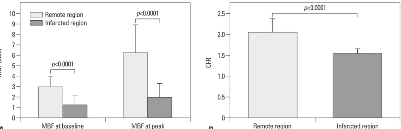
Comparisons of MBF and CFR among infarct, remote and normal regions
In the infarcted region, the mean resting MBF was 1.27±
0.86 dB/s, and it was 1.94±1.33 dB/s during dobutamine in- fusion. MBF in the remote regions was 2.94±1.06 at base- line and 6.22±2.68 dB/s at stress. Both at baseline and fol- lowing low dose dobutamine administration, MBF in the remote region was significantly higher than in the infarcted region, and mean CFR of the remote region was also signif- icantly higher than in the infarcted region (2.05±0.31 vs.
1.55±0.11, p<0.01) (Table 3, Fig. 1). There was no difference in MBF between the remote and normal regions at baseline, while MBF in the normal region was significantly increased from that in the remote region at peak stress (p<0.05) (Fig.
2). Mean CFR was also higher than the remote region (2.94±
1.06 vs. 2.05±0.31, p<0.01) (Fig. 2).
CFR in the remote region is related to infarct size Mean CFR in the remote region was 2.05. After defining the normal lower limit of CFR as 2.05, the patients were di- vided into two groups: five patients had impaired CFR (CFR <2.05, Group I) and 13 patients had preserved CFR (CFR >2.05, Group II). No differences were observed be- tween the two groups in terms of age, gender, cardiac risk factors, time from symptom onset to reperfusion, and site of infarct. Medications throughout the hospital stay and during the follow-up period were also similar between the two groups. Peak cTnI was higher in Group I patients than Group II patients (36.40±18.35 vs. 21.38±10.04, p<0.05).
Impaired CFR was associated with a greater degree of myocardial damage. Furthermore, impaired CFR in the re- mote region was linked to impaired CFR in the infarcted
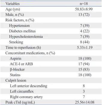
Table 1. Baseline Characteristics of the Study Patients (n=18)
Variables n=18
Age (yrs) 58.83±8.99 Male, n (%) 13 (72) Risk factors, n (%)
Hypertension 7 (39) Diabetes mellitus 4 (22) Hypercholesterolemia 7 (39) Smoking 8 (44) Time to reperfusion (h) 5.33±1.19 Concomitant medications, n (%)
Aspirin 18 (100) ACE-I or ARB 17 (94) β-blocker 15 (83) Statins 18 (100) Culprit lesion
Left anterior descending 8 Left circumflex 3 Right coronary artery 7
Peak cTnI (ug/mL) 25.56±14.08
Values are presented as number (%) or mean (SD). ARB, angiotensin II receptor blocker; ACE-I, angiotensin-converting enzyme inhibitor.
Table 2. Hemodynamic Data during Dobutamine Stress MCE
Variables n=18
Baseline
HR (beats/min) 71±5
SBP (mm Hg) 122±11
DBP (mm Hg) 74±9
RPP (mm Hg/min) 8566±711 Peak
HR (beats/min) 120±6*
SBP (mm Hg) 137±10*
DBP (mm Hg) 78±9
RPP (mm Hg/min) 16413±15479*
HR, heart rate; SBP, systolic blood pressure; DBP, diastolic blood pressure;
RPP, rate-pressure product; MCE, myocardial contrast echocardiography.
RPP was calculated by multiplying HR x SBP.
*p<0.05 compare to baseline.
Table 3. MBF and CFR in Patients with AMI
Variables Infarcted region Remote region p value
Baseline
A (dB) 2.70±0.89 4.72±0.93 <0.001
β (S-1) 0.44±0.18 0.61±0.16 0.004
MBF (dB/s) 1.27±0.86 2.94±1.06 <0.001
Peak
A (dB) 3.28±0.93 5.29±1.01 <0.001
β (S-1) 0.55±0.25 1.14±0.41 <0.001
MBF (dB/s) 1.94±1.33 6.22±2.68 <0.001
CFR 1.55±0.11 2.05±0.31 <0.001
MBF, myocardial blood flow; CFR, coronary flow reserve; AMI, acute myocardial infarction.
A represents the plateau acoustic intensity and β represents the rate of acoustic intensity increase and reflects microbubble velocity. MBF was calculated as the product of A×β. CFR was calculated as MBF at stress/MBF at rest.
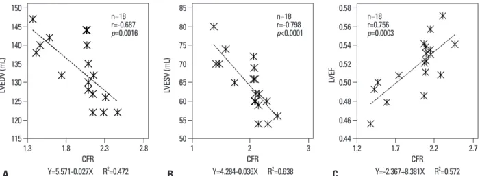
p=0.0016) (Fig. 3). LVESV, LVEF, and CFR all correlated with each other (r=-0.798, p<0.0001; r=0.756, p=0.0003, respectively) (Fig. 3). Impaired CFR led to a significant in- crease in LVEDV at six months following PCI.
DISCUSSION
It is well known that PCI is the most effective means to re- store patency and adequate reflow in the infarct-related ar- tery in patients with AMI. However, detailed studies have shown that up to 25% of patients undergoing angioplasty lack myocardial reperfusion despite revascularization of the infarct-related artery.9 As a result, cardiac mortality remains high, particularly in patients who develop heart failure. This study was designed to assess the relationship between CFR in the remote region and LVR in patients with a first AMI who underwent successful revascularization. CFR was cal- culated as the ratio of myocardial blood flow following ad- region. The characteristics of Group I and II are presented
in Table 4.
CFR in the remote region
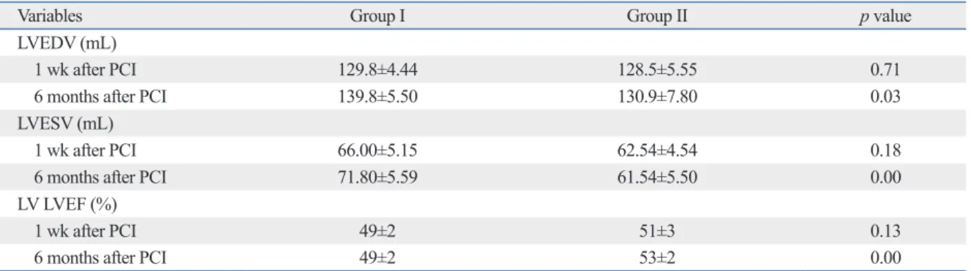
At one week post-PCI, LVEDV in Group I was higher than in Group II, however, there was no statistically significant difference (129.8 mL vs.128.5 mL, p>0.05). LVEDV in Group I increased compared with Group II (139.8 mL vs.
130.9 mL, p<0.05) at six months following PCI. During the six-month follow-up period, LVEDV and LVESV in Group I increased (129.8 mL vs. 139.8 mL, and 66 mL vs. 71.8 mL, respectively) and LVFF was not improved (49% vs. 49%).
LVEDV in Group II slightly increased (128.5 mL vs. 130.9 mL), while LVESV decreased (62.5 mL vs. 61.5 mL) and LVEF increased (51% vs. 53%); however, none of these comparisons reached statistical significance. Changes in LV volumes and LVEF at each time point are described in Table 5. Furthermore, there was a significant negative correlation between CFR and LVEDV in the remote region (r=-0.687,
Fig. 1. MBF and CFR in the infarcted and remote regions. (A) Both at baseline and after low dose dobutamine administration, MBF in remote region was sig- nificantly higher than in the infarcted region. (B) Comparison of CFR in infarcted and remote regions. Mean CFR of the remote region was significantly higher than the infarcted region. MBF, myocardial blood flow; CFR, coronary flow reserve.
Fig. 2. MBF and CFR in the remote and normal regions. (A) No difference was observed in MBF between the remote regions in normal subjects at baseline.
At peak stress, MBF in normal subjects was significantly increased compared to the remote region. (B) Correlation between CFR in the remote and normal regions. Mean CFR was higher than in the remote region. MBF, myocardial blood flow; CFR, coronary flow reserve.
A
A
B
B
0
0
0
0 1
2
2
0.5
0.5 3
4
4
1.0
1.5 1.0 5
6
6 1.5
2.0 7
8
8 2.0
3.0 2.5 9
10 10
12
2.5
3.5
MBF (db/s)MBF (db/s) CFRCFR
MBF at baseline
MBF at baseline
Remote region
Remote region MBF at peak
MBF at peak
Infarcted region
Normal region p<0.0001
p=0.3876
p<0.0001
p=0.0089
p<0.0001
p<0.0001 Remote region
Infarcted region
Remote region Normal region
it is associated also with extensive necrosis in the shared mi- crovascular. Finally, CFR and LVEDV were negatively cor- related. Impaired CFR in the remote regions was propor- tionally associated with increased LVEDV. Taken together, our data provide evidence of a strong correlation between CFR in the remote region and LVR following reperfusion.
Early CFR measured in the remote region can independent- ly predict late LVR following AMI.
CFR in the remote region measured using MCE with do- butamine stress is an effective parameter for predicting LVR in patients with AMI.13,14 Following successful PCI, the average CFR was 2.05 in the remote myocardium, low- er than that in the normal subjects, although the remote myocardium was not supplied directly by the infarct-related coronary artery. We observed that both LVEDV and LVESV ministration of dobutamine to basal blood flow.10
There were four main findings in the present study. Firstly, no study subjects were excluded due to side effects of dobu- tamine stress during MCE. The use of MCE for non-invasive quantification of microvascular flow is feasible,11,12 Previous data have suggested a good agreement among MCE, SPECT, and CMR for the evaluation of myocardial perfusion. Sec- ondly, although the infarct-related artery was successfully revascularized, CFR still decreased compared to the normal group, not only in the infarcted region, but also in the re- mote region. Thirdly, CFR in the remote region correlated with both infarct size and severity. Patients with the greatest impaired CFR had the highest peak cTnI, while cTnI corre- lated with infarct size. In addition, CFR in the remote region was linked to CFR in the infarcted region. It is possible that
Table 5. Temporal Changes in Echocardiographic Parameters from Both Groups
Variables Group I Group II p value
LVEDV (mL)
1 wk after PCI 129.8±4.44 128.5±5.55 0.71
6 months after PCI 139.8±5.50 130.9±7.80 0.03
LVESV (mL)
1 wk after PCI 66.00±5.15 62.54±4.54 0.18
6 months after PCI 71.80±5.59 61.54±5.50 0.00
LV LVEF (%)
1 wk after PCI 49±2 51±3 0.13
6 months after PCI 49±2 53±2 0.00
LVEDV, left ventricular end diastolic volume; LVESV, left ventricular end systolic volume; LVEF, left ventricular ejection fraction.
Table 4. Comparison of Clinical Variables
Variables Group I, n=5 Group II, n=13 p value
Gender (m/f) 4/1 9/4 1.00
Mean, age 56.60±12.56 59.70±7.67 0.31
Infarct-related artery, n (%)
LAD 3 (60) 5 (38)
RCA 2 (40) 5 (38)
LCX 0 3 (24) 0.79
Diabetes mellitus 1 3 1.00
Hyperlipidemia 2 5 1.00
Smoking 2 6 1.00
Hypertension 2 5 1.00
Concomitant medications
ACE-I/ARB 5 12 1.00
β-blocker 4 11 1.00
Statins 5 13 1.00
Time to reperfusion (h) 5.20±1.30 5.38±1.19 0.83
Peak cTnI, ug/mL 36.40±4.62 21.38±8.84 0.04
CFR in the infarcted region 1.43±0.19 1.59±0.09 0.006 LAD, left anterior descending artery; RCA, right coronary artery; LCX, left circumflex artery; ARB, angiotensin II receptor blocker; ACE-I, angiotensin- converting enzyme inhibitor; TnI, cardiac troponin I; CFR, coronary flow reserve.
that myocardial damage in the infarcted region may lead to compensatory changes in metabolism imbalance in the re- mote myocardium, such as consumption of adenosine tri- phosphate and creatine phosphate, and decreasing levels of glycogen, both confirmed in our laboratory.
In conclusion, effective microvascular reflow within the remote region following reperfusion is a key determinant for preventing LVR. Although we could not clarify the mecha- nisms of microvascular dysfunction in the remote myocardi- um, we suggest that impaired CFR in the remote myocardi- um is a more reliable parameter for estimating microvascular dysfunction.
Study limitations
There are some limitations in the present study. The num- ber of patients was small, the follow-up period was short, and the role of diastolic dysfunction or other factors was not fully examined. A large-scale study is needed to deter- mine the relationship of CFR and LVR following AMI.
ACKNOWLEDGEMENTS
This work was supported by the Department of Medical Ul- trasonics of the second Affiliated Hospital of Harbin Medi- cal University. We also thank Dr. Li Xue (the forth Affiliated Hospital of Harbin Medical University) and Guoqing Du (the second Affiliated Hospital of Harbin Medical Universi- ty) for providing amendment opinions.
We thank Medjaden Bioscience Limited for assistance in the preparation of this manuscript.
in patients with impaired CFR were significantly greater than the patients with preserved CFR. Therefore, adequacy of perfusion in the non-infarct region was also important in maintaining LV function, and CFR in the remote region correlated inversely with LVEDV and LVESV. Higher val- ues of CFR within the remote region were predictive for lack of remodeling over six months.
We did not perform MCE with dobutamine stress imme- diately following the onset of AMI since microvascular dysfunction is a complex and dynamic process that begins during the ischemic period and extends up to 48 hours fol- lowing reperfusion. The abrupt release of cytotoxic factors, local inflammation and vasoconstriction, peripheral emboli- zation of atherosclerotic debris, platelet plugs and intimal edema can immediately influence microvascular perfusion following reperfusion. Microvascular dysfunction in the non-infarcted myocardium is representative of more exten- sive necrosis and deterioration of cardiac function. Several possible mechanisms have been proposed,15-19 including im- pairment of endothelium-dependent dilation, sharing of mi- crovascular systems between the infarcted and non-infarct- ed myocardium, and vasoconstriction mediated by systemic or local neurohumoral reflexes. Furthermore, cardiospecific overexpression of the angiotensin II type 1 receptor and a decrease in microvessel density in the non-infarcted myo- cardium have also been observed, which lead to microvas- cular dysfunction. Experimental studies have confirmed that acute myocardial infarction activates alpha-adrenergic ac- tivity to increase vasoconstriction in remote regions nor- mally perfused by coronary arteries. These reasons could explain the observed reduced CFR. Other studies suggest
Fig. 3. Correlation analyses of CFR in the remote region. (A) There was a significant negative correlation between CFR in the remote region and LVEDV at six months. Impaired CFR leads to a significant increase in LVEDV at follow up. (B) Correlation between CFR and LVESV. There was a significant negative correla- tion between CFR in the remote region and LVESV. (C) Correlation between CFR and LVEF. There was a positive correlation between CFR and LVEF at follow up.
CFR, coronary flow reserve; LVEDV, left ventricular end diastolic volume; LVESV, left ventricular end systolic volume; LVEF, left ventricular ejection fraction.
CFR CFR CFR
Y=5.571-0.027X R2=0.472 Y=4.284-0.036X R2=0.638 Y=-2.367+8.381X R2=0.572
115 50 0.44
120 55 0.46
125 60 0.48
130 65 0.50
135 70 0.52
140 75 0.54
145 80 0.56
150 85 0.58
LVEDV (mL) LVESV (mL) LVEF
1.3 1.8 2.3 2.8 1 2 3 1.2 1.7 2.2 2.7
A B C
n=18r=-0.687 p=0.0016
n=18r=-0.798 p<0.0001
n=18r=0.756 p=0.0003
rect correlation with coronary flow velocity reserve by Doppler flow wire. Eur Heart J 2009;30:444-52.
11. Armstrong WF, Zoghbi WA. Stress echocardiography: current methodology and clinical applications. J Am Coll Cardiol 2005;
45:1739-47.
12. Bolognese L, Carrabba N, Parodi G, Santoro GM, Buonamici P, Cerisano G, et al. Impact of microvascular dysfunction on left ventricular remodeling and long-term clinical outcome after pri- mary coronary angioplasty for acute myocardial infarction. Circu- lation 2004;109:1121-6.
13. Lepper W, Hoffmann R, Kamp O, Franke A, de Cock CC, Kühl HP, et al. Assessment of myocardial reperfusion by intravenous myocardial contrast echocardiography and coronary flow reserve after primary percutaneous transluminal coronary angioplasty [correction of angiography] in patients with acute myocardial in- farction. Circulation 2000;101:2368-74.
14. Anantharam B, Janardhanan R, Hayat S, Hickman M, Chahal N, Bassett P, et al. Coronary flow reserve assessed by myocardial contrast echocardiography predicts mortality in patients with heart failure. Eur J Echocardiogr 2011;12:69-75.
15. Neizel M, Futterer S, Steen H, Giannitsis E, Reinhardt L, Loss- nitzer D, et al. Predicting microvascular obstruction with cardiac troponin T after acute myocardial infarction: a correlative study with contrast-enhanced magnetic resonance imaging. Clin Res Cardiol 2009;98:555-62.
16. Wita K, Lelek M, Filipecki A, Turski M, Wróbel W, Tabor Z, et al.
Microvascular damage prevention with thrombaspiration during primary percutaneous intervention in acute myocardial infarction.
Coron Artery Dis 2009;20:51-7.
17. Coser A, Franchi E, Marini M, Cemin R, Benini A, Beltrame F, et al. Intravenous contrast echocardiography after myocardial infarc- tion: relationship among residual myocardial perfusion, contractile reserve and long-term remodelling. J Cardiovasc Med (Hager- stown) 2007;8:1012-9.
18. Grayburn PA, Choi JW. Advances in the assessment of no-reflow after successful primary percutaneous coronary intervention for acute ST-segment elevation myocardial infarction: now that we can diagnose it, what do we do about it? J Am Coll Cardiol 2008;
51:566-8.
19. Park SM, Hong SJ, Kim YH, Ahn CM, Lim DS, Shim WJ. Pre- dicting myocardial functional recovery after acute myocardial in- farction: relationship between myocardial strain and coronary flow reserve. Korean Circ J 2010;40:639-44.
REFERENCES
1. Funaro S, La Torre G, Madonna M, Galiuto L, Scarà A, Labbadia A, et al. Incidence, determinants, and prognostic value of reverse left ventricular remodelling after primary percutaneous coronary intervention: results of the Acute Myocardial Infarction Contrast Imaging (AMICI) multicenter study. Eur Heart J 2009;30:566-75.
2. Geshi T, Nakano A, Uzui H, Okazawa H, Yonekura Y, Ueda T, et al. Relationship between impaired microvascular function in the non-infarct-related area and left-ventricular remodeling in patients with myocardial infarction. Int J Cardiol 2008;126:366-73.
3. Bodí V, Sanchis J, Núñez J, López-Lereu MP, Mainar L, Bosch MJ, et al. [Abnormal myocardial perfusion after infarction in pa- tients with persistent TIMI grade-3 flow. Only an acute phenome- non?]. Rev Esp Cardiol 2007;60:486-92.
4. Pacella JJ, Villanueva FS. Effect of coronary stenosis on adjacent bed flow reserve: assessment of microvascular mechanisms using myocardial contrast echocardiography. Circulation 2006;114:
1940-7.
5. Laser A, Ingwall JS, Tian R, Reis I, Hu K, Gaudron P, et al. Re- gional biochemical remodeling in non-infarcted tissue of rat heart post-myocardial infarction. J Mol Cell Cardiol 1996;28:1531-8.
6. Wei K. Detection and quantification of coronary stenosis severity with myocardial contrast echocardiography. Prog Cardiovasc Dis 2001;44:81-100.
7. Mathias W Jr, Kowatsch I, Saroute AN, Osório AF, Sbano JC, Dourado PM, et al. Dynamic changes in microcirculatory blood flow during dobutamine stress assessed by quantitative myocardial contrast echocardiography. Echocardiography 2011;28:993-1001.
8. Cerqueira MD, Weissman NJ, Dilsizian V, Jacobs AK, Kaul S, Laskey WK, et al. Standardized myocardial segmentation and no- menclature for tomographic imaging of the heart. A statement for healthcare professionals from the Cardiac Imaging Committee of the Council on Clinical Cardiology of the American Heart Associ- ation. Int J Cardiovasc Imaging 2002;18:539-42.
9. Wita K, Filipecki A, Lelek M, Bochenek T, Elżbieciak M, Wróbel W, et al. Prediction of left ventricular remodeling in patients with STEMI treated with primary PCI: use of quantitative myocardial contrast echocardiography. Coron Artery Dis 2011;22:171-8.
10. Kurita T, Sakuma H, Onishi K, Ishida M, Kitagawa K, Yamanaka T, et al. Regional myocardial perfusion reserve determined using myocardial perfusion magnetic resonance imaging showed a di-