189 Copyright © 2013 The Korean Society of Cardiology
Korean Circulation Journal
Introduction
Supraventricular tachycardia can be caused by diverse aberrant pathways. Dual atrioventricular (AV) nodal physiology, responsible for atrioventricular nodal reentrant tachycardia (AVNRT), has been re- ported in up to 12 percent of patients with the Wolff-Parkinson-Wh- ite syndrome. However, supraventricular tachycardia, due to coex- isting dual AV nodal pathways and an accessory pathway (AP) in a single patient, is very rare.
We report a patient with supraventricular tachycardia, which was attributed to AVNRT due to dual AV nodal pathways and atrioven- tricular reentrant tachycardia (AVRT) due to a left lateral AP. Furth- ermore, a radiofrequency (RF) ablation of the left lateral AP was not successful through a conventional endocardial approach, but it was successful through an intracoronary sinus approach.
Case Report
http://dx.doi.org/10.4070/kcj.2013.43.3.189 Print ISSN 1738-5520 • On-line ISSN 1738-5555
Successful Ablation of Resistant Left Lateral Accessory Pathway and Coexisting Atypical Atrioventricular Nodal Reentrant Tachycardia
June Namgung, MD
Division of Cardiology, Department of Internal Medicine, Inje University College of Medicine, Ilsan Paik Hospital, Goyang, Korea
A 41-year-old male was presented with drug-resistant supraventricular tachycardia. Electrophysiological study confirmed that the supra- ventricular tachycardia was caused by dual atrioventricular nodal pathways and a left lateral accessory pathway (AP). The left lateral AP was resistant to traditional endocardial ablation, but was successfully eliminated by radiofrequency ablation via the intracoronary sinus approach. (Korean Circ J 2013;43:189-192)
KEY WORDS: Accessory atrioventricular bundle; Tachycardia, atrioventricular nodal reentry; Radiofrequency catheter ablation.
Received: April 12, 2012
Revision Received: August 1, 2012 Accepted: August 23, 2012
Correspondence: June Namgung, MD, Division of Cardiology, Department of Internal Medicine, Vision 21 Cardiac & Vascular Center, Inje University College of Medicine, Ilsan Paik Hospital, 170 Juhwa-ro, Insanseo-gu, Goy- ang 411-706, Korea
Tel: 82-31-910-9645, Fax: 82-31-910-7829 E-mail: [email protected]
• The author has no financial conflicts of interest.
This is an Open Access article distributed under the terms of the Creative Commons Attribution Non-Commercial License (http://creativecommons.
org/licenses/by-nc/3.0) which permits unrestricted non-commercial use, distribution, and reproduction in any medium, provided the original work is properly cited.
Case
A 41-year-old man had a 5-year history of palpitations with exer- tion, due to a paroxysmal supraventricular tachycardia. The paroxy- smal supraventricular tachycardia was intractable to medical tr- eatment, and he was referred for electrophysiological study and RF ablation. The documented electrocardiography of supraventricular tachycardia revealed a heart rate of 168 bpm and no definitive P waves. After obtaining an informed consent, all antiarrhythmic me- dications were discontinued, and an electrophysiological study was performed. Under local anesthesia and using sterile technique, two quadripolar electrode catheters (St. Jude Medical, Inc., Minnetonka, MN, USA) were positioned to record the activity of the His bundle and right ventricular (RV) apex. The high right atrium (RA), low RA, and coronary sinus (CS) were mapped with a deflectable duo-de- capolar catheter (St. Jude Medical, Inc., St. Paul, MN, USA), inserted via the left femoral vein. Intracardiac electrograms were recorded us- ing a Prucka CardioLab
TMelectrophysiology system (General Electric Health Care System, Inc., Milwaukee, WI, USA).
The retrograde conduction pattern was eccentric and the earliest retrograde atrial activation was recorded in the distal CS during RV pacing (Fig. 1A). A narrow QRS tachycardia was repeatedly inducible with programmed atrial and ventricular stimulation (Fig. 1B). The me- chanism of tachycardia was confirmed to be AVRT, based on the ad- vancement of atrial activation, induced by the ventricular extra-sti- mulation at the time of the refractoriness.
Ablation of the left lateral AP, during a sinus rhythm, was first at-
tempted via a transseptal approach. RF energy was delivered to this
site at a temperature of 55°C, but it did fail to eliminate the bypass
190 AVRT and Atypical AVNRT
http://dx.doi.org/10.4070/kcj.2013.43.3.189 www.e-kcj.org
tract. Despite the repeated RF applications around the left lateral re- gion, the tachycardia remained inducible with programmed atrial sti- mulation. Extensive RF applications from the left lateral to the pos-
terolateral region were ineffective, as well. After injection of the contrast medium under fluoroscopy in left anterior oblique 45° and right anterior oblique 30° projections, the ablation catheter was po-
I aVF V1 HRA
HISd HISm HISp CS9, 10 CS7, 8 CS5, 6 CS3, 4 CS1, 2 RVA
I aVF V1 HRA
ABLd ABLp HISd HISm HISp CS9, 10 CS7, 8 CS5, 6 CS3, 4 CS1, 2 RVA
Spontaneous SVT induction Activation sequence during RV pacing
A B
Fig. 1. Simultaneous recordings for surface leads I, aVF, V1 and intracardiac electrograms from high right atrium, proximal and distal coronary sinus, His bundle, and right ventricular apex. Earliest atrial (A) activation during right ventricular (V) pacing (A) or spontaneous supraventricular tachycardia induction are recorded from the CS1, 2 electrodes (B). The atrial activation sequence is eccentric and earliest at the distal coronary sinus. HIS: His bundle, RVA: right ventricular apex, HRA: high right atrium, CS: coronary sinus, ABL: ablation.
I aVF V1 HRA
HISd HISm HISp CS9, 10 CS7, 8 CS5, 6 CS3, 4 ABLd ABLp CS1, 2 RVA
I aVF V1 HRA
HISd HISm HISp CS9, 10 CS7, 8 CS5, 6 CS3, 4 ABLd ABLp CS1, 2 RVA
At successful ablation site VA blocking after RF application
C D
A
B
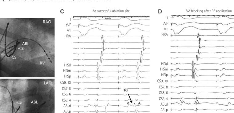
Fig. 2. Ablation of resistant left lateral accessory pathway via the intracoronary sinus approach. A and B: the fluoroscopic images, right anterior oblique
and left anterior oblique, showing the position of ablation catheter at distal coronary sinus through intracoronary sinus. C: the local electrogram from distal
ABL catheter during right ventricle (RV) pacing has the same ventricular (V) and atrial (A) timing compared to that of proximal CS catheter. D: after radio-
frequency energy (RF) application at this point, the electrogram shows the retrograde conduction through HIS during RV pacing. HIS: His bundle, RVA: right
ventricular apex, HRA: high right atrium, CS: coronary sinus, ABL: ablation.
191 June Namgung
http://dx.doi.org/10.4070/kcj.2013.43.3.189 www.e-kcj.org
sitioned in the intra CS (Fig. 2A and B). RF energy delivered at this site, using a maximum power of 30 Watts (Fig. 2C) and a maximum electrode-tissue interface temperature of 50°C eliminated the ret- rograde conduction over the AP (Fig. 2D).
Upon programmed atrial stimulation, atypical AVNRT was initi- ated with antegrade conduction through the fast pathway and ret- rograde conduction through the slow pathway (fast-slow type) (Fig.
3). The induced tachycardia had a tachycardia cycle length (TCL) of 340 msec, a short AH (140 msec) interval, a long HA interval (210 msec), AH/HA ratio <1, and a long VA interval, over 60 msec (185 msec).
Single ventricular extra stimuli with a coupling interval of 360 msec were unable to reset the tachycardia. Following a cessation of ven- tricular entrainment pacing, the tachycardia resumes with a V-A-V response, and the values of post-pacing interval minus TCL were gr- eater than 115 msec (Fig. 4). Thus, a fast-slow type AVNRT was con- firmed. The tachycardia was reproducibly induced upon a decremen- tal atrial pacing and terminated with atrial overdrive pacing. A 7-Fr, 4-mm-tip deflectable ablation catheter (Boston Scientific Corp., Natick, MA, USA) was positioned at the roof of the CS ostium, and slow pathway ablation was attempted in the inferior region of the Koch’s triangle, where the local A/V was <0.5. An accelerated junc-
tional rhythm was immediately observed and AVNRT could not be induced with either decremental atrial pacing or programmed atrial stimulation. A post-RF ablation electrophysiology study with isopro- terenol did not induce any supraventricular tachycardia. The patient remained asymptomatic three years later.
Discussion
This case describes a patient who had dual AV nodal pathways and resistant AP localized in the left lateral area. We performed ca- theter ablation of coexisting AP and a slow pathway. The clinically documented supraventricular tachycardia was AVRT with antero- grade conduction over the normal AV conduction system, and ret- rograde conduction via the left lateral AP. AVNRT was subsequently induced by programmed atrial pacing after the elimination of AP and the slow pathway ablated.
The AP may coexist with dual AV nodal pathways. The dual AV no- dal physiology can be observed in more than 10% of patients with Wolff-Parkinson-White syndrome.
1)However, supraventricular ta- chycardia, due to coexisting AP and dual AV nodal pathways in a sin- gle patient, is less common. In our patient, the dual AV nodal path- Fig. 3. Initiation of atypical atrioventricular nodal reentrant tachycardia (AVNRT) with programmed atrial stimulation (550/280 msec); AH jump and the induction of atypical AVNRT with antegrade conduction (downward arrow) by fast pathway and retrograde conduction (upward arrow) by slow pathway.
The tachycardia cycle length is 342 msec, AH interval is 140 msec, HA interval is 210 msec, AH/HA ratio <1 and VA interval is 185 msec. HRA: high right atri- um, HIS: His bundle, ABL: ablation, CS: coronary sinus, RV: right ventricular.
I aVF V1 HRA
HISd HISm HISp ABLd ABLp CS9, 10 CS7, 8 CS5, 6 CS3, 4 CS1, 2 RV
192 AVRT and Atypical AVNRT
http://dx.doi.org/10.4070/kcj.2013.43.3.189 www.e-kcj.org
ways were unmasked after successful elimination of AP. The slow pa- thway could be documented and ablated after abolishing the con- duction over the AP. The current case suggests the importance of investigating another mechanism of supraventricular tachycardia, even after a successful ablation of the AP.
Another instructive finding in the current case was the success- ful elimination of the left lateral AP, through the intra CS approach.
The left lateral AP was resistant to a conventional endocardial RF ab- lation via a transseptal approach. Left-sided AV APs, the anatomical substrates for the AVRT composed of muscular bundles, usually co- urse through the AV fat pad between the CS and the annulus fibro- sus and hug the hinge point of the mural leaflet of the mitral valve, which can be mapped and ablated from the left atrium, left ventricle, or CS.
2)3)Ablation of left free wall AP requires that the tip of the abl- ation catheter be positioned at a target site along either the atrial or ventricular surface of the mitral annulus. These pathways can be ap- proached from the left cardiac chambers via a transseptal or trans- aortic approach. Earlier studies reported that the successful rates of endocardial ablation of AP were 87-99%.
4)5)There may be a variety of reasons for a lengthy or ablation failure of AP. For example, epi- cardial insertion of the AP is an important cause of difficult endo- cardial AP ablation, particularly in the left lateral anatomic location.
Haissaguerre et al.
6)reported a successful epicardial catheter abla- tion of the left lateral AP, through the CS, when endocardial ap-
proaches were unsuccessful. As in this case, RF ablation via inside the CS would be an effective alternative approach if AP is resistant to the traditional endocardial RF ablation.
References
1. Chen YJ, Chen SA, Chiang CE, et al. Dual AV node pathway physiology in patients with Wolff-Parkinson-White syndrome. Int J Cardiol 1996;
56:275-81.
2. Becker AE, Anderson RH, Durrer D, Wellens HJ. The anatomical sub- strates of wolff-parkinson-white syndrome. A clinicopathologic corre- lation in seven patients. Circulation 1978;57:870-9.
3. Jackman WM, Wang XZ, Friday KJ, et al. Catheter ablation of accessory atrioventricular pathways (Wolff-Parkinson-White syndrome) by ra- diofrequency current. N Engl J Med 1991;324:1605-11.
4. Calkins H, Langberg J, Sousa J, et al. Radiofrequency catheter ablation of accessory atrioventricular connections in 250 patients. Abbreviated therapeutic approach to Wolff-Parkinson-White syndrome. Circulation 1992;85:1337-46.
5. Lesh MD, Van Hare GF, Schamp DJ, et al. Curative percutaneous cathe- ter ablation using radiofrequency energy for accessory pathways in all locations: results in 100 consecutive patients. J Am Coll Cardiol 1992;
19:1303-9.
6. Haissaguerre M, Gaita F, Fischer B, Egloff P, Lemetayer P, Warin JF. Ra- diofrequency catheter ablation of left lateral accessory pathways via the coronary sinus. Circulation 1992;86:1464-8.
Fig. 4. Ablation of atypical atrioventricular nodal reentrant tachycardia. A: delivery of a ventricular premature depolarization when the HIS is refractory cannot advance the atrial activation. B: during atypical atrioventricular nodal reentrant tachycardia, following cessation of ventricular entrainment pacing, the tachycardia resumes with a V-A-V response. The values of post-pacing interval (PPI) minus tachycardia cycle length (TCL) were greater than 115 msec and the ΔHA=40 msec. HRA: high right atrium, HIS: His bundle, CS: coronary sinus, ABL: ablation, RVA: right ventricular apex.
I aVF V1 HRA
HISd HISm HISp CS9, 10 CS7, 8 CS5, 6 CS3, 4 ABLd ABLp CS1, 2 RVA