LPS로 자극한 RAW 264.7 세포에서
염증성세포활성물질 생산에 미치는 導赤散 물 추출의 억제 효과
김지은1⋕, 김성배1, 강옥화1, 신인식1, 강석훈1, 이승호2, 권동렬1*
1 : 원광대학교 약학대학 한약학과, 원광한약연구소, 원광생명공학연구소, 2 : 김천대학교 한방재활학과
Inhibitory Effect of Water Extract from Dojuksan on LPS-induced Proinflammatory Cytokines Production in RAW 264.7 Cells
Ji-Eun Kim 1⋕ , Sung-Bae Kim 1 , Ok-Hwa Kang 1 , In-Sik Shin 1 , Suk-Hoon Kang 1 , Seung-Ho Lee 2 , Dong-Yeul Kwon 1*
1 : Department of Oriental Pharmacy, College of Pharmacy, Wonkwang Oriental Medicines Research Institute, and Institute of Biotechnology, Wonkwang University, Iksan, Jeonbuk, 570-749,
2 : Department of Rehabilitative Therapy in Oriental Medicine, Gimcheon University, Gimcheon, 740-704, Gyuungbuk, Korea.
ABSTRACT
Objectives : DojukSan is known to be effective for treating a urinary diseases and stomatitis. However, there has been a lack of studies regarding the effects of Dojuksan on the inflammatory activities and effector inflammatory disease mechanism about macrophage before is not known. To elucidate the molecular mechanisms of Dojuksan water extract (DJS) on pharmacological and biochemical actions in inflammation, we examined the effect of DJS on pro-inflammatory mediators in lipopolysaccharide (LPS)-stimulated macrophages.
Methods : In the present study, pro-inflammatory cytokine production was determined by performing enzyme-linked immunosorbent assay, reverse transcription polymerase chain reaction, and western blot analysis to measure the activation of MAPKs. Cells were treated with 200 ng/mL of LPS 1 h prior to the addition of DJS. Cell viability was measured by MTS assay. The investigation focused on whether DJS inhibited nitric oxide (NO) and prostaglandin E2 (PGE
2) productions, as well as the expressions of inducible NO synthase (iNOS), cyclooxygenase-2 (COX-2), interleukin-6 (IL-6) and mitogen-activated protein kinases (MAPKs) in LPS-stimulated RAW 264.7 cells.
Results : We found that DJS inhibited LPS-induced NO, PGE
2and IL-6 productions as well as the expressions of iNOS and COX-2. Furthermore, DJS suppressed the LPS-induced phosphorylation of p38 MAPK and c-Jun NH2-protein kinase (JNK).
Conclusions : These results suggest that DJS has inhibitory effects on LPS-induced PGE
2, NO, and IL-6 production, as well as the expressions of iNOS and COX-2 in the murine macrophage. These inhibitory effects occur through blockades on the MAPKs phosphorylation.
Key words : Dojuksan (DJS), lipopolysaccharide (LPS), inflammation, RAW 264.7 Cells
서 론
1)
導赤散의 方名은 心火를 引導하여 下行한다는 뜻에서 取한
것으로 名醫 錢乙 (AD1032 -1113)의 저서 ⟪小兒藥證直結
⟫에 처음 수록된 처방으로 小兒의 心熱을 治療하는데 사용되 었다
1). 表陰之氣가 내려오지 못하고 熱邪의 막힘을 받아서 일
*교신저자 : 권동렬 전북 익산시 신용동 344-2 원광대학교 약학대학 한약학과 본초학교실 ·Tel : 063-850-6802 ·E-mail : [email protected]
#제1저자 : 김지은 전북 익산시 신용동 344-2 원광대학교 약학대학 한약학과 본초학교실 ·Tel : 063-850-6802 ·E-mail : [email protected]
·접수:2013년 4월 22일 ·수정:2013년 5월 16일 ·채택:2013년 5월 23일
어나는 병증으로 心과 小腸에 열이 있어 나타는 증상을 치료 하는 병증으로 心熱로 인한 病의 症狀인 口內炎, 自律神經 失 調症, 膀胱炎, 腎盂炎, 血尿, 小便不利 등에 사용되어 왔다
2,3)
. 導赤散은 腎臟疾患 및 泌尿器系에 대한 연구가 이루어지 고 있고
4), 加味導赤散을 이용하여 利尿, 解熱, 鎭痙 작용과 항 스트레스 작용, 高血壓과 같은 心血管係 질환, 肥滿, 면역 계 및 아토피 등의 연구가 보고되고 있다
5-10). 그러나 대부분 다른 약재들을 加減한 연구들이 주를 이루고 있고, 導赤散만 을 이용한 생리활성 기전연구는 미흡한 실정이다. 임상에서 염증작용에 많이 사용되고 있음에도 불구하고 연구가 많이 이 루어지지 않음을 착안하여 본 연구를 진행하게 되었다.
염증은 외부 자극에 대한 생체조직의 방어반응의 하나로서 유해물질이나 화학적 자극에 의한 손상으로 일어난다
11). 염증 발생 시 염증부위에서 면역세포들이 침투되고 이들 세포들에 의해 여러 종류의 화학물질 및 cytokine을 생산 분비하여 생체 방어 및 염증반응을 일으킨다. 이 과정에서 염증을 유발하는 물질들을 염증인자라 한다. 염증인자 중에는 iNOS에 의해 만 들어지는 NO와 COX-2에 의해 만들어지는 PGE
2가 있다. NO 는 급성, 만성 염증반응을 조절하는데 nitric oxide synthase (NOS)의 촉매에 의해 생산되며 특히 iNOS은 염증반응에 관 여한다
12).
또한 PGE
2는 COX에 의해서 Arachidonic acid로부터 생 산된다. 본 연구에서는 항염효과가 있을 것으로 예상되는 導 赤散 물추출물이 LPS로 활성화된 RAW 264.7 대식세포에서 의 염증 매개물질 억제효과를 얼마나 효율적으로 저해할 수 있는지 관찰하였다. 그 지표로서 세포에서 방출하는 NO, PEG
2생성량과 iNOS, COX-2의 발현정도 뿐만 아니라 염증 성 사이토카인인 IL-6 생성량을 알아 본 바 유의한 결과를 얻었기에 보고하는 바이다.
재료 및 방법
1. 재료
1) 시약LPS (E. coli lipoplysaccaride), RPMI-1640 배지는 Sigma (MO, USA)로부터 구입하였고, FBS는Hyclone (Hyclone Labs Logan, UT)에서 구입하였다. PGE
2, IL-6 ELISA Kit 를 R&D시스템스 (MN, USA)로부터 구입하였으며, COX-2, iNOS, MAPKs (ERK, JNK, p38) 단일 monoclonal antibodies 와 streptavidin-horseradish peroxidase 복합의 2차 항체 는 Santa Cruz Biotechnology Inc. (Santa Cruz, CA)에서 구입하였다.
2) 세포 배양
설치류 대식 세포주 (murine macrophage cell line, RAW 264.7)는 한국 세포주 은행으로부터 구입하여 사용하였 다 (Korea Research Institute of Bioscience and BiotechnologyAmerican Tissue Culture Collection).
RAW 264.7 세포는 항생제 및 항균제 (100 U/ml의 페니실 린 G, 100 U/ml의 스트렙토마이신 (streptomycin)를 첨가 하고 10%의 열처리 우태아 혈청 (heat inactivated FBS)을
첨가한 완전한 RPMI 1640 배지에서 5% CO
2의 습한 대기, 37℃의 온도조건으로 배양하였다.
3) Primer 준비
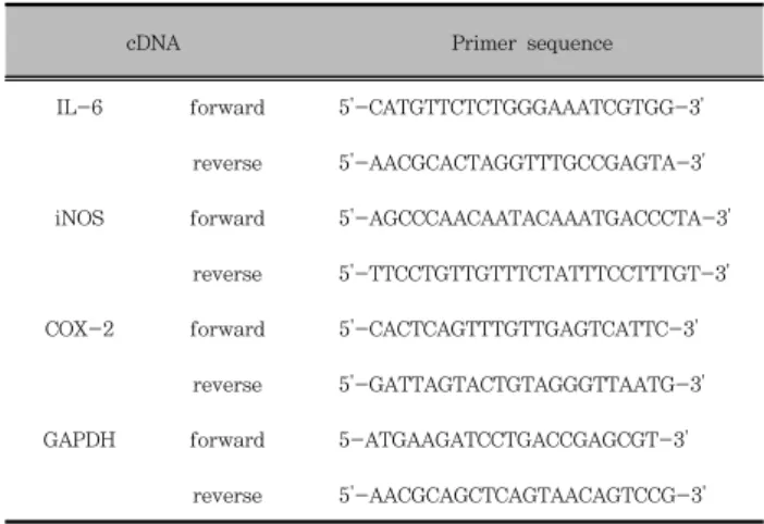
RT-PCR을 위해 여러 개의 primer를 사용하였다. Primer 의 구성은 다음과 같다 (Table 1).
Table 1. Primer sequences for RT-PCR
cDNA Primer sequence
IL-6 forward 5'-CATGTTCTCTGGGAAATCGTGG-3' reverse 5'-AACGCACTAGGTTTGCCGAGTA-3' iNOS forward 5'-AGCCCAACAATACAAATGACCCTA-3'
reverse 5'-TTCCTGTTGTTTCTATTTCCTTTGT-3' COX-2 forward 5'-CACTCAGTTTGTTGAGTCATTC-3'
reverse 5'-GATTAGTACTGTAGGGTTAATG-3' GAPDH forward 5-ATGAAGATCCTGACCGAGCGT-3'
reverse 5'-AACGCAGCTCAGTAACAGTCCG-3'
2. 방법
1) 시료의 조제방법
導赤散 물 추출물은 증류수 (100 g/L)로 2시간 동안 고온 으로 추출하여 준비하였다. 그 추출물은 0.45 µm 필터로 여 과한 후 감압 동결 건조 (lyophilized)하였으며, 4℃에서 보 관했다. 동결건조한 추출물은 실험하기 위하여 인산완충 식염 수 (phosphate-buffered saline. PBS)에 녹였다. 導赤散 물 추출물은 生地黃 6g, 木通 6g, 甘草 6g, 竹葉 6g의 조성으로 되어 있으며, 대학한약국 (Iksan, Korea)에서 구입하였다 (Table 2).
Table 2. The composition of Dojuksan
Herbal Name Pharmacognostic Name Weight (g)
生地黃 Rehmanniae Radix 6
木通 Akebiae Caulis 6
甘草 Glycyrrhizae Radix et Rhizoma 6
竹葉 Phyllostachys Folium 6
2) MTS 분석
세포 생존력은 MTS assay를 이용하였다. 導赤散을 처리한 세포에 20 µL의 MTS (3-(4,5-dimethylthiazol-2-yl) -5-(3-carboxymethoxyphenyl)-2-(4-sulfophenyl)-2H- tetrazolium), 5 mg/mL 용해액을 첨가한 후 37℃에서 1시 간 보존한 후 microplate reader를 이용하여 490 nm에서 흡광도를 측정하였다.
3) NO 생성량 측정
NO 생성에 대한 導赤散 물추출물 (DJS)의 효과를 알아보
기 위해 물 추출물 (250, 500 µg/mL)의 농도로 전처리하고 여기에 LPS (200 ng/mL)를 각각 주입한 다음 RAW 264.7 세포를 24시간 배양하였다. 그 상층액에 Griess reagent (1% sulfanilamide, 0.1% N-(1-naphthyl)-ethylene diamine dihydrochloride in 2.5% phosphoric acid solution)와 동 량으로 주입한 후 10분간 실온에서 방치한 후 570 ㎚에서 흡 광도에서 측정하였다.
4) ELISA
RAW 264.7 세포를 5×10
5cells/mL로 조절한 후 48 well plate에 접종하고, 18시간 배양하였다. 이후 배지를 교 체하고 導赤散 물추출물 (250, 500 µg/mL)을 30분 동안 처 리한 후, 그람-음성 박테리아 내독소인 LPS (200 ng/mL)로 RAW 264.7 세포를 자극한 후 24시간 배양 하였고 세포 부 유액을 원심분리하여 상층액을 수집하고, 상층액 내 IL-6 생 성량을 ELISA kit (R&D Systems Inc., Minneapolis, MN, USA)를 이용하여 사용자 매뉴얼에 기재된 방법대로 정량해 분석하였다.
5) PGE
2
측정RAW 264.7 세포를 5×10
5cells/mL로 조절한 후 48 well plate에 접종하고, 18시간 배양하였다. 이후 배지를 교체 하고 導赤散 물추출물 (250, 500 µg/mL)을 각각 30분 동안 처리한 후, LPS (200 ng/mL)로 RAW 264.7 세포를 자극한 후 세포들을 침전시켜 상층액을 수집하고, 상층액 내 PGE
2생 성량을 EIA kit (R&D Systems Inc., Minneapolis, MN, USA)를 이용하여 분석하였다.
6) RNA 분리 및 RT-PCR
RAW 264.7 세포를 100 ㎜ 세포배양 접시에 5×10
6cells/mL로 분주한 다음, 하루 밤 동안 안정화 시켰다. 이 세포에 導赤散을 처리한 후, LPS로 처리하고 24시간 후에 세 포를 모아 PBS로 세척하여 이지 블루 (easy blue, Intron) 1 mL를 가하여 실온에서 교반하였다. 클로로포름 200 µL를 넣고 다시 교반하여 13,000 rpm, 4℃에서 10분간 원심분리 한 다음, 상층액 400 µL에 이소프로판올을 가하여 다시 원심 분리하여 RNA를 얻었다. 여기서 얻어진 RNA 에 MuLV 역 전사효소 (reverse transcriptase), 1 mM dNTP 0.5 µg을 넣어 cDNA를 만들었다. 여기에 IL-6, iNOS, COX-2, GAPDH primer를 넣고 유전자 증폭기 (thermal cycler)를 이용하여 증폭시켰다. 만들어진 RNA를 1.5% agarose gel에 전기 영동시켜 UV 검출기로 확인하였다.
7) Western blot analysis
導赤散 물추출물과 LPS로 처리하고 24시간 후 세포를 수 집하여 2-3회 PBS (phosphate buffered saline)로 세척 한 후 200 µL의 lysis buffer을 첨가하여 20분간 lysis시킨 후 13,000 rpm에서 10분간 원심 분리하여 세포막 성분 등을 제 거하였다. 단백질 농도는 BSA (bovine serum albumin)를 표준화하여 Bio-Rad Protein Assay Kit를 사용하여 정량하 였다. 20-30 µg의 lysate를 8-12% mini gel SDS-PAGE 로 변성 분리하여, 이를 PVDF (polyvinylidene difluoride)
membrane (BIO-RAD, Richmond, CA, USA)에 200 mA 로 2시간 동안 transfer하였다. 그리고 membrane의 blocking 은 5% skim milk가 함유된 TTBS (0.1% Tween20 + TBS) 용액에서 상온에서 2시간 동안 실시하였다. Monoclonal antibodies를 TTBS 용액에서 희석하여 상온에서 2시간 반응 시킨 후 TTBS로 3회 세정하였다. 2차 항체로는 HRP (horse radish peroxidase)가 결합된 anti-mouse IgG (Amersham Pharmacia Biotech, Little Chalfont, UK)를 희석하여 상 온에서 30분 간 반응시킨 후, TTBS로 3회 세정하여 ECL Prime기질 (Amersham Biosciences, Piscataway, NJ, USA) 과 1-3분 간 반응 후 chemical Luminescent image LAS-4000mini (GE Healthcare Bio-Sciences AB, Uppsala, Sweden) 감광하였다.
8) 통계분석
모든 실험은 3회 이상 반복으로 이루어졌으며, 실험결과는 각 항목에 따라 평균치 ± 표준편차 (SEM)를 구하여 그 유의 성은 Student’s t-test 분석법을 이용하여 신뢰수준 95% p
< 0.05에서 통계적 유의차를 평가하였다.
결 과
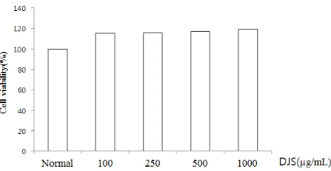
1. 導赤散 물추출물이 세포생존율에 미치는 영향
導赤散 물추출물 (DJS)이 세포독성에 영향을 주는지 알아 보기 위해 MTS 분석법을 이용하여 세포생존율을 측정하였다.
DJS를 100, 250, 500, 1000 µg/mL의 농도로 처리 하였을 때 모든 농도에서 세포독성은 나타나지 않았다 (Figure 1).
Figure 1. Effect of DJS on cell viability in RAW 264.7 cells. Cell viability was evaluated with the MTS assay. Data represent the mean ± S.E.M. of duplicate determinations from three separate experiments.
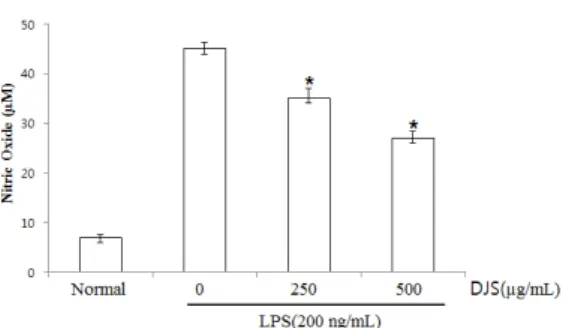
2. NO 생성에 대한 導赤散 물추출물의 억제 효과
NO 생성에 대한 導赤散 물추출물 (DJS)의 효과를 알아보
기 위해 Griess 시약을 이용하여 세포 배양액 중에 존재하는
NO
2−의 형태로 측정하였다. Figure 2 에서 보면, NO 생성
량은 LPS에 의해 현저히 증가 되었다. 반면, DJS 250, 500
µg/mL의 처리농도에서 농도 의존적이고 유의적으로 NO 생
성이 억제되는 것으로 나타났다 (Figure 2).
Figure 2. Effect of DJS on LPS-induced NO production in RAW 264.7 cells. RAW 264.7 cells were pretreated with the indicated concentration of DJS for 30 minutes before being incubated with LPS (200 ng/mL) for 24 hours. The culture supernatant was subsequently isolated and analyzed for LPS treated group. Statistical significance: *P<0.05, when compared to the LPS treated group.
Significant differences between treated groups were determined using the Student’s t-test. Values shown are the mean ± S.E.M.
of duplicate determinations from three separate experiments.
3. iNOS mRNA와 iNOS protein 발현에 대한 導赤散 물추출물의 억제 효과
NO는 iNOS에 의해 생성되어 염증상태에서 혈관 투과성, 부종 등의 염증반응을 촉진시키고, 염증매개체의 생합성을 촉 진해 염증을 악화시킨다고 알려져 있다. 따라서 NO를 생성하 게 하는 iNOS의 protein과 mRNA 발현에서 DJS의 항염증 효과를 알아 보기위해 Western blot과 RT-PCR을 수행하였 다. 그 결과, LPS 처리에 의해 형성되는 iNOS mRNA 발현 은 DJS 250, 500 µg/mL에서 현저한 억제 양상을 보였으며 (Figure. 3A), iNOS protein 역시 DJS 250, 500 µg/mL에 서 농도 의존적으로 억제되는 것을 확인하였다 (Figure. 3B).
Figure 3. Effect of DJS on LPS-induced iNOS mRNA and iNOS protein expression in RAW 264.7 cells. (A) iNOS mRNAs were assessed by RT-PCR in RAW 264.7 cells. Cells were pretreated with the indicated concentrations of DJS for 30 minutes before being incubated with LPS (200 ng/mL) for 24 hours. The β-actin mRNA was assayed in parallel to confirm equivalency of the cDNA preparation. (B) RAW 264.7 cells were pretreated with the indicated concentrations of DJS for 30 minutes before being incubated with LPS (200 ng/mL) for 24 hours. Equal amounts of protein (20 µg) were separated by sodium dodecyl sulfate-polyacrylamide gel electrophoresis and immunoblotted with iNOS and β-actin antibodies. Equal loading of protein was verified by β-actin.
4. PGE 2 생성에 대한 導赤散 물추출물의 억제 효과
대식세포 매개 PGE
2는 COX-1과 COX-2로부터 생성되는 데, 소량의 경우 생체대사에 필수적이지만 유도성 COX-2의
과발현에 의한 과량의 PGE
2는 강력한 염증 매개물질로 작용 하는 것으로 보고되어 있다
13-15). 따라서 본 연구에서는 PGE
2를 대상으로 DJS의 약리학적인 효능 평가를 실시하였 다. 그 결과, Normal에서는 PGE
2생성양이 매우 낮게 측정 되었으나, LPS에 자극에 PGE
2농도가 현저히 증가 되었다.
반면, DJS 250, 500 µg/mL의 농도에서 농도 의존적으로 PGE
2생성이 억제되는 것을 확인할 수 있었다 (Figure 4).
Figure 4. Effect of DJS on LPS-induced PGE2 production in RAW 264.7 cells. RAW 264.7 cells were pretreated with the indicated concentration of DJS for 30 minutes before being incubated with LPS (200 ng/mL) for 24 hours. The culture supernatant was subsequently isolated and analyzed for LPS treated group.
*P<0.05 when compared to the LPS treated group. Significant differences between treated groups were determined using the Student’s t-test. Values shown are the mean ± S.E.M. of duplicate determinations from three separate experiments.
5. COX-2 mRNA와 COX-2 protein 발현에 대한 導赤散 물추출물의 억제 효과
導赤散 물추출물 (DJS)의 COX-2 protein과 COX-2 mRNA 발현에 대한 효과를 알아보기 위해 Western blot과 RT-PCR을 수행하였다. 그 결과, LPS 처리에 의해 증가된 COX-2 mRNA 발현은 500 µg/mL에서 78%로 억제되는 양 상을 보였으며 (Figure 5A), COX-2 protein 발현은 250, 500 µg/mL 에서 억제되는 것을 확인하였다 (Figure 5B).
Figure 5. Effect of DJS on LPS-induced COX-2 mRNA and COX-2 protein expression in RAW 264.7 cells. (A) COX-2 mRNA were assessed by RT-PCR in RAW 264.7 cells. Cells were pretreated with the indicated concentrations of DJS for 30 minutes before being incubated with LPS (200 ng/mL) for 24 hours. The β-actin mRNA was assayed in parallel to confirm equivalency of the cDNA preparation. (B) RAW 264.7 cells were pretreated with the indicated concentrations of DJS for 30 minutes before being incubated with LPS (200 ng/mL) for 24 hours. Equal amounts of protein (20 µg) were separated by sodium dodecyl sulfate-polyacrylamide gel electrophoresis and immunoblotted with COX-2 antibodies. Equal loading of protein was verified by β-actin.
6. Pro-inflammatory cytokine IL-6 생성 대 한 導赤散 물추출물의 억제 효과
IL-6와 같은 pro-inflammatory cytokine은 염증단계에서 중추적 역할은 한다고 알려져 있다. 대식세포주인 RAW 264.7 세포로부터 염증성 사이토카인 IL-6의 생성을 ELISA를 이용 하여 실험하였다. 그 결과, LPS 자극원을 처리하였을 때 IL-6 생성이 현저히 증가하였고, DJS를 250, 500 µg/mL로 처리하 였을 때 IL-6 생성 억제 효과가 나타났다 (Figure 6).
Figure 6. Effect of DJS on LPS-induced IL-6 production in RAW 264.7 cells. Cells were pretreated with the indicated concentrations of DJS for 30 minutes before being incubated with LPS (200 ng/mL) for 24 hours. Production of IL-6 was measured by ELISA.
Cells were pretreated with the indicated concentrations of DJS for 30 minutes before being incubated with LPS (200 ng/mL) for 24 hours. Data are mean ± S.E.M. values of duplicate determinations from three separate experiments.
*P
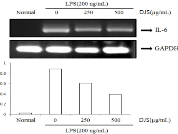
< 0.05.7. IL-6 mRNA 발현에 대한 導赤散 물추출물 의 억제 효과
대식세포주인 RAW 264.7 세포로부터 IL-6 mRNA 발현 정도를 RT-PCR을 통해 알아 본 결과, DJS를 250, 500 µg/mL로 처리하였을 때 IL-6 mRNA 발현이 농도 의존적으 로 억제되었다 (Figure 7).
Figure 7. Effect of DJS on LPS-induced IL-6 mRNA expression in RAW 264.7 cells. The IL-6 mRNA expression was assessed by RT-PCR. Cells were pretreated with the indicated concentrations of DJS for 30 minutes before being incubated with LPS (200 ng/mL) for 24 hours. The GAPDH mRNA was assayed in parallel to confirm equivalency of the cDNA preparation.
8. MAPKs의 인산화에 대한 導赤散 물추출물의 억제 효과
MAPKs는 세포의 성장과 분화 및 사이토카인과 스트레스 제어에 중요한 역할을 한다. 따라서 導赤散 물추출물 (DJS) 의 억제 메커니즘이 MAPKs 경유하는지 알아보기 위해 MAPKs의 인산화를 Western blot을 통해 확인하였다. 그 결 과 LPS에 의해 활성화 된 RAW 264.7 대식세포에 DJS를 250, 500 µg/mL를 처리한 경우 JNK 1/2와 p38의 인산화 를 억제하는 것을 확인하였다 (Figure 8).
Figure 8. Effect of DJS on LPS-induced phospholyration (P-) of MAPKs in RAW 264.7 cells. RAW 264.7 cells were treated with the indicated concentrations of DJS for 30 minutes before being incubated with LPS (200 ng/mL) for 30 minutes. Whole-cell lysates were analyzed by western blot analysis. The experiment was repeated three times, and similar results were obtained.
고 찰
본 연구에서 사용된 導赤散은 消炎, 凉血, 養陰하는 効能이 있는 生地黃, 木通, 竹葉, 甘草 의 네 가지 약재로 구성된 처
방이다
16-17). 生地黃은 氣味가 甘寒而潤하고 心腎經하여 淸心
熱한다. 凉血자滋陰하고 甘寒質潤한 性味는 腎經에 들어가 腎
陰을 滋하여 潤調시키고, 苦寒한 性味는 心, 肝血分에 들어가
淸營凉血하므로 邪熱을 泄熱하게 된다
18-21). 木通은 성미가
苦寒하고 心과 小腸经에 들어가 淸心降火, 利水通淋하는데 生
地黃과 木通 두 약이 배합하여 滋陰制火而不戀邪, 利水通淋
而不傷陰한다
22,23). 竹葉은 氣味가 甘淡하여 淸心除煩, 引熱下
行하고, 甘草는 味는 甘하고 性은 平하며. 脾, 胃, 肺經에 들
어간다. 또한, 清热解毒하고 여러 藥과 조화를 통하여 淋痛을
치료하며, 木通과 生地黃의 寒凉한 氣味가 傷胃하는 것을 방
지한다
24). 따라서 위의 네 가지 약재가 배합되어 清热利水,
養陰하는 효능을 발휘하므로, 導赤散을 清热劑-清臟腑热의
방제로 볼 수 있다
25,26). 네 가지 약물들의 사전에 보고되었던
항염증에 관한 연구로 木通에서의 급성 신장염의 보호효과에
관하여 연구되어 있고 生地黃의 항염 및 항산화 효과에 대해
연구 되어있다
27,28). 또한 甘草의 추출용매별 항염증 효과와
마우스에서 궤양성 대장염 억제효과와 竹葉이 알콜성 고지혈
증과 간 손상의 예방 효과와 생쥐의 복강암에 미치는 영향에
대해 보고되었다
29-32). 최근에 양방에서 보면 요로계의 가벼
운 염증이나 농축요로 인한 고 삼투압에 자극을 받아 발생하
는 증상, 구강궤양, 급성비뇨기계감염증 등의 질환에 자주 쓰
이고 있다
33-35).
본 연구에서는 導赤散 물추출물이 NO와 COX-2의 저해 및 pro-inflammatory cytokine의 억제기전과 관련이 있을 것으로 예상됨에 따라, 導赤散이 염증반응의 주체가 되는 대 식세포 계열인 RAW 264.7 세포에서 LPS로 유도된 IL-6와 같은 pro-inflammatory cytokine과 NO의 생성억제 효과, 그리고 iNOS의 단백질 억제와의 관련성을 조사하기 위해 Western blot 분석법을 이용하여 세포질 내의 iNOS 단백질 량을 조사하였다. 또한, COX-2의 단백질 발현 억제효과 및 PGE
2생성 억제효과 등을 통해 알아보았다. 대식세포 계열인 RAW 264.7 세포에 LPS로 자극을 주고 導赤散을 처리하여 확인해 본 결과, NO의 생성억제 효과 강하게 나타났으며, iNOS, COX-2 및 PGE
2생성 억제 효과가 또한 강하게 나타 났다. 그리고 IL-6 생성과 mRNA 발현에서도 큰 억제효과를 나타냈다.
최근 LPS에 의한 대식세포의 활성은 toll-like receptor (TLR) 4의 발현을 조절한다는 알려져 있다
36). 그람-음성균 의 세포 외막에 존재하는 lipopolysaccharide (LPS)는 대표적 인 내독소 중의 하나로서, RAW 264.7과 같은 macrophage 와 monocyte에서 tumor necrosis factor-α (TNF-α), interleukin-6 (IL-6), interleukin-1β (IL-1β)와 같은 염증성 cytokine의 분비를 촉진한다고 알려져 있다
37,38). 또 한, LPS 자극에 의해 대식세포는 iNOS를 발현하고, NO를 생성하게 되며 이에 의한 세포독성은 염증반응, 세포의 돌연 변이 및 종양 발생 등에도 관여하는 것으로 알려져 있다. 염 증반응과 관련된 조직 손상에서 NO와 iNOS의 발현이 증가 되어 있음이 보고되어 있다
39). 따라서 염증반응이 진행되는 동안 유의적으로 증가하는 NO와 iNOS의 생성을 효과적으로 억제하는 억제제개발에 대한 연구가 최근 이루어지고 있다.
지금까지 잘 알려진 염증반응의 세포내 분자전달신호체계 중에 Mitogen-activated protein (MAP) kinase (MAPKs) 는 염증반응에 관련된 신호전달기전이며, 그 중에서 extracellularregulated protein kinase (ERK), c-Jun NH2-protein kinase (JNK)/stress-activated protein kinase (SAPK), serine/threonine protein kinase인 p38 MAPK 등이 주요한 전달 기전이다
40). 활성화된 MAPKs는 다른 kinase, 전사인자 등을 활성화하여 표적유전자의 발현 을 변화시키고, 유전자 발현의 변화는 세포의 여러 소기관과 효소에 영향을 주게 된다. 따라서 이러한 전염증성 사이토카 인의 활성을 억제함으로써 염증반응의 분자신호 전달기전인 MAPK superfamily에 속하는 세 가지 효소들인 ERK, JNK, p38 MAPK 등의 염증매개체들의 신호전달을 통해 억 제하는지 알아보았다. 그 결과 導赤散 물추출물은 MAPKs의 인산화는 JNK와 p38 MAPK의 인산화는 억제하는 반면 ERK 1/2의 인산화에는 아무런 영향을 주지 못했다. 이는 LPS로 유도한 대식세포의 염증반응은 JNK와 p38 MAPK의 신호전달 경로를 경유하는 반면 ERK 1/2의 경로와는 다르다 는 것을 보여준다. 결론적으로, 導赤散 물추출물은 세포내의 전염증성 인자들을 현저하게 억제하는 효과가 있음을 확인할 수 있었다. 본 실험결과를 토대로 導赤散 물추출물이 대식세 포를 매개로한 염증반응의 작용기전 연구에 중요한 기초 자료 가 될 수 있고, 향후 임상적인 응용을 위해서 導赤散의 추가 적인 후속연구가 진행되어 새로운 효능을 가지는 약물로 개발 되기를 기대하는 바이다.
결 론
설치류의 대식세포주인 RAW 264.7 세포를 LPS로 자극 하였을 때 導赤散의 항염증 효과를 조사하여 다음과 같은 결 론을 얻었다.
1. 導赤散은 LPS로 유도된 대식세포에서 NO와 PGE
2생성을 농도 의존적으로 억제하였다.
2. 導赤散은 iNOS와 COX-2의 발현을 억제하였고 염증성 사이토카인 IL-6 생성과 IL-6 mRNA발현도 억제하였다.
3. 導赤散은 MAPKs 인산화를 저해 하는지 알아보기 위해 Western blot을 수행한 결과 MAPKs (JNK 1/2, p38), 인산화가 저해되었다.
이와 같은 결과로 보아 導赤散은 대식세포에 작용하여 MAPKs의 인산화 저해를 통해 NO, PGE
2, IL-6의 생성과 iNOS, COX-2 발현을 억제함으로써 항염증에 효과가 있음을 알 수 있다.
감사의 글
This study was supported by the Grant of the Traditional Korean Medicine R&D project, Ministry of Health & Welfare, Republic of Korea (No. B120069).
References