玄參 물추출물이 LPS로 유발된 대식세포의 염증인자에 미치는 영향
한효상*#
중부대학교 보건행정학과
Effect of Scrophulariae Radix buergeriana Water Extract on the Proinflammatory Mediators in RAW 264.7 Cells Induced by LPS
Hyo-Sang Han
*#Department of Health Administration, College of Tourism Health, Joongbu University, Geumsan 32713, Korea
ABSTRACT
Objective : The purpose of this study was to investigate the effects of Scrophulariae Radix Water Extract (SR) on the production of inflammatory mediators in RAW 264.7 mouse macrophages cells induced by lipopolysaccharide (LPS).
Method : We examined effect of Scrophulariae Radix Extract on the cell viability of mouse macrophages cells.
Futhermore, After 24 hours treatment we investigated anti-inflammatory effect of Scrophulariae Radix Extract by the production of Bio-Plex cytokine assay, concentrations of various cytokines such NO, interleukin(IL)-1α, IL-3 and interferon inducible protein-10(IP-10).
Result : No significant changes have been found in the mouse macrophge cell viability by the Scrophulariae Radix Extract at the concentration of 25, 50, 100 and 200 ㎍/㎖. The water extract of Scrophulariae Radix significantly inhibited the production of NO in the LPS-induced macrophage at the concentration of 25, 50, 100 and 200 ㎍/㎖. The water extract of Scrophulariae Radix significantly inhibited the production of IL-1α, IL-3 and IP-10 in the LPS-induced macrophage at the concentration of 50, 100 and 200 ㎍/㎖.
Conclusion : The water extract of Scrophulariae Radix significantly inhibited the production of NO, IL-1α, IL-3 and IP-10 at the concentration of 50 ㎍/㎖ or higher in the LPS-induced macrophages with no changes in the cell viability of them. These results suggest that water extract of Scrophulariae Radix has anti-inflammatory effect related with its inhibition of proinflammatory cytokines such as IL-1α, IL-3 and IP-10 in the LPS-induced macrophages.
1)
Key words : Scrophulariae Radix, macrophage, anti-inflammatory effect, cytokine, nitric oxide
Ⅰ. 서 론
염증의 반응은 그 원인과 반응조직의 차이에 관계없이 아주 유사한 변화를 보이는데 이런 현상은 조직 손상 직후에 일어 나는 변화로 손상에 의하여 생체 내 국소부위에 유리되는 공통 적인 물질요인의 존재를 추정하게 하며, 염증의 반응을 중개 하는 물질인 화학적 매개체로는 프로스타글란딘(prostaglandin, PG), 산화질소(nitric oxide, NO), 염증을 유발하는 여러 가지 사이토카인(cytokine) 등이 있다1). 또한 염증반응을 유도하는 주요한 화학 전달 매개물질로는 크게 즉시형 혈관투과성 항진에
관여하는 amine류(serotonin, histamine 등)와 kinin류 (bradykinin 등), 지연형 반응에 주로 작용하는 사이토카인 (cytokine)류 등으로 분류 할 수 있다2). 따라서 IL-1β, IL-6, IP-10 등과 같은 염증 매개물질을 억제하는 물질을 발견한다면, 각종 면역질환 및 인체질환의 치료에 도움이 될 것이다3,4).
玄參는『神農本草經』에 처음 收載된 이래 養血滋陰, 瀉火 解毒의 효능이 있어, 熱病傷陰, 舌絳煩渴, 溫毒發斑, 津傷便秘, 骨蒸勞嗽, 目赤, 咽痛, 瘰癧, 白喉, 癰腫瘡毒 등을 치료하는 약물로 사용되어 왔다5). 玄參의 기원은 대한민국약전6)에
*#Corresponding author and First author : Hyo-Sang Han. Department of Health Administration, College of Tourism Health, Joongbu University, Geumsan 32713, Korea.
·Tel : +82-41-750-6292 ·E-mail : [email protected]
·Received:1 March 2017 ·Revised:8 April 2017 ·Accepted:20 May 2017
“현삼 Scrophularia buergeriana Miquel 또는 중국현삼 (中國玄參) Scrophularia ningpoensis Hemsley (현삼과 Scrophulariaceae)의 뿌리”라고 收載되어 있다.
玄參의 성분으로는 뿌리에는 alkaloid, 당류, sterol, l-asparagin 등의 amino acid, oleic acid,linoleic acid, stearic acid 등의 지방산, 微量의 精油, carotene 등이 함유 되어 있다5).
약리작용으로는 항불안 작용7), 항균작용8), 혈압 강하 작용9), 항알레르기 작용10), 아토피 발진 억제 작용11), 갑상선기능항 진증 억제 효과12), 항산화효과13), 항치매작용14), 항당뇨효과와 항염증효과15), 간세포 보호 효과16), 상처치유효과17) 등이 보고 되어 있다. 또한, 玄參의 항염 및 면역작용에 대하여 변 등18)은 현삼메탄올 추출물이 LPS로 유도된 Raw 264.7 cell에서의 TNF-α, IL-1β, IL-6 및 nitric oxide 생성에 미치는 영향을 보고하였다.
玄參의 약리중 대표적인 항염 효과에 대해 각각의 질환별 해당 면역매개인자들에 대한 연구는 보고되었으나 玄參 물추 출물을 이용하여 다양한 염증매개인자들에 대한 연구는 부족 한 것으로 판단되었다.
이에 본 연구에서는 玄參의 항염효능에 대하여 알아보기 위하여 玄參을 물추출하여 제조한 시료(SR = Scrophulariae Radix)를 대상으로 mouse macrophage RAW 264.7 cells의 cell viability와 lipopolysaccharide(LPS)로 유발된 nitric oxide(NO) 그리고 interleukin(IL)-1α, IL-3, interferon inducible protein-10(IP-10) 등의 cytokine 생성증가에 대한 영향을 조사하여 유의한 결과를 얻었기에 이에 보고하는 바이다.
Ⅱ. 재료 및 방법
1. 재료
1) 약재실험에 사용된 玄參(Scrophularia buergeriana Miquel의 塊根)은 한국 서울의 동양허브 주식회사로부터 2015 년 6 월에 구입(NO; 2015-006)하였으며, 약재는 가천대학교 한의과대학 본초학교실에서 감정하였고 모든 약재는 실험 전에 초음파 세 척기(Branson, USA)를 이용하여 불순물을 제거하고 실험에 사용하였다.
2) Cell line
실험에 사용된 대식세포는 마우스 대식세포(mouse macrophage line)인 Raw 264.7 세포로서 한국세포주은행 (KCLB, Korea)으로부터 구입하여 사용하였다.
3) 시약 및 기기
본 실험을 위해서 FBS(Sigma, USA), ethyl alcohol (Samchun Chemical, Korea), penicillin(Sigma, USA), streptomycin(Sigma, USA), DMEM(Sigma, USA), methyl alcohol(Samchun Chemical, Korea), DMSO(Sigma, USA), 1×PBS(Sigma, USA), EDTA(Sigma, USA), Trypsin- EDTA(Sigma, USA), MTT assay kit(Sigma, USA), Fluo-4
calcium assay kit(Molecular Probes, USA), NO assay kit (Sigma, USA), Bio-Plex cytokine assay kit(Panomics, USA) 등이 사용되었다. 각 시약의 품질은 분석용 등급 이상 의 것으로 하여 사용하였다. 본 실험에 사용된 기기는 CO2
incubator (Nuaire, USA), rotary vacuum evaporator (Eyela, Japan), research microscope (Becton dickinson, USA), centrifuge (Hanil, Korea), fume hood (Hanil, Korea), clean bench (Jeio thec, Korea), ultrasonic cleaner (Branson, USA), microplate reader (Bio-Rad, USA), vortex mixer (Vision Scientific Co, Korea), water bath (Intron Biotech, Korea), ice-maker (Vision Scientific Co, Korea), Bio-Plex 200(Bio-Rad, USA), spectrofluorometer(Dynex, UK) 등이다.
2. 방법
1) 시료의 제조玄參 50 g을 정확하게 중량을 측정한 후, 1 차 증류수 2,000 ㎖와 함께 환류추출기에 넣은 뒤 탕액이 끓기 시작한 후 2 시간 동안 가열하여 추출하였다. 추출액을 filter paper (Advantec No.2, Japan)로 감압 여과한 뒤 여과액을 rotary vacuum evaporator를 이용하여 농축액을 얻었다. 이 농축액을 동결건조기를 이용하여 건조한 분말을 시료로 사용하였다. 동결 건조 추출물은 10.4 g을 얻었으며, 수율은 20.8%였다.
2) 세포 배양
Raw 264.7 세포를 FBS, penicillin(100 U/㎖), streptomycin (100 ㎍/㎖)이 첨가된 DMEM 배지로 37℃, 5% CO2 incubator 에서 배양되었다. Raw 264.7 세포를 75 cm2 flask (Falcon, USA)에서 충분히 증식된 후 배양 3일 간격으로 배양세포 표 면을 phosphate buffered saline (PBS) 용액으로 씻어준 후 50 ㎖ flask 당 1 ㎖의 0.25% trypsin-EDTA용액을 넣고 실온에서 1분간 처리한 다음 trypsin용액을 버리고 37℃에서 5분간 보관하여 세포를 탈착하여 계대 배양하였다. 탈착된 세 포는 10% FBS가 첨가된 DMEM 배양액 10 ㎖에 부유시킨 다음 새로운 배양용기(50 ㎖ culture flask)에 옮겨 1 : 2의 split ratio로 CO2 배양기(37℃, 5% CO2)에서 배양하였다.
3) 세포독성 검사(cytotoxicity assay)
준비된 시료가 Raw 264.7 세포에 나타내는 세포독성유발 정도를 알아보기 위하여 Mosmann19)의 방법을 응용하여 MTT assay를 실시하였다. 96 well plate에 1×10⁴ cells/well의 세포를 100 ㎕씩 넣고 37℃, 5% CO2 incubator에서 24 시간 동안 배양한 후 배지를 버리고 배양세포 표면을 phosphate buffered saline(1×PBS) 용액으로 씻어주었다. 같은 양의 배지와 PBS에 녹인 시료 (25, 50, 100, 200 ㎍/㎖)를 각 well에 처리하고 24시간 동안 배양하였다. 배양이 끝난 후 PBS에 녹인 1 ㎎/㎖ MTT (Sigma, USA)를 100㎕씩 각 well에 처리하여 알루미늄호일로 차광시킨 후 2시간 동안 같은 조건에서 배양하였다. 배양액을 모두 제거한 후 DMSO를 100㎕ 처리하고 37℃에서 2시간 방치 후 microplate reader
(Molecular Devices, USA)를 이용하여 490 ㎚에서 흡광도를 측정하였다. Cell viability는 다음 공식으로 계산되었다.
Cell viability(%) = 100 × AT / AC AC : absorbance of control
AT : absorbance of tested extract solution.
4) 일산화질소(Nitric oxide) 생성 측정
LPS를 단독처리(1 ㎍/㎖)하거나 혹은 다양한 농도의 시료 (25, 50, 100, 200 ㎍/㎖)와 함께 배지에 담아 각 well에 처 리하고 24시간 동안 37℃, 5 % CO2 Incubator에서 배양한 후 세포배양 상등액 100 ㎕을 채취하여 여기에 그리스 시약 (Griess reagent) 100 ㎕을 혼합하여 15분 동안 반응시킨 후 Microplate Reader(Bio-Rad, USA)를 이용하여 540 ㎚에서 흡광도를 측정하였다. 세포의 일산화질소(nitric oxide) 생성은 다음 공식으로 계산하였다.
Productions of Nitric oxide(%) = 100 × AT / AC AC : absorbance of control
AT : absorbance of tested extract solution.
5) 사이토카인(cytokine) 분비 측정
사이토카인 분비와 관련된 시료의 영향을 알아보기 위해 Politch 등20)의 방법을 응용하여 Bio-Plex Cytokine Assay를 다음과 같이 시행하였다. 96 well plate에 1×10⁵ cells/㎖의 cell을 100 ㎕씩 넣고 37℃, 5 % CO2 Incubator에서 24 시 간동안 배양한 후 배지를 버리고 배양세포 표면을 1×PBS 용 액으로 씻어준 뒤 각 well에 LPS를 단독처리(1 ㎍/㎖)하거나 혹은 다양한 농도의 시료(25, 50, 100, 200 ㎍/㎖)와 함께 배지에 담아 처리하고 24시간 동안 배양하였다. 배양이 끝나면 상등액(cell culture supernatant)을 채취하여 filter plate (96 well type)에 미리 준비되어 있던 antibody-conjugated capture beads와 결합시킨다. 결합된 capture beads가 담긴 filter plate의 각 well을 150 ㎕의 wash buffer로 세척한다.
세척이 끝난 뒤 각 well에 detection antibody를 추가한 후 30 분간 배양한다. 배양이 끝나면 wash buffer로 3회 세척한 뒤 각 well에 streptavidin-PE를 분주하고 상온에서 300~500 rSB의 조건으로 30 분간 진동배양한다. 배양이 끝나면 wash buffer로 3회 세척한 뒤 각 well에 120 ㎕의 reading buffer를 분주하고 상온에서 300~500 rSB의 조건으로 5 분간 진동배 양한 후 bio-plex array reader(Bio-Plex 200)를 이용, 측정 코자 하는 사이토카인류의 발현에 대한 시료의 영향을 계산, 비교하였다.
3. 통계처리
본 실험에서 얻은 결과에 대해서는 mean ± SD로 나타내 었으며, 대조군과 각 실험군과의 평균의 차이는 Student's t-test와 ANOVA test로 분석하여 p-value값이 0.05 미만 일 때 통계적으로 유의한 차이가 있는 것으로 판정하였다.
Ⅲ. 결 과
1. 세포독성에 대한 효과
玄參 물추출물이 대식세포의 증식에 미치는 영향을 알아보기 위하여 玄參 물추출물을 24 시간 동안 처리한 결과 25 ㎍/㎖
이상의 모든 농도에서 세포생존률이 증가하였다(Fig.1)
Fig. 1. Effect of SR on cell viability in Raw 264.7 cells. Cells were incubated for 24 hrs. Values are the mean ± SD of three independent experiments.
SR : Scrophulariae Radix water extract.
Normal : Treated with media only. * represents P < 0.05 compared to the normal.
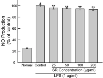
2. LPS로 유발된 NO 생성증가에 대한 효과
玄參 물추출물이 LPS로 유발된 대식세포의 NO 생성증가에 대한 효과를 알아보기 위하여 LPS(1 ㎍/㎖)와 함께 玄參 물 추출물을 24 시간 동안 처리한 결과 25 ㎍/㎖ 이상의 모든 농 도에서 LPS에 의한 NO 생성증가를 유의하게(P<0.01) 억제 하였다(Fig. 2).
Fig. 2. Effect of SR on the nitric oxide production in Raw 264.7 cells treated with lipopolysaccharide (LPS; 1 ㎍/㎖). Cells were incubated for 24 hrs. Values are the mean ± SD of three independent experiments.
SR : Scrophulariae Radix water extract.
Normal : Treated with media only.
Control : Treated with LPS only. # represents P < 0.05 compared to the normal. ** represents P < 0.01 compared to the control.
3. 사이토카인 생성에 대한 효과
(1) IL-1α생성에 대한 효과玄參 물추출물이 LPS로 활성화된 대식세포에서 IL-1α 생성 증가에 대한 영향을 알아보기 위하여 LPS(1 ㎍/㎖)와 함께 玄參 물추출물을 24 시간 동안 처리한 결과 25 ㎍/㎖ 이상의 모든 농도에서 LPS에 의한 IL-1α 생성증가를 유의하게 억제 하였다(Fig. 3).
Fig. 3. Effect of SR on the IL-1α production in Raw 264.7 cells treated with lipopolysaccharide (1 ㎍/㎖). Cells were incubated for 24 hrs. Values are the mean ± SD of three independent experiments.
SR : Scrophulariae Radix water extract.
Normal : Treated with media only.
Control : Treated with LPS only. # represents P < 0.05 compared to the normal. * represents P < 0.05 compared to the control.
** represents P < 0.01 compared to the control.
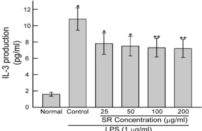
(2) IL-3 생성에 대한 효과
玄參 물추출물이 LPS로 활성화된 대식세포에서 IL-3 생성 증가에 대한 영향을 알아보기 위하여 LPS(1 ㎍/㎖)와 함께 玄參 물추출물을 24 시간 동안 처리한 결과 25 ㎍/㎖ 이상의 모든 농도에서 LPS에 의한 IL-3 생성증가를 유의하게 억제 하였다(Fig. 4).
Fig. 4. Effect of SR on the IL-3 production in Raw 264.7 cells treated with lipopolysaccharide (1 ㎍/㎖). Cells were incubated for 24 hrs. Values are the mean ± SD of three independent experiments.
SR : Scrophulariae Radix water extract.
Normal : Treated with media only.
Control : Treated with LPS only. # represents P < 0.0 compared to the normal. ** represents P < 0.01 compared to the control.
(3) ICP-10 생성에 대한 효과
玄參 물추출물이 LPS로 활성화된 대식세포에서 ICP-10 생성 증가에 대한 영향을 알아보기 위하여 LPS(1 ㎍/㎖)와 함께 玄參 물추출물을 24 시간 동안 처리한 결과 25 ㎍/㎖ 이 상의 모든 농도에서 LPS에 의한 IP-10 생성증가를 유의하게 억제하였다(Fig. 5).
Fig. 5. Effect of SR on the ICP-10 production in Raw 264.7 cells treated with lipopolysaccharide (1 ㎍/㎖). Cells were incubated for 24 hrs. Values are the mean ± SD of three independent experiments.
SR : Scrophulariae Radix water extract.
Normal : Treated with media only.
Control : Treated with LPS only. # represents P < 0.05 compared to the normal. * represents P < 0.05 compared to the control.
** represents P < 0.01 compared to the control.
Ⅳ. 고 찰
玄參은 뿌리로 불규칙하게 구부러져 있고 긴 원기둥모양 ~ 방추형으로 길이 4 ~ 20 ㎝, 지름 1 ~ 3 ㎝이다. 바깥면은 황갈색 ~ 갈색이고 거친 세로주름이 있으며 옆으로 긴 껍질 눈과 드문드문 잔뿌리 자국이 있다. 질은 단단하며 꺾기 힘들고 꺾인 면은 검은색 ~ 흑갈색을 띤다6).
玄參의 성분으로는 Halpagoside, 8-o-p- methoxycinnamate, phytosterol, stacyose, buergeriside A1, buergerisidee C1, e-pmethoxycinnamic acid, e-isoferulic acid, 4-O-E-p-methoxycinnamoyl-alpha- L-rhamnopyranoside ester, p-methoxycinnamic acid 등을 함유한다고 알려져 있다.21)
이와 같이 玄參은 항산화, 항불안효과, 항당뇨효과, 상처치 유효과 등을 보이고 있음을 알고 있지만, 한의학 임상에서 쓰 이는 玄參의 물추출에 의한 玄參의 항염효능에 관한 연구는 아직 보고되지 않아, 이에 대한 검증을 할 필요가 있다고 사 료되었다.
이에 본 연구에서는 玄參을 물추출하여 얻은 시료(SR)를 대상으로 마우스 대식세포인 RAW 264.7 cells의 세포생존율과 LPS로 유발된 NO, IL-1α, IL-3, IP-10의 다양한 cytokines의 생성증가에 대한 영향을 측정하였다.
염증의 가장 주된 증상은 통증(pain), 발열(heat), 발적 (redness), 종창(swelling), 그리고 기능 상실(loss of function) 이다. 조직에 염증이 일어나면 가장 먼저 혈관반응이 나타나서
모세혈관이 확장되고 혈류가 증가하며 여기에 이어서 혈관 벽의 투과성이 증가되어 혈장성분과 단백질 성분이 혈관 벽을 통해서 간질조직으로 삼출되고, 이때에 세균이나 손상된 국소조직에서 유래하는 화학인자의 유도에 의해서 호중구, 다형 핵 백혈구와 단핵구가 아메바 운동을 통해 간질조직으로 나오며 이들에서 유리된 대식세포가 삼출되어 염증 자극물을 탐식하게 되며, 염증이 오래되면 림프구 및 형질세포도 많이 나타나게 된다22). LPS는 그람음성 박테리아에서 유래하는 내독소(endotoxin)의 일종으로 대식세포(macrophage) 등의 면역세포를 자극하여 일산화질소(nitric oxide, NO)와 사이토카인(cytokine) 등 각 종의 전염증성인자들(proinflammatory mediators)의 생성 증가를 유도하며23), 면역세포 특히 대식세포(macrophage)로 하여금 염증 촉발 물질의 급격한 생성증가를 유발한다24).
본 연구에서는 마우스의 대식세포 RAW 264.7 cells에 玄參 물추출물을 25, 50, 100, 200 ㎍/㎖의 농도로 처리한 뒤에 24 시간동안 37℃에서 배양한 후, 세포의 증식을 MTT assay를 이용하여 확인한 결과 玄參 물추출물은 25 ㎍/㎖ 이상의 모든 농도에서 마우스 대식세포에 유의한 독성을 유발하지 않았으며 이는 玄參 물추출물이 대식세포에 유의한 독성을 유발하지 않는 것으로 볼 수 있다.
최근에 세포사이의 작용을 매개하는 메신저 물질로 중요한 연구 대상이 되고 있는 NO는 신경계통 조직에서 신경전달물질, 신경조절물질 또는 이차전령물질(second messenger)로 작 용하는 것으로 알려진 free radical이다25). 또한 NO는 혈소판 내에서 혈소판의 응집을 억제하며 대식세포에서 세포독성을 매개하는 작용을 하며 일부 인체조직에서는 혈관확장을 매개 하는 물질로도 알려져 있다26).
玄參 물추출물이 LPS로 유발된 대식세포의 NO 생성증가에 대한 영향을 알아보기 위하여 LPS와 함께 玄參 물추출물을 24 시간 동안 RAW 264.7 cells에 처리한 결과 25 ㎍/㎖이상의 모든 농도에서 LPS에 의한 NO 생성증가를 유의하게 억제하 였다. 이와 같이 玄參 물추출물이 LPS로 유발된 대식세포의 NO 생성증가를 억제하는 것은 玄參 물추출물이 지나친 NO 생성에 의한 염증악화를 억제할 수 있는 효과를 가지고 있음을 의미한다.
사이토카인(cytokine)이란 인체 내 핵을 가진 모든 세포 형 에서 생산될 수 있는 조절 단백 물질로, 숙주 방어와 손상 치유 과정에 관여하는 세포들과 조혈 세포에 작용하여 면역반응과 조혈작용을 조절하는 인자들을 말한다. 현재 약 35종의 인터 루킨계, 48종의 케모카인계, 두 그룹으로 나누어지는 인터페 론계, 집락자극인자(colony stimulating factor)계, 성장인자 (growth factor)계 및 종양괴사인자(Tumor necrosis factor, TNF)계 등이 사이토카인이라 불리며 현재까지 200여종 이상의 단백물질이 사이토카인에 속하게 된다27). IL-1α는 T, B cell 등의 증식과 분화에 관계되며, IL-1β는 IL-2 유도, 뼈의 재 흡수, 조직의 분해대사의 작용이 있다28).
玄參 물추출물이 LPS로 유발된 대식세포의 IL-1α 생성증 가에 대한 영향을 알아보기 위하여 LPS와 함께 玄參 물추출 물을 24 시간 동안 RAW 264.7 cells에 처리한 결과 25 ㎍/㎖
이상의 모든 농도에서 LPS에 의한 IL-1α 생성증가를 유의하게 억제하였다.
IL-3는 조혈과정을 자극하는 cytokine으로써, multilineage
colony stimulating factor(multi-CSF)로 알려져 있으며, CD4+T 세포의 산물로서 미성숙 골수 전구세포에 작용하여, 성숙한 세포로 분화를 촉진한다29).
玄參 물추출물이 LPS로 유발된 대식세포의 IL-3 생성증 가에 대한 영향을 알아보기 위하여 LPS와 함께 玄參 물추출 물을 24 시간 동안 RAW 264.7 cells에 처리한 결과 25 ㎍/㎖
이상의 모든 농도에서 LPS에 의한 IL-3 생성증가를 유의하게 억제하였다.
IIP-10은 CXC chemokine family의 하나로 다양한 염증 세포를 염증 부위로 불러들일 뿐 아니라 Th1 lymphocyte, NK cell, macrophage와 같은 leukocyte에서 CXCR3 receptor 에 연결시키는 기능을 한다30,31).
玄參 물추출물이 LPS로 유발된 대식세포의 IP-10 생성증 가에 대한 영향을 알아보기 위하여 LPS와 함께 玄參 물추출 물을 24 시간 동안 RAW 264.7 cells에 처리한 결과 25 ㎍/㎖
이상의 모든 농도에서 LPS에 의한 IP-10 생성증가를 유의하게 억제하였다.
이와 같이 玄參 물추출물이 마우스 대식세포의 세포생존율을 감소시키지 않으면서도 LPS로 인해 유발된 대식세포의 각종 염증매개물질들(inflammatory mediators) 생성증가를 유의 하게 억제하는 것은 玄參 물추출물이 대식세포의 염증매개물질 과다배출로 인한 다양한 염증질환을 완화할 수 있는 항염효능이 있음을 의미하는 것이다.
이상의 결과, 玄參 물추출물은 대식세포에 유의한 세포독 성을 유발하지 않으면서도 LPS로 유발된 대식세포의 NO, IL-1α, IL-3, IP-10의 생성증가를 50 ㎍/㎖ 이상의 농도에서 유의하게 억제시키는 등 대식세포와 관련된 염증반응을 조절할 수 있는 항염효능이 있는 것으로 판단된다. 향후 玄參을 이용한 염증질환치료제개발을 위하여 보다 세심한 연구가 요구되어 지는 바이다.
Ⅴ. 결 론
본 연구에서는 玄參 물추출물(SR)을 대상으로 마우스 대식 세포인 RAW 264.7 cells의 세포생존율과 LPS로 유발된 NO, 세포내 칼슘, 그리고 IL-1α, IL-3, IP-10의 다양한 사이 토카인의 생성증가에 대한 효과를 측정하여 다음과 같은 결과를 얻었다.
1. 玄參 물추출물은 25, 50, 100 및 200 ㎍/㎖의 모든 농 도에서 마우스 대식세포에 유의한 독성을 유발하지 않 았다.
2. 玄參 물추출물은 LPS에 의해서 유발된 대식세포의 NO 생성증가를 25, 50, 100 및 200 ㎍/㎖의 모든 농도에서 유의하게 억제시켰다.
3. 玄參 물추출물은 LPS에 의해서 유발된 대식세포의 IL-1α, IL-3, IP-10 생성증가를 25, 50, 100 및 200
㎍/㎖의 모든 농도에서 유의하게 억제시켰다.
이상의 결과, 玄參 물추출물은 대식세포에 유의한 세포독 성을 유발하지 않으면서도 LPS로 유발된 대식세포의 NO, 세 포내 칼슘, IL-1α, IL-3, IP-10,의 생성증가를 50 ㎍/㎖ 이 상의 농도에서 유의하게 억제시켰다. 이러한 결과는 玄參 물 추출물이 대식세포와 관련된 과잉염증반응을 조절할 수 있는 항염효능이 있음을 의미한다.
References
1. Daehanbyeongrihakhoe. Byeongrihak. Seoul : Komoonsa 1995 : 71-104.
2. Moon TC, Chung KC, Son KH, Kim HP, Kang SS, Chang HW. Screening of Cyclooxygenase-2(COX-2) Inhibitors from Natural Products. Yakhak Hoeji.
1998 ; 42(2) : 214-9.
3. Matsuda H, Morikawa T, Ando S, Toguchida I, Yoshikawa M. Structural requirements of flavonoids for nitric oxide production inhibitory activity and mechanism of action. Bioorg Med Chem. 2003 ; 11(9) : 1995-2000.
4. Calixto JB, Campos MM, Otuki MF, Santos AR.
Anti-inflammatory compounds of plant origin. Part Ⅱ.
modulation of pro-inflammatory cytokines, chemokines and adhesion melecules. Planta Med. 2004 ; 70(2) : 93-103.
5. Kim IR, Kim HC, Kuk YB, Park SJ, Park YK, Park JH, Seo BI, Seo YB, Shin MK, Lee YJ, LeeYC, Lee JH, Leem KH, Cho SI, Chung JK, Joo YS, Choi HY,. Boncho-Hak. Seoul : Young-Lim Press. 2007 : 233-4.
6. Korea Food and Drug Administration. The Korean Pharmacopoeia Eleventh Edition. Seoul : Korea Food and Drug Administration. 2015 : 118.
7. Choi YH, Jung JW. Anxiolytic-like Effects of Scrophularia buergeriana Miquel Using the Elevated Plus-Maze in Mice : Involvement of GABAergic Nervous System. Korean J Oriental Physiology &
Pathology. 2010 ; 24(3) : 476-83.
8. Yook KD. Antimicrobial activity and cytotoxicity test of Scrophularia ningpoensis hemsl extracts against Klebsiella pneumoniae. Journal of The Korea Society of Computer and Information. 2016 ; 21(5) : 135-9.
9. Park KH, Ham IH, Choi HY. Hypotensive Effect of Scrophularia buergeriana and Gene Analysis of Scrophularia Species. J of Herbology. 2008 ; 23(2) : 213-23.
10. Song JS, Lee JC, Choi JH, Kim JH, Park SY. Effects of Scrophulariae Radix (SR) on Allergic Contact Dermatitis (ACD) induced by DNCB in mice. The Journal of Korean Oriental Medical Ophthalmology
& Otolaryngology & Dermatology. 2011 ; 24(3) : 1-16.
11. Yeo EJ, HAN JK, Kim YH. Inhibitory Studies of Scrophulariae Radix on Development of Dermatitis in NC/Nga Mice. J Korean Oriental Pediatrics.
2007 ; 21(2) : 13-34.
12. Park CU, Cho CS, Kim CJ. The Effects of Radix Scrophulariae on the Hyperthyroidism of Rats. J Korean Oriental Med. 2006 ; 27(3) : 178-186.
13. Park YK, Kang BS. Effects of Scrophulariae radix on Antioxidation in Rat’s live. J of Herbology. 1998
; 13(1) : 201-20.
14. Kim SR, Kang SY, Lee KY, Kim SH, Markelonis GJ, Oh TH, Kim YC. Anti-amnestic activity of E-P- methoxycinnamic acid from Scrophularia buergeriana.
Cognitive Brain Research. 2003 ; 17(2) : 454-61.
15. Ahmed B, Al-Rehaily AJ, A1-Howiriny TA, El- Sayed KA, Ahmad MS. Scropolioside-D2 and harpagoside-B: two new iridoid glycosides and antiinflammatory activity. Biol. Pharm. Bull. 2003
; 26(4) : 462-7.
16. Lee EJ, Kim SR, Kim J, Kim YC. Hepatoprotective Phenylpropanoids from Scrophularia buergeriana roots against CCl4-Induced toxicity: Action mechanism and structure-activity relationship.
Planta Med. 2002 ; 68(5) : 407-11.
17. Tohda C, Kakihara Y, Komatsu K, Kuraishi, Y.
lnhibitory effects of methanol extracts of herbal medicines on substance P-induced itch-scratch response. Biol. Pharm. Bull. 2000 ; 23(5) : 599-601.
18. Byun, SH, Yang CH, Kim SC. Inhibitory effect of Scrophulariae Radix extract on TNF-α, IL-1β, IL-6 and Nitric Oxide production in Lipopolysaccharide - activated Raw 264.7 cells. J of Herbology. 2005 ; 20(2) : 7-16.
19. Mosmann T. Rapid colorimetric assay for cellular growth and survival: application to proliferation and cytotoxicity assays. J Immunol Methods. 1983
; 65(1-2) : 55-63.
20. Politch JA, Tucker L, Bowman FP, Anderson DJ.
Concentrations and significance of cytokines and other immunologic factors in semen of healthy fertile men. Hum Reprod. 2007 ; 22(11) : 2928-35.
21. Kim DH, Kim HM, Ryu JH, Eom JY, Kim SC, Yang JH, Cho MK, Lim JP, Hong SH. Hanbangyakrihak.
Seoul : Shinilbukseu. 2007 : 693-5.
22. Lee YT. Choesinmyeonyeokhak. Seoul : Jipmundang.
1989 : 355-38.
23. Cohen J. The immunopathogenesis of sepsis.
Nature. 2002 ; 420(6917) : 885-91.
24. Cho HY, Noh KH, Cho MK, Jang JH, Lee MO, Kim SH, Song YS. Anti-oxidative and Anti-inflammatory Effects of Genistein in BALB/c Mice Injected with LPS. J Kor Soc Food Sci Nutr. 2008 : 37(9) : 1126-35.
25. Nagata S, Tsuchiva M, Asano S, Kaziro Y, Tamazaki T, Yamamoto O, Hirata Y, Kubota N, Oheda M, Nomura H, Ono M. Molecular cloning and expression of cDNA for human granulocyte colony-stimulating factor. Nature. 1986 ; 319(6052) : 415-8.
26. Feldman PL, Griffith OW, Sheuhr DJ. The surprising life of nitric oxide. Chem Eng News. 1993 : 26-38.
27. Kim HS. The Cytokines:An Overview. Yeungnam Univ. J of Med. 2010 ; 27(1) : 1-7.
28. Nazarenko I, Marhaba R, Reich E, Voronov E, Vitacolonna M, Hildebrand D, Elter E, Rajasagi M, Apte RN, Zö, ller M. Tumorigenicity of IL-1alpha- and IL-1beta-deficient fibrosarcoma cells. Neoplasia.
2008 ; 10(6) : 549-62.
29. Abul KA, Andrew HL. Cellular and Molecular Immunology. Saunders. 2003 : 243-74.
30. Agostini C, Cassatella M, Zambello R, Trentin L, Gasoerini S, Perin A, Piazza F, Siviero M, Facco M, Dziejman M, Chilosi M, Qin SX, Luster A, Semenzato G. Involvement of IP-10 chemokine in sarcoid granulomatous reactions. J Immunol. 1998
; 161(11) : 6413-20.
31. Agostini C, Facco M, Siviero M, Carollo D, Galvan S, Cattelan AM, Zambello R, Trentin L, Semenzato G. CXC chemokines IP-10 and mig expression and direct migration of pulmonary CD8+/CXCR3+ T cells in the lungs of patients with HIV infection and T-cell alveolitis. Am J Respir Crit Care Med.
2000 ; 162(4 Pt 1) : 1466-73.