476
©The Korean Society of Food Science and Technology
단풍취 열수 추출물의 HepG2 인간간세포의 LPS/D-Gal에 의해 유발된
급성 간 손상에 대한 간보호 효과
이선엽
1·한준희
1·최다혜
1·홍 민
1·권태형
1·이용진
1·유근형
1,*
1(재)춘천바이오산업진흥원 기술개발실Hepatoprotective effect of Ainsliaea acerifolia water extract on
LPS/D-GalN-induced acute liver injury in human HepG2 cells
Sun-Yeop Lee1, Joon-Hee Han1, Da-Hye Choi1, Min Hong1, Tae-Hyung Kwon1, Yong-Jin Lee1, and Keun-Hyung Yu1,*
1Department of Research and Development, Chuncheon Bioindustry Foundation
Abstract This study investigated the antioxidant and hepatoprotective effects of Ainsliaea acerifolia water extract (AAWE) on HepG2 cells. Five types of caffeoylquinic acid (CQA) were detected in AAWE, namely, 4,5-di-O-caffeoylquinic acid (4,5-DCQA; 11.16 mg/g), 3,4-di-O-caffeoylquinic acid (3,4-DCQA; 5.23 mg/g), 5-O-caffeoylquinic acid (5-CQA; 4.88 mg/ g), 3,5-di-O-caffeoylquinic acid (3,5-DCQA; 3.51 mg/g), and 4-O-caffeoylquinic acid (4-CQA; 3.31 mg/g). AAWE exerted ABTS+ antioxidant effects, evidenced by polyphenol content and 2,2'2'-azino-bis(3-ethylbenzothiazoline-6-sulfonic acid) (ABTS) and 2,2-diphenyl-1-picryl-hydrazyl-hydrate (DPPH radical scavenging) activities. AAWE (300 µg/mL) treatment significantly decreased the activities of gamma glutamyl transferase (GGT), aspartate transaminase (AST), and lactate dehydrogenase (LDH) as compared to control and exerted protective effects against the increase in liver function index induced by lipopolysaccharide (LPS)/galactosamine (D-GalN) in HepG2 cells. In addition, the secretion of tumor necrosis factor (TNF)-α by HepG2 cells induced by LPS/D-GalN significantly increased in all treatment groups compared to that in the control. However, AAWE (100-300 µg/mL) treatment significantly decreased the secretion of TNF-α compared to that in the control. These results suggest that AAWE treatment reduces hepatotoxicity by increasing antioxidant activities, reducing GGT, AST, and LDH activities, and inhibiting TNF-α secretion.
Keywords: Ainsliaea acerifolia, water extract, hepatoprotective effect, LPS/D-GalN
서
론
체외에서 유입되거나 체내에서 생성된 호르몬 등의 각종 물질 들을 가공·처리하고 탄수화물대사, 아미노산 및 단백질 대사, 지방 대사, 담즙산 및 빌리루빈 대사, 비타민 및 무기질 대사, 호 르몬 대사, 해독 작용 및 살균 작용 등의 주요 기능을 담당하는 간은 인체의 화학공장으로 비유된다. 하지만 ‘침묵의 장기’라 불 리는 간은 70~80%가 파괴될 때까지 자각 증상이 없기 때문에 간 기능이 손상되어도 몸의 이상이 쉽게 늦기지 못한다. 일반적으로 간질환은 간 조직의 섬유화가 간경화로 진행된 후 최종적으로 간 암으로 발전되는 일련의 과정을 거치게 된다. 간질환은 바이러스, 음주, 약물, 지방간, 자가 면역 및 독성물질 등 다양한 원인에 의 해서 나타난다고 알려져 있다(Murakami 등, 1998).D-galactosamine (D-GalN)은 galactose의 대사장애를 통해
uri-dine triphosphate (UTP), uriuri-dine diphosphate (UDP) 및 uridine monophosphate (UMP) 등의 농도 감소로 RNA의 합성을 저해하 는 한편 실험동물의 복강에 투여할 경우 간세포의 지방 변화, 염 증세포 침윤 및 간세포의 괴사 등이 나타나 간염과 같은 유사한 병변을 유도하는 것으로 보고되고 있다(Keppler 등, 1968; Farber 등, 1973; Yu 등, 2015). 또한 D-GalN은 대장 점막투과성에 변화 를 일으켜 endotoxin의 흡수를 증가시키고, 이는 간세포가 세포 막을 보수하는 기전을 방해하여 간 독성을 유발한다고 알려져 있 다(Chojkier 등, 1985). 결국 D-GalN은 급성중독시에는 간 괴사를 유발하고, 만성중독 시에는 간경변과 세포성 종양을 유발한다 (Keppler 등, 1968; Lesch 등, 1969). 단풍취 (Ainsliaea acerifolia)는 초롱꽃목 국화과의 산에서 흔히 자생하는 다년생 초본으로 한국, 중국, 일본 등지에 분포하고 있 으며, 어린순을 식용으로 사용해왔다. 민간요법에서는 류머티즘 관절염, 장염치료를 위해 이용되었고 단풍취 지상부의 2차 대사 산물인 caffeoylquinic acid는 항산화, 항당뇨, 항바이러스, 항혈전, 간보호, 신경보호와 같은 생리활성이 있는 것으로 보고된 바 있 다(Park 등, 2010; Choi 등, 2006). 따라서 본 연구에서는 간보호 활성이 우수한 것으로 알려진 단 풍취 열수 추출물을 이용하여 lipopolysaccharide (LPS)를 함유한 D-GalN 독성유발에 따른 단풍취의 간 기능 개선효과를 연구하였 으며, 향후 이를 통해 단풍취의 활용가치를 높이고 건강기능식품 *Corresponding author: Keun-Hyng Yu, Department of Research
and Development, Chuncheon Bioindustry Foundation, Chuncheon, Gangwon 24232, Korea
Tel: +82-33-258-6991 Fax: +82-33-258-6172 E-mail: [email protected]
Received August 10, 2020; revised August 25, 2020; accepted September 6, 2020
으로서의 가능성을 알아보고자 하였다.
재료 및 방법
단풍취 열수 추출물 제조
강원도 농업기술원 산하 평창분소로부터 시료를 공급받아 속 슬렛 추출장치(MS-EAM, MISUNG, Yangju, Korea)를 이용하여 10배의 증류수를 가하고 80oC에서 8시간씩 환류냉각 추출 하였 으며, 추출한 시료는 감압농축(N-1110, EYELA, Tokyo, Japan)하 고 동결건조(PVTF20R, ILSHINBIOBASE, Dongduchun, Korea)하 여 실험재료로 사용하였다.
Caffeoylquinic acid (CQA) 분석
단풍취 열수 추출물을 3차 증류수를 이용하여 1 mg/mL 농도 로 용해한 후 10분간 sonicating하고 3,200×g에서 20분간 원심분 리하였다. 원심분리 후 상등액을 0.45 µm syringe filter로 여과한 후 분석시료로 사용하였다. 5종의 caffeoylquinic acid (CQA) 분석 을 위해 4-O-caffeoylquinic acid (4-CQA), 5-O-caffeoylquinic acid (5-CQA), 3,4-di-O-caffeoylquinic acid (3,4-DCQA), 3,5-di-O-caf-feoylquinic acid (3,5-DCQA) 및 4,5-di-O-caffeoylquinic acid (4,5-DCQA) 등의 표준품은 Sigma-Aldrich (St. Louis, MO, USA)에서 구매하여 사용하였다. 정량을 위한 검량선을 작성하기 위해 각 표준물질이 100 µg/mL이 되도록 혼합 후 희석하여 5, 10 및 25 µg/mL 농도로 제작하여 이용하였다. HPLC 분석조건은 Table 1 과 같이 시행하였으며, 분석에 사용한 HPLC는 UFLC LC-20AD (Shimadzu, Koyto, Japan)를 사용하였고 검출기는 UV/VIS SPD-20A (Shimadzu)를 이용하여 단풍취 열수 추출물 내 5종의 CQA 함량을 분석하였다.
총 폴리페놀 함량
단풍취 열수 추출물의 총 폴리페놀 함량은 Folin-Denis 방법을 변형하여 측정하였다. 추출물을 3차 증류수에 1 mg/mL 농도로 용해하고 0.2 µm syringe filter를 이용하여 filtering한 후 96 well plate에 20 µL 분주하고 Folin & Ciocalteu’s phenol reagent (Sigma-Aldrich)를 20 µL 첨가한 후 10% Na2CO3 (Sigma-Aldrich) 를 20 µL 첨가하였다. 96 well plate에 140 µL의 3차 증류수를 가 하고 상온에서 1시간 반응시킨 후 700 nm에서 흡광도를 측정하 였다. 총 폴리페놀의 함량은 tannic acid (Sigma-Aldrich)를 이용하 여 검량선을 작성하고 700 nm에서 측정한 흡광도 값을 대입하여 총 폴리페놀의 함량을 산출하였다.
ABTS cation radical 소거능
7 mM ABTS (2,2'-azino-bis-3-ethylbenzothiazoline-6-sulphonic acid, 5 mL)와 140 mM potassium peroxodisulfate (88 µL)를 혼합
하고 빛이 차단된 실온에서 하루 이상 방치하여 양이온 라디칼 을 안정화 시킨 후 stock solution으로 사용하였다. Stock solution 을 에탄올로 희석(1:88) 후, 734 nm에서 흡광도가 0.70±0.02가 되 도록 조절하여 ABTS solution을 제조하였다. ABTS radical 소거 능 측정을 위해 ABTS solution 150 µL와 0.2 µm syringe filter로 filtering하여 각각의 농도로 희석한 단풍취 열수 추출물을 150 µL 씩 혼합하여 10분간 반응시킨 후 734 nm에서 흡광도 측정하였으 며, ABTS radical 소거능 측정을 위해 시약은 Sigma-Aldrich에서 구매하여 사용하였다.
DPPH radical 소거능
DPPH radical 소거활성은 Blois 방법(Blois, 1958)에 따라 측정 하였다. 150 µM DPPH (2,2-diphenyl-1-picrylhydrazyl)용액 100 µL와 각각의 농도로 희석한 단풍취 열수 추출물을 100 µL씩 혼 합하여 실온에서 10분간 반응시킨 후 517 nm에서 흡광도를 측정 하였으며, DPPH radical 소거능 측정을 위해 시약은 Sigma-Ald-rich에서 구매하여 사용하였다. 세포독성 단풍취 열수 추출물의 세포독성 평가를 위해 HepG2 (ATCC BB-8065) 세포는 American Type Culture Collection으로부터 구입 하여 사용하였다. HepG2 세포를 24 well plate에 2.0×10⁵ cells/
well 농도로 seeding하고 24시간 동안 CO2 incubator에서 배양한 후 phosphate buffered saline (PBS, Invitrogen, Carlsbad, CA, USA)를 이용하여 cell을 2회 washing하였으며, 0.2 µm syringe filter로 filtering하여 각각의 농도로 희석한 단풍취 열수 추출물 500 µL씩 24 well plate에 분주하고 24 및 48시간 동안 CO2 incubator에서 추가 배양하였다. 배양 24 및 48시간에 CCK-8 (Dojindo, Kumamoto, Japan)를 50 µL씩 24 well plate에 분주한 후 CO2 incubator에서 4시간 배양하고 450 nm에서 ELISA reader
(Molecular Devices, Sunnyvale, CA, USA)를 이용하여 흡광도를 측정하였다.
간세포 보호효과
HepG2 세포를 24 well plate에 2.0×10⁵ cells/well 농도로 seed-ing하고 24시간 동안 CO2 incubator에서 배양한 후 PBS를 이용하 여 cell을 2회 washing하였으며, 0.2 µm syringe filter로 filtering한 후 각각의 농도로 희석한 단풍취 열수 추출물을 500 µL씩 24 well plate에 분주하고 48시간 동안 CO2 incubator에서 추가 배양하였다. 배양 48시간에 PBS를 이용하여 cell을 2회 washing하고 D-GalN (D-galactosamine, Sigma-Aldrich) 50 mM과 LPS (lipopolysaccharide, Sigma-Aldrich) 10 µg/mL를 함유한 LPS/D-GalN를 500 µL씩 24 well plate에 분주한 후 CO2 incubator에서 48시간 배양하였다. 배 양 후 well plate를 3,200×g에서 10분간 원심분리한 후 상등액을
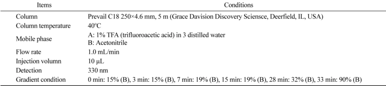
Table 1. The operating condition of HPLC system
Items Conditions
Column Prevail C18 250×4.6 mm, 5 m (Grace Davision Discovery Sciensce, Deerfield, IL, USA) Column temperature 40oC
Mobile phase A: 1% TFA (trifluoroacetic acid) in 3 distilled water B: Acetonitrile
Flow rate 1.0 mL/min Injection volumn 10 µL Detection 330 nm
회수하고 생화학분석기(Konelab 20B, Thermo, Vantaa, Finaland)를 이용하여 GGT (gamma (γ)-glutamyl transferase), AST (aspartate aminotransferase) 및 LDH (lactate dehydrogenase)를 측정하였다.
Tumor necrosis factor (TNF)-α 측정
HepG2 세포를 24 well plate에 2.0×10⁵ cells/well 농도로 seed-ing하고 24시간 동안 CO2 incubator에서 배양한 후 PBS를 이용하 여 cell을 2회 washing하였으며, 0.2 µm syringe filter로 filtering한 후 각각의 농도로 희석한 단풍취 열수 추출물을 500 µL씩 24 well plate에 분주하고 48시간 동안 CO2 incubator에서 추가 배양하였 다. 배양 48시간에 PBS를 이용하여 cell을 2회 washing하고 LPS/ D-GalN을 500 µL씩 24 well plate에 분주한 후 CO2 incubator에 서 48시간 배양하였다. 배양 후 well plate를 3,200×g에서 10분간 원심분리한 후 상등액을 회수하고 human TNF-α immunoassay kit (R&D System, Minneapolis, MN, USA)을 이용하여 TNF-α의 함 량을 정량하였다. TNF-α 단클론항체가 코팅된 96 well plate에 각 각의 시료를 200 µL씩 분주하고 실온에서 2시간 반응시켰다. 2시 간 후 washing buffer를 이용하여 3회 washing하고 TNF-α 다중 클론항체를 96 well plate에 200 µL씩 분주하고 실온에서 1시간 반응시켰다. 1시간 후 washing buffer를 이용하여 3회 washing하 고 동량의 hydrogen peroxide와 tetramethylbenzidine 혼합액을 96 well plate에 200 µL씩 분주한 후 30분간 반응시켰다. 30분 후 2 N sulfuric acid를 50 µL씩 각 well에 분주하고 450 nm에서 ELISA reader (Molecular Devises)를 이용하여 흡광도를 측정하였다. TNF-α의 함량은 TNF-α 표준곡선을 작성하여 계산하였다.
통계처리
실험결과는 실험군당 평균(mean)과 표준편차(SD)로 나타내었 고, GraphPad prism 4 (GraphPad software, La Jolla, CA, USA) 를 이용하여 일원배치 분산분석(one-way analysis of variance)을 실시한 후 p<0.05 수준에서 Tukey’s multiple comparison test에 의해 각 실험군 평균치 간의 유의성을 검정하였다.
결과 및 고찰
Caffeoylquinic acid (CQA) 분석
단풍취 열수 추출물의 CQA 분석결과 30분 이내에 5종의 CQA
가 완전히 분리되어 나오는 것을 확인하였다(Fig. 1). 용리순서는 5-CQA, 4-CQA, 4,5-DCQA, 3,5-DCQA, 3,4-DQCA의 순으로 용 리되었으며, Table 2와 같이 CQA의 함량은 4,5-DQCA 11.16 mg/g, 3,4-DCQA 5.23 mg/g, 5-CQA 4.88 mg/g, 3,5-DCQA 3.51 mg/g 및 4-CQA 3.31 mg/g 순으로 나타났다. Nugroho 등(2019)은 단풍 취 메탄올 추출물의 CQA 분석결과 3,5-DCQA의 함량이 145.6 mg/g으로 가장 높은 함량을 나타냈으며, 이 화합물이 acetylcho-linesterase (AChE)에 대한 활성을 나타내는 것으로 보고하였다. 총 폴리페놀 함량 단풍취 열수 추출물이 함유하고 있는 총 폴리페놀 함량은 74.03 mg/g인 것으로 확인되었다(Table 3). Lee 등(2015)은 단풍취 에탄 올 추출물의 경우 18.1 mg/g의 페놀화합물을 함유하고 있으며, 에 탄올 추출물을 분확한 용매에 따라 ethylacetate (EtOAc) 분획물 에서는 22.4 mg/g, n-butyl alcohol (n-BuOH)에서는 20.1 mg/g, H2O 층에서는 4.3 mg/g의 페놀성 화합물의 함량을 확인한 바 있다. ABTS cation radical 소거능
ABTS cation radical 소거능은 시료의 항산화능에 의해 청녹색 의 ABTS radical 색상이 탈색되는 정도를 측정하는 방법(Re 등, 1999; Lee 등, 2015) 으로 단풍취의 ABTS+ radical 소거능을 측 정하였다(Table 3). 추출물의 농도 변화에 따른 ABTS+ radical 소
거능 측정결과 추출물의 농도가 50, 100, 200 및 400 µg/mL의 범 위에서 단풍취 열수 추출물의 ABTS+ radical 소거능은 각각 12.22, 25.05, 40.89, 68.55%의 소거율을 보였다. Yoon (2018)은 단풍취 열수 및 에탄올 추출물의 ABTS+ radical 소거능 측정 결과 각 각 IC50는 258.9 및 199.42 µg/mL으로 보고하였으나, Lee 등(2015)은 단풍취 70% 에탄올 추출물 31.3 µg/mL에서 46.3%의 ABTS+ rad-ical 소거능이 있는 것으로 보고하고, 이 때 IC50는 34.3 µg/mL인 것으로 보고하였다. 단풍취 추출물의 항산화 활성이 다르게 나타 난 것은 시료의 채집시기 및 재배지역 등이 영향을 미친 것으로 판단되며, 높은 폴리페놀 함량에서 기인된 것으로 판단된다. DPPH radical 소거능 다양한 천연소재로부터 항산화 활성을 탐색할 때 이용되는 DPPH 라디칼 소거능은 자유 라디칼 자체가 홀수전자를 가지고 있어 517 nm에서 강한 흡광도를 나타내며, 항산화 물질과 반응
하여 짙은 보라색이 탈색되는 원리를 이용한 방법이다. 단풍취 열수 추출물 농도 50, 100, 200 및 400 µg/mL의 범위에서에서 14.97, 26.84, 41.18, 55.83%의 DPPH radical 소거능을 나타냈다 (Table 3). Lee 등(2015)은 단풍취 에탄올 추출물 및 각 유기용매 분획에 대한 radical 소거능을 평가한 결과 125 µg/mL의 농도에 서 에탄올 추출물 66.3%, EtOAc 분획 77.6%, n-BuOH 분획 68.4%의 소거능을 나타냈다고 보고하였다. 대부분 천연물 내에 존재하는 것으로 알려진 페놀성분은 free radical을 효과적으로 제 거하지만 radical의 기질에 따라 선택적으로 작용하는 페놀성분이 존재하기 때문에 ABTS+와 DPPH radical 소거 활성이 차이를 보 이는 것으로 판단된다. 세포독성 단풍취 열수 추출물의 농도별 첨가에 따른 HepG2 세포의 생 존율은 단풍취 열수 추출물을 24시간 처리하였을 때 모든 농도 에서 세포독성이 없는 것으로 나타났다 (Fig. 2). Seo(2017)는 Raw 264.7 cell에 단풍취 에탄올 추출물을 15, 30, 60 µg/mL 농도로 24 시간 처리하였을 때 모든 처리 농도에서 독성이 없다고 보고한 바 있으며, Yoon(2018)은 3T3-L1 cell에 단풍취 에탄올 추출물을 100, 200, 300 µg/mL 농도로 24시간 처리하였을 때 모든 농도에 서 세포독성이 없는 것을 확인하였다. 따라서 향후 단풍취 열수 추출은 각종 식품소재나 기능성 원료로 사용이 가능할 것으로 판 단된다. 간세포 보호효과 HepG2 세포에 LPS/D-GalN를 처리하여 간독성을 유발하여 단 풍취 추출물의 간세포 보호효과를 알아보았다(Table 4). 일반적으 로 간, 신장 및 췌장에 분포하며, 질병 방어기능을 하고 간질환 이나 알코올성 간질환에서 증가하는 것으로 알져진 GGT의 활성 을 측정한 결과 LPS/D-GalN을 처리하였을 때 정상군과 비교하 여 모든 처리군에서 유의하게 GGT 활성이 증가하였으나 단풍취 열수 추출물 300 µg/mL 처리군에서 정상군 수준의 GGT 활성을 나타내었다. 간과 심장에 고농도로 존재하며 간염, 간 경변 등의 Table 2. Content of caffeoylquinic acids in Ainsliaea acerifolia water extract
Sample Content (mg/g)
4-CQA 5-CQA 3,4-DCQA 3,5-DCQA 4,5-DCQA Total CQA
AAWE1) 3.31±0.42) 4.88±0.8 5.23±0.5 3.51±0.3 11.16±0.6 28.09±1.7 1)Ainsliaea acerifolia water extract.
2)Mean±SD.
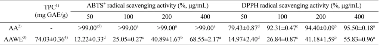
Table 3. Antioxidant assays of Ainsliaea acerfolia water extract TPC1)
(mg GAE/g)
ABTS+ radical scavenging activity (%, μg/mL) DPPH radical scavenging activity (%, μg/mL)
50 100 200 400 50 100 200 400
AA2) - >99.00a5) >99.00a >99.00a >99.00a 79.43±0.87d 92.31±0.47c 94.40±0.09b 95.50±0.18a AAWE3) 74.03±0.364) 12.22±0.33d 25.05±0.27c 40.89±1.67b 68.55±2.17a 14.97±2.40d 26.84±0.87c 41.18±1.59b 55.83±0.96a 1)Total phenolic contents were expressed in terms of mg gallic acid equivalents.
2)Ascorbic acid (positive control). 3)Ainsliaea acerifolia water extract. 4)Mean±SD.
5)Different letters means significantly different at p<0.05.
Fig. 2. Percentage of HepG2 cells viability at various concentration of Ainsliaea acerifolia water extract (AAWE) for 24 hr. Values represent the mean±SD (n=3). NS means not significantly different at p<0.05.
Table 4. Effects of Ainsliaea acerifolia water extract on the GGT, AST, and LDH activities in HepG2 cell treated with 50 mM D-GalN and 10 µg/mL LPS Group GGT AST LDH Normal1) 5.75±0.328)b9) 45.33±0.52d 345.93±3.80d Control2) 7.05±0.48a 62.57±0.85a 716.90±20.97a SM-503) 6.24±0.69b 36.27±0.57e 431.61±6.30c AAWE-504) 7.16±0.45a 56.29±0.55b 714.45±15.70a AAWE-1005) 6.63±0.20ab 54.61±1.13c 699.53±18.60a AAWE-2006) 6.52±0.37ab 53.62±0.61c 655.87±7.17b AAWE-3007) 5.79±0.33b 53.62±0.88c 658.03±6.40b 1)Not treated. 2)50 mM D-GalN and 10 µg/mL LPS.
3)Silymarin (positive control) 10 µg/mL, 50 mM D-GalN, and 10 µg/
mL LPS.
4)Ainsliaea acerifolia water extract 50 µg/mL, 50 mM D-GalN, and 10
µg/mL LPS.
5)Ainsliaea acerifolia water extract 100 µg/mL, 50 mM D-GalN, and
10 µg/mL LPS.
6)Ainsliaea acerifolia water extract 200 µg/mL, 50 mM D-GalN, and
10 µg/mL LPS.
7)Ainsliaea acerifolia water extract 300 µg/mL, 50 mM D-GalN, and
10 µg/mL LPS.
8)Mean±SD.
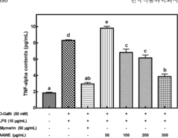
지표로 널리 사용되는 AST의 활성을 측정한 결과 LPS/D-GalN 을 처리하였을 때 정상군과 비교하여 모든 처리군에서 유의하게 AST 활성이 증가하였으며, 단풍취 열수 추출물을 처리한 모든 처리군에서 유의하게 AST 활성이 감소하였다. 심장질환, 간질환, 악성종양 및 백혈병, 용혈성 및 악성빈혈 등에서 증가하며, 급성 간염에서 GGT, AST와 동일하게 간세포로부터 유리되어 증가하 고 황달을 동반한 독성간염에서 약 10배 정도 증가하는 것으로 알려진 LDH 활성을 측정한 결과 LPS/D-GalN을 처리하였을 때 정상군과 비교하여 모든 처리군에서 LDH 활성이 증가하였고 단 풍취 열수 추출물을 200 µg/mL 이상 처리한 처리군에서 대조군 과 비교하여 유의적으로 효소활성이 감소하는 것을 확인하였다. 국화과 식물에 내 존재하는 CQA는 caffeic acid와 quinic acid가 결합한 화합물이지만 여기서는 다른 phenylpropanoid계 화합물인 pcoumaric acid와 ferulic acid가 결합한 화합물까지 포함하며, caffeoylquinic acid는 결합하는 caffeic acid의 수에 따라 monocaf-feoylquinic acid류, dicaffeoylquinic acid류, tricaffeoylquinic acid류 등이 있다(Park 등, 2009; Park 등, 2017). 이러한 화합물은 강력 한 항산화 물질로 진통작용, 소염 효과, 항바이러스 효과, 간 보 호 효과, 항산화 효과, 혈소판 응집 억제 효과 등이 보고되어 있 다(Zhao 등, 2006). Park 등(2017)은 3,5-DCQA가 HepG2 세포에 hydrogen peroxide에 처리에 의해 유도된 산화적 스트레스에 대 한 보호 효과를 세포막 손상 시 배양액 내로 유출되는 간 기능 지표 효소 활성을 측정한 결과 GGT, AST, LDH 효소활성이 유 의적으로 감소한다고 보고하였다. 이와 같이 곰취 추출물의 항산 화 및 간 보호 효과는 곰취 추출물의 주요 성분인 CQA에 의한 것으로 보고된 바 있으며(Choi 등, 2005), Yu 등(2015)은 곰취 발 효 추출물이 간 조직 내 SOD 활성을 증가시키고 지질과산화 반 응을 억제시킴으로써 간 손상을 완화시키는 데 효과적이라고 보 고하였다. 본 연구결과에서 보는 바와 같이 단풍취 열수 추출물 에 존재하는 CQA에 의한 간 보호 효과가 있는 것으로 판단되나 추가적인 연구가 필요할 것으로 판단된다. TNF-α 농도에 미치는 영향 단풍취 열수 추출물이 간세포인 HepG2 세포에 LPS/D-GalN의 처리하였을 때 TNF-α의 농도변화 알아보기 위해 ELISA를 통하 여 TNF-α의 양을 정량하였다. Monokine인 TNF-α는 여러 면역 활성을 중재하는 단백질로서 주로 대식세포에 의해 분비되며, 염 증 반응 시 사이토카인은 간과 여러 장기의 손상 시 중요한 역 할을 하며(Sakaguchi 등, 1999) 이 중 TNF-α는 괴사 진행 과정 의 주요 인자로 TNF-α를 억제하는 것이 항염증 치료에서 중요 한 바이오마커로 알려져 있다(Suzuki 등, 1996; Park 등, 2008). Fig. 3에서 보는 바와 같이 LPS/D-GalN을 처리하였을 때 정상군 과 비교하여 모든 처리군에서 TNF-α 분비가 유의적으로 증가하 는 것을 확인 할 수 있었으며, 단풍취 열수 추출물을 100 µg/mL 이상 처리하였을 때 TNF-α의 분비가 유의하게 감소하는 것을 확 인할 수 있었다. Seo 등(2017)은 단풍취 에탄올 추출물이 RAW 264.7 세포에 LPS에 의해 유도된 TNF-α의 분비를 억제하는 것 으로 보고 하였다. 이러한 결과로 볼 때 단풍취 열수 추출물은 GalN에 의해 유도된 염증반응에서 TNF-α 분비 억제를 통해 염 증반응을 완화하는 것으로 판단된다.
요
약
본 연구에서는 간보호 활성이 우수한 것으로 알려진 단풍취의 활용가치를 높이고 향후 건강기능성식품 소재로써의 가능성을 알 아보고자 LPS/D-GalN 독성유발에 따른 단풍취 열수 추출물의 간 기능 개선효과를 연구하였다. 단풍취 열수 추출물 내에 존재하는 5종의 Caffeoylquinic acid (CQA) 분석결과 4,5-di-O-caffeoylquinic acid (4,5-DCQA) 11.16 mg/g, 3,4-di-O-caffeoylquinic acid (3,4-DCQA) 5.23 mg/g, 5-O-caffeoylquinic acid (5-CQA) 4.88 mg/g, 3,5-di-O-caffeoylquinic acid (3,5-DCQA) 3.51 mg/g 및 4-O-caf-feoylquinic acid (4-CQA) 3.31 mg/g 순으로 나타났다. 단풍취 열 수 추출물이 함유하고 있는 총 폴리페놀 함량은 74.03 mg/g이었 으며, ABTS cation radical 소거능은 추출물의 농도 50, 100, 200 및 400 µg/mL의 범위에서 각각 12.2, 25.0, 40.8, 68.55%를 나타 냈고 DPPH radical 소거능은 50 µg/mL의 농도에서 54.38%를 나타 냈다. HepG2 세포에 LPS/D-GalN을 처리하여 간독성을 유발한 결과 정상군을 제외한 모든 처리군에서 GGT, AST 및 LDH 활 성이 증가하였고, 단풍취 열수 추출물 300 mg/mL을 처리하였을 때 대조군과 비교하여 GGT, AST 및 LDH 활성이 유의적으로 감 소하는 것으로 나타났다. LPS/D-GalN을 처리하였을 때 정상군과 비교하여 모든 처리군에서 TNF-α 분비가 유의적으로 증가하는 것을 확인 할 수 있었으며, 단풍취 열수 추출물을 100 µg/mL 이 상 처리하였을 때 TNF-α의 분비가 유의하게 감소하였다. 이상의 결과로 볼 때 단풍취 열수 추출물은 항산화 활성 증가 및 GGT, AST, LDH의 활성을 감소시키고 TNF-α 분비를 억제시킴으로써 LPS/D-GalN으로 인한 간 손상을 예방하는 것으로 판단된다.References
Blois MS. Antioxidant activity determination by the use of a stable free radical. Nature 181: 1199-1200 (1958)
Choi J, Park JK, Lee KT, Park KK, Kim WB, Lee JH, Jung HJ, Park HJ. In vivo antihepatotoxic effects of Ligularia fischeri var. spiciformis and the identification of the active component, 3,4-dicaffeoylquinic acid. J. Med. Food 8: 348-352 (2005)
Choi SZ, Yang MC, Choi SU, Lee KR. Cytotoxic terpenes and lign-ans from the roots of Ainsliaea acerifolia. Arch. Pharm. Res. 29: 203-208 (2006)
Chojkier M, Fierer J. D-Galactosamine hepatotoxicity is associated with endotoxin sensitivity and mediated by lymphoreticular cells in mice. Gastroenterology 88: 115-121 (1985)
Farber JL, Gill G, Konishi Y. Prevention of galactosamine-induced
Fig. 3. Effects of Ainsliaea acerifolia water extract (AAWE) on the TNF-α secretion in HepG2 cell treated by 50 mM D-galactosamine and 10 µg/mL lipopolysasccaride. Values represent the mean±SD (n=3). Different letters above a bar means significantly different at p<0.05.
liver necrosis by uridine. Am. J. Pathol. 72: 53-62 (1973)
Keppler D, Lesch R, Reutter W, Decker K. Experimental hepatitis induced by D-galactosamine. Exp. Mol. Pathol. 9: 279-290 (1968)
Lee EW, Kim TW, Kim HS, Park YM, Kim SH, Im MH, Kwak JH, Kim TH. Antioxidant and α-glucosidase inhibitory effects of Ain-sliaea acerifolia. Korean J. Food Preserv. 22: 275-280 (2015) Lesch R, Reutter W, Keppler D, Decker K. Liver restitution after
acute galactosamine hepatitis: autoradiographic and biochemical studies in rats. Exp. Mol. Pathol. 12: 58-69 (1969)
Murakami T, Kim T, Nakamura H. Hepatitis, cirrhosis, and hepatoma. J. Magn. Reson. Imaging 8: 346-358 (1998)
Nugroho A, Choi J, Song BM, Park HJ. Effect of Ainsliaea acerifo-lia extract on anti-acetylcholinesterase and peroxynitrite scaveng-ing and analysis of caffeoylquinic acids. Korean J. Plant Res. 32: 270-274 (2019)
Park, HJ. Chemistry and pharmacological action of caffeoylquinic acid derivatives and pharmaceutical utilization of chwinamul (Korean Mountainous vegetable). Arch. Pharm. Res. 33: 1703-1720 (2010)
Park HJ, Nugroho A, Lee J, Kim JD, Kim WB, Lee KR, Choi JS. HPLC analysis of caffeoylquinic acids in the extract of Cacalia firma and peroxynitrite scavenging effect. Korean J. Pharmacogn. 40: 365-369 (2009)
Park JY, Park CM, Kim JJ, Song YS. Hepatoprotective Activity of dandelion (Taraxacum officinale) water extract against D-galac-tosamine-induced hepatitis in rats. J. Korean Soc. Food Sci. Nutr. 37: 177-183 (2008)
Park SY, Kim GY, Jhoo JH. Protective Effect of 3,5-Dicaffeoylquinic Acid Isolated from Ligularia fischeri against Oxidative Damage in HepG2 Cells. J. Korean Soc. Food Sci. Nutr. 46: 1286-1292
(2017)
Re R, Pellegrini N, Proteggente A, Pannala A, Yang M, Rice-Evans C. Antioxidant activity applying an improved ABTS radical cat-ion decolorizatcat-ion assay. Free Radic. Biol. Med. 26: 1231-1237 (1999)
Sakaguchi T, Nakamura S, Suzuki S, Oda T, Ichiyama A, Baba S, Okamoto T. Participation of platelet-activating factor in the lipopolysaccharide-induced liver injury in partially hepatectomized rats. Hepatology 30: 959-967 (1999)
Seo MG, Kang YM, Chung KS, Cheon SY. Anti-inflammatory Effects of Ethanol Extract of Various Korean Compositae Herbs in LPS-induced RAW 264.7 Macrophages Kor. J. Herbol. 32: 17-24 (2017)
Seo MG. Anti-inflammatory effects of Ainsliaea acerifolia in LPS-induced Raw 264.7 macrophages. MS Thesis, Sangji University, Korea, p 9-10 (2017)
Suzuki S, Nakamura S, Serizawa A, Sakaguchi T, Konno H, Muro H, Kosugi I, Baba S. Role of Kupffer cells and the spleen in modulation of endotoxin-induced liver injury after partial hepatec-tomy. Hepatology 24: 219-225 (1996)
Yoon US. Anti-obesity effect of Maple-leaf Ainsliaea (Ainsliea aceri-folia Sch.Bip). MS Thesis, Andong University, Korea, p 35-40 (2018)
Yu KH, Lee SY, Yang HM, Ham HA, Lee SU, Chae SW, Lee YJ. Effect of fermented water extracts from Ligularia fischeri on hepatotoxicity induced by D-galactosamine in rats. J. Korean Soc. Food Sci. Nutr. 44: 1422-1430 (2015)
Zhao Y, Zhao J, Li XP, Zhou CX, Sun HD, Hao XJ, Xiao PG. Advances in caffeoylquinic acid research. Zhongguo Zhong Yao Za Zhi 31: 869-874 (2006)