女貞實이 LPS로 유발된 RAW 264.7 cell의 염증에 미치는 영향
1
상지대학교 한의과대학 한방부인과학교실,2
가천대학교 한의과대학 한방부인과학교실 이용현1
, 임은미2
ABSTRACT
Anti-Inflammatory Effect of Ligustri Lucidi Fructus Water Extract in RAW 264.7 Cells Induced by LPS
Yong-Hyun Lee 1 , Eun-Mee Lim 2
1 Dept. of Gynecology, College of Oriental Medicine, Sang-Ji University
2 Dept. of Gynecology, College of Oriental Medicine, Ga-Chon University Purpose: This study was carried out to investigate the anti-inflammatory effects of Ligustri Lucidi Fructus water extract (LF) in the lipopolysaccharide (LPS)-induced mouse macrophages RAW 264.7 cell.
Methods: Ligustri Lucidi Fructus was extracted with distilled water (2,000 ml) for 2 hours. In order to evaluate cytotoxicity of LF, 3-(4,5-dimethylthiazol-2-yl) -2,5-diphenyltetrazolium bromide (MTT) assay was performed. To investigate anti-inflammatory effects of LF, the concentration of nitric oxide (NO) was measured with NO assay, cytokine was measured by Bio-Plex cytokine assay, and intracellular calcium (Ca) was measured with Fluo-4 Ca assay in RAW 264.7 cell. And when p-value is below 0.05, it is judged to have the significant difference statistically (P<0.05).
Results:
1. LF showed no cytotoxicity.
2. LF inhibited significantly the production of NO at the concentration of 25, 50 and 100
μg/ml.
3. LF inhibited significantly the production of interleukin (IL)-4, macrophage inflammatory protein (MIP)-1
α, granulocyte colony stimulating factor (G-CSF) at the concentration of 25, 50, 100 and 200
μg/ml.
4. LF inhibited significantly the production of granulocyte macrophage-colony stimulating factor (GM-CSF), vascular endothelial growth factor (VEGF) at the concentration of 50, 100 and 200
μg/ml, the interferon (IFN)-
γat 25, 50 and 100
μg/ml respectively.
5. LF inhibited significantly the production of IL-1
βat the concentration of 50 and 200
μg/ml, the IL-5 at 25 and 100
μg/ml, the IL-12p70, MIP-1
βat 50 and 100
μg/ml, the regulated on activation, normal T cell expressed and secrete d (RANTES) at 100 and 200
μg/ml respectively.
6. LF inhibited significantly the production of IL-10, interferon gamma-induced protein (IP)-10 at the concentration of 200
μg/ml.
7. LF inhibited significantly the production of intracellular Ca at the concentration of 25, 50, 100 and 200
μg/ml.
Conclusions: These results suggest that LF has anti-inflammatory effect and immuno-modulating activity.
Key Words: Ligustri Lucidi Fructus, NO, Cytokine, Ca, Anti-inflammation
5)
Corresponding author(Eun-Mee Lim) : Dept. of Gynecology, Gil Oriental Medical, Ga-Chon University, 1200-1, Guwol 1-dong, Namdong-gu, Incheon, Republic of Korea
Tel : 070-7120-5010 E-mail : [email protected]
Ⅰ. 서 론
염증은 활성화된 면역세포에 의해 일 어나는 선천 면역 계통의 복합적인 반응 으로 1) 감염, 외상, 화학적․기계적․방사 선 및 열에 의한 손상, 과민반응 등에 의 해 발생한다 2) . 국소 순환계의 순환장애와 혈액 성분의 혈관 외로 삼출 및 조직 증 식이 동반되는 일련의 과정으로 나타나 게 되며 발적, 종창, 발열, 동통 및 국소 기능장애 등이 나타난다 3) . 이러한 염증 반응은 조직에 가해진 손상을 회복하기 위한 일련의 과정으로 생명체가 외부자 극에 대한 자기보호를 목적으로 혈관, 신경, 체액 및 세포를 이용하여 손상을 국소화시키고 제거하는 것을 의미한다 4) .
염증 반응에 활성화된 대식세포는 nitric oxide(NO), prostaglandin E2(PGE2), tumor necrosis factor-α(TNF-α), interleukin-1 (IL-1), interleukin-6(IL-6) 등 여러 염 증매개인자들을 방출한다 5) . 염증매개인 자들은 조직 손상의 복구와 보호에 필요 한 것이기도 하지만 6,7) , 과량 생산되면 정상조직에도 과도한 면역 반응과 염증 반응을 일으키게 된다 8,9) .
여정실(女貞實; Ligustri Lucidi Fructus;
Ligustrum lucidum Aiton 또는 동속식물 의 성숙한 果實)은 물푸레나무과(Oleaceae) 에 속하는 상록 소교목인 당광나무의 성 숙한 과실을 건조한 것으로 苦, 甘, 凉, 無毒하고 肝·腎 二經에 작용한다. 그리고 補肝益腎, 淸熱明目 등의 효능을 지니고 있어 頭暈, 耳鳴, 明暗不明, 腰膝痠軟, 鬚 髮早白, 骨蒸勞熱 등의 병증을 치료한다 10) .
女貞實에 관한 실험으로는 이 등 11) 은 여정자가 NO, TNF-α의 생성을 증가함
으로써 대식세포의 탐식기능을 촉진시킴 을 밝혔고, 송 12) 은 여정자가 활성화된 대식세포로부터 NO, TNF-α, IL-6의 생 성을 억제하고 IL-12의 생성을 증가함으 로써 염증성 세포활성물질들의 생성을 억제시킴을 밝혔으며, 전 13) 은 여정자가 NO, PGE2, nitric oxide synthases(iNOS), cyclooxygenase-2(COX-2), TNF-α, IL-1β, IL-6의 생성을 억제함으로써 항염증 효과 가 있음을 밝혔고, 나 14) 는 여정자가 Hydrogen peroxide(H 2 O 2 )에 의한 NO, iNOS, COX-2 의 생성을 억제하여 염증 반응에 대한 억제 효능이 있음을 밝혀 항염 작용에 대한 선행연구가 진행된 바 있다. 하지 만 현재까지 女貞實에 관한 여러 연구가 있지만 다양한 염증매개인자들에 대한 연구는 미비한 실정이다.
이에 저자는 女貞實의 滋陰, 補肝益腎하 는 扶正의 효능이 면역기능을 증강시킴으 로서 염증에 대하여도 효능이 있을 것으로 사료되어 다양한 염증매개인자들에 대한 연 구를 위해 女貞實을 열수추출하여 제조한 LF를 대상으로 RAW 264.7 cell의 cell viability와 LPS로 유발된 RAW 264.7 cell 의 NO 생성, IL과 chemokine 및 growth factor 등의 cytokines 생성, 세포내 Ca 생성에 미치는 영향을 조사하여 유의한 결과를 얻었기에 이에 보고하는 바이다.
Ⅱ. 재료 및 방법
1. 재 료 1) 약 재
실험에 사용된 女貞實은 한국 대구의
옴니허브 주식회사로부터 2012년 6월에
구입(NO; 2012-0602)하였으며, 모든 약
재는 초음파 세척기(Branson, USA)를 이용하여 불순물을 제거하고 사용하였다.
2) Cell lines
실험에 사용된 세포는 mouse macrophage RAW 264.7 cell line으로 한국 세포주 은 행(KCLB, Korea)에서 구입하였다.
3) 시약 및 기기 (1) 시 약
본 실험을 위해서 FBS(Sigma, USA), ethyl alcohol(Samchun Chemical, Korea), penicillin(Sigma, USA), streptomycin(Sigma, USA), DMEM(Sigma, USA), methyl alcohol(Samchun Chemical, Korea), DMSO (Sigma, USA), 1×PBS(Sigma, USA), EDTA(Sigma, USA), trypsin-EDTA(Sigma, USA), MTT assay kit(Sigma, USA), Fluo-4 Ca assay kit(Molecular Probes, USA), NO assay kit(Sigma, USA), Bio-Plex cytokine assay kit(Panomics, USA) 등 이 사용되었다.
(2) 기 기
본 실험에 사용된 기기는 centrifuge (Hanil, Korea), CO 2 incubator(NUAIRE, USA), rotary vacuum evaporator(Eyela, Japan), air compressor(Tamiya, Japan), homogenizer(O-mni, USA), research microscope (Olympus, Japan), fume hood(Hanil, Korea), clean bench(Jeio thec, Korea), ultrasonic cleaner(Branson, USA), deep freezer(Ilshin Lab Co, Korea), microplate reader(Bio-Rad, USA), thermo aluminum bath(Fine PCR, USA), vortex mixer (Vision Scientific Co, Korea), water bath(In-Tron biotech., Korea), ice-maker (Vision Scientific Co, Korea), Bio-Plex 200(Bio-Rad, USA), spectrofluorometer (Dynex, UK) 등이다.
2. 방 법 1) 시료의 제조
女貞實 30 g을 정확하게 중량을 측정한 뒤, 환류추출기에 1차 증류수 2,000 ml와 함 께 넣은 뒤 끓는 시점으로부터 2시간 동안 가열하여 추출한 다음 추출액을 filter paper(Advantec No.2, Japan)로 감압 여 과한 여과액을 rotary vacuum evaporator 를 이용하여 농축액을 얻었다. 이 농축 액을 동결건조기로 건조하여 얻은 분말 을 LF로 사용하였다. 동결건조 분말은 2.60 g을 얻었으며, 수율은 8.67%였다.
2) 세포 배양
RAW 264.7 cell은 37℃, 5% carbon dioxide(CO 2 ) 조건에서 10% FBS, penicillin (100 U/ml, streptomycin(100
μg/ml)이 첨가된 DMEM 배지로 배양되었다. 세 포들은 75 cm 2 flask에서 충분히 증식된 후 배양 3일 간격으로 배양세포 표면을 PBS 용액으로 씻어준 후 50 ml flask 당 1 ml의 0.25% trypsin-EDTA용액을 넣고 실온에서 1분간 처리한 다음 trypsin용액을 버리고 37℃에서 5분간 보관하여 세포를 탈착하여 계대 배양하였다. 탈착된 세포는 10% FBS가 첨가된 DMEM 배양액 10 ml 에 부유시킨 다음 새로운 50 ml의 배양 용기에 옮겨 1 : 2의 split ratio로 CO 2 배 양기(37℃, 5% CO 2 )에서 배양하였다.
3) 세포생존율 검사
세포독성 유발 효과를 알아보기 위하
여 264.7 cell에 MTT assay를 다음과 같
이 실시하였다. 96 well plate에 RAW
264.7 cell을 1×10⁵ cells/well이 되게 분
주하고 37℃, 5% CO 2 incubator에서 24
시간동안 배양한 후 배지를 버리고 배양
세포 표면을 1×PBS 용액으로 씻어주었
다. 같은 양의 배지와 PBS에 녹인 다양
한 농도의 LF를 각 well에 처리하고 24 시간 동안 배양하였다. 배양이 끝난 후 PBS에 녹인 1 mg/ml MTT를 100 μl씩 각 well에 처리하여 알루미늄호일로 차광 시킨 후 2시간 동안 같은 조건에서 배양 하였다. 배양액을 모두 제거한 후 DMSO 를 100
μl 처리하고 37℃에서 2시간 방치 후 microplate reader를 이용하여 490 nm 에서 흡광도를 측정, 세포 생존율을 비 교하였다.
4) NO 생성 측정
NO의 기질인 L-arginine은 L-citrulline 과 NO로 변하는데, 이는 빠르게 안정된 Nitrogendioxide(NO 2 ), Nitrite, Nitrate로 변한다. 그리스 시약(griess reagent: 0.5%
의 sulfanilamide, 2.5%의 phosphoric acid 및 0.5%의 naphthylethyleneamine)은 MINO2 와 화학 반응하여 보라색의 azo-dye를 형성하고 이것은 NO의 농도와 일치하기 때문에, azo-dye의 농도로부터 Nitrite의 농도를 측정하기 위해 microplate reader 를 이용하여 540 nm에서 흡광도를 측정 하여 NO의 생성정도를 비교하였다. 이 를 위해 다음과 같이 실험하였다. LPS 1
μg/ml를 단독 혹은 다양한 농도의 LF와 함께 배지에 담아 각 well에 처리하고 24 시간 동안 37℃, 5% CO 2 incubator에서 배양한 후 세포배양 상등액 100
μl를 채 취하여 여기에 그리스 시약 100 μl을 혼 합하여 15분 동안 반응시킨 후 microplate reader를 이용하여 540 nm에서 흡광도를 측정, NO 생성을 비교하였다.
5) Cytokines 생성 측정
면역단백질 분비와 관련된 LF의 영향 을 알아보기 위해 Politch 등 15,16) 을 참조 하여 실험하였다. 96 well plate에 RAW 264.7 cell을 1×10⁵ cells/well이 되게 분
주하고 37℃, 5% CO 2 incubator에서 24시 간동안 배양한 후 배지를 버리고 배양세 포 표면을 1×PBS 용액으로 씻어준 뒤 각 well에 LPS 1
μg/ml를 단독 혹은 다양한 농도의 LF와 함께 배지에 담아 처리하고 24시간 동안 배양하였다. 배양이 끝나면 상등액을 채취하여 96 well filter plate에 미리 준비되어 있던 antibody-conjugated capture beads와 결합시켰다. 결합된 capture beads가 담긴 filter plate의 각 well을 150 μl의 wash buffer로 세척하였다. 세 척이 끝난 뒤 각 well에 detection antibody 를 추가한 후 30분간 배양하였다. 배양 이 끝나면 wash buffer로 3회 세척한 뒤 각 well에 streptavidin-phycoerythrin을 분주하고 상온에서 300~500 rpm의 조 건으로 30분간 진동배양한다. 배양이 끝 나면 wash buffer로 3회 세척한 뒤 각 well에 120
μl의 reading buffer를 분주하고 상온에서 300~500 rpm의 조건으로 5분 간 진동배양한 후 Bio-Plex array reader를 이용, 측정하고자 하는 cytokine의 양을 조사, 비교하였다.
6) 세포내 Ca 측정
RAW 264.7의 세포내 Ca 방출에 미치는 LF의 영향을 알아보기 위하여 Fluo-4 Ca assay를 실시하였다. 96 well plate에 RAW 264.7 cell을 1×10⁵ cells/well이 되게 분 주하고 37℃, 5% CO 2 incubator에서 24 시간 동안 배양한 후 배지를 제거하고 배양세포 표면을 1×PBS 용액으로 씻어 준 뒤 각 well에 LPS 1 μg/ml를 단독 혹 은 다양한 농도의 LF와 함께 배지에 담 아 처리하고 24시간 동안 37℃, 5% CO 2
incubator에서 배양하였다. 배양이 끝난
후 각 well의 배지를 제거하고 100 ml의
Fluo-4 dye solution을 각 well에 처리한
후, 30분 동안 37℃, 5% CO 2 incubator 에서 배양, 상온에서 30분 동안 추가 배양 하였다. 배양이 끝난 후, spectrofluorometer (485 nm excitation filter; 535 nm emission filter)를 이용하여 각 well의 형광강도를 측정, 비교하였다.
3. 통계처리
본 실험에서 얻은 결과에 대해서는 평 균치±표준편차(mean±SD)로 나타내었으 며, 대조군과 각 실험군과의 평균의 차 이는 Student's t-test와 ANOVA test로 분석하여 p-value 값이 0.05 미만일 때 통계적으로 유의한 차이가 있는 것으로 판정하였다.
Ⅲ. 결 과
1. 세포생존율의 변화
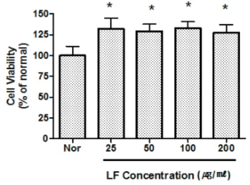
LF가 RAW 264.7 cell의 세포생존율에 미치는 영향을 측정한 결과 세포독성을 나타내지 않았다(Fig. 1).
2. LPS로 유발된 NO 생성의 변화 LF는 NO 생성을 25, 50, 100
μg/ml농도에서 유의하게 억제하였다(Fig. 2).
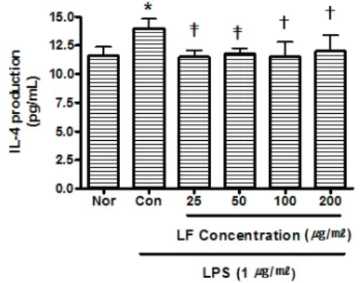
3. LPS로 유발된 cytokine 생성의 변화 1) IL-4
LF는 IL-4 생성을 25 μg/ml 이상의 모 든 농도에서 유의하게 억제하였다(Fig. 3).
2) MIP-1
αLF는 MIP-1α 생성을 25 μg/ml 이상의 모든 농도에서 유의하게 억제하였다(Fig. 4).
3) G-CSF
LF는 G-CSF 생성을 25 μg/ml 이상의 모
든 농도에서 유의하게 억제하였다(Fig. 5).
Fig. 1. Effect of LF on Cell Viability in RAW 264.7 Cell.
LF : Ligustri Lucidi Fructus water extract Nor : Normal. Treated with media only.
Cells were incubated with LF at the concentration of 25, 50, 100, 200
μ
g/ml for 24 hrs.Results are represented as mean±SD of three independent experiments.
* : represents P<0.01 compared to the normal.
Fig. 2. Effect of LF on NO Production of RAW 264.7 Cell Treated with LPS.
LF : Ligustri Lucidi Fructus water extract Nor : Normal. Treated with media only.
Con : Control. Treated with LPS (1
μ
g/ml) only.LPS-induced Cells were incubated with LF at the concentration of 25, 50, 100, 200
μ
g/ml for 24 hrs.Results are represented as mean±SD of three independent experiments.
* : represents P<0.05 compared to the normal.
†: represents P<0.05 compared to the control.
‡: represents P<0.01 compared to the control.
Fig. 3. Effect of LF on IL-4 Production of RAW 264.7 Cell Treated with LPS.
LF : Ligustri Lucidi Fructus water extract Nor : Normal. Treated with media only.
Con : Control. Treated with LPS (1
μ
g/ml) only.LPS-induced Cells were incubated with LF at the concentration of 25, 50, 100, 200
μ
g/ml for 24 hrs.Results are represented as mean±SD of three independent experiments.
* : represents P<0.05 compared to the normal.
†: represents P<0.05 compared to the control.
‡: represents P<0.01 compared to the control.
Fig. 4. Effect of LF on MIP-1
αProduction of RAW 264.7 Cell Treated with LPS.
LF : Ligustri Lucidi Fructus water extract Nor : Normal. Treated with media only.
Con : Control. Treated with LPS (1
μ
g/ml) only.LPS-induced Cells were incubated with LF at the concentration of 25, 50, 100, 200
μ
g/ml for 24 hrs.Results are represented as mean±SD of three independent experiments.
* : represents P<0.05 compared to the normal.
†: represents P<0.05 compared to the control.
‡: represents P<0.01 compared to the control.
Fig. 5. Effect of LF on G-CSF Production of RAW 264.7 Cell Treated with LPS.
LF : Ligustri Lucidi Fructus water extract Nor : Normal. Treated with media only.
Con : Control. Treated with LPS (1
μ
g/ml) only.LPS-induced Cells were incubated with LF at the concentration of 25, 50, 100, 200
μ
g/ml for 24 hrs.Results are represented as mean±SD of three independent experiments.
* : represents P<0.05 compared to the normal.
†: represents P<0.05 compared to the control.
‡: represents P<0.01 compared to the control.
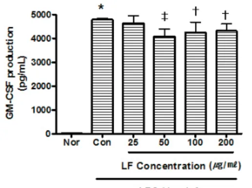
4) GM-CSF
LF는 GM-CSF 생성을 50
μg/ml 이상 의 모든 농도에서 유의하게 억제하였다 (Fig. 6).
5) VEGF
LF는 VEGF 생성을 50
μg/ml 이상의 모든 농도에서 유의하게 억제하였다(Fig. 7).
6) IFN-
γLF는 IFN-
γ생성을 25, 50, 100
μg/ml 의 농도에서 유의하게 억제하였다(Fig. 8).
7) IL-1
βLF는 IL-1
β생성을 50, 200
μg/ml의
농도에서 유의하게 억제하였다(Fig. 9).
Fig. 6. Effect of LF on GM-CSF Production of RAW 264.7 Cell Treated with LPS.
LF : Ligustri Lucidi Fructus water extract Nor : Normal. Treated with media only.
Con : Control. Treated with LPS (1
μ
g/ml) only.LPS-induced Cells were incubated with LF at the concentration of 25, 50, 100, 200
μ
g/ml for 24 hrs.Results are represented as mean±SD of three independent experiments.
* : represents P<0.05 compared to the normal.
†: represents P<0.05 compared to the control.
‡: represents P<0.01 compared to the control.
Fig. 7. Effect of LF on VEGF Production of RAW 264.7 Cell Treated with LPS.
LF : Ligustri Lucidi Fructus water extract Nor : Normal. Treated with media only.
Con : Control. Treated with LPS (1
μ
g/ml) only.LPS-induced Cells were incubated with LF at the concentration of 25, 50, 100, 200
μ
g/ml for 24 hrs.Results are represented as mean±SD of three independent experiments.
* : represents P<0.05 compared to the normal.
†: represents P<0.05 compared to the control.
‡: represents P<0.01 compared to the control.
Fig. 8. Effect of LF on IFN-
γProduction of RAW 264.7 Cell Treated with LPS.
LF : Ligustri Lucidi Fructus water extract Nor : Normal. Treated with media only.
Con : Control. Treated with LPS (1
μ
g/ml) only.LPS-induced Cells were incubated with LF at the concentration of 25, 50, 100, 200
μ
g/ml for 24 hrs.Results are represented as mean±SD of three independent experiments.
* : represents P<0.05 compared to the normal.
†: represents P<0.05 compared to the control.
‡: represents P<0.01 compared to the control.
Fig. 9. Effect of LF on IL-1
βProduction of RAW 264.7 Cell Treated with LPS.
LF : Ligustri Lucidi Fructus water extract Nor : Normal. Treated with media only.
Con : Control. Treated with LPS (1
μ
g/ml) only.LPS-induced Cells were incubated with LF at the concentration of 25, 50, 100, 200
μ
g/ml for 24 hrs.Results are represented as mean±SD of three independent experiments.
* : represents P<0.05 compared to the normal.
†: represents P<0.05 compared to the control.
‡: represents P<0.01 compared to the control.
8) IL-5
LF는 IL-5 생성을 25, 100 μg/ml의 농 도에서 유의하게 억제하였다(Fig. 10).
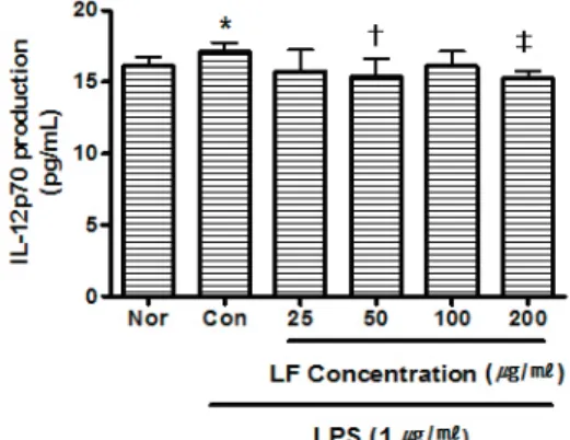
9) IL-12p70
LF는 IL-12p70 생성을 50, 200 μg/ml 의 농도에서 유의하게 억제하였다(Fig. 11).
10) MIP-1
βLF는 MIP-1β 생성을 50, 100 μg/ml 의 농도에서 유의하게 억제하였다(Fig. 12).
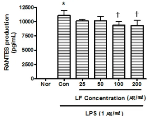
11) RANTES
LF는 RANTES 생성을 100, 200 μg/ml 의 농도에서 유의하게 억제하였다(Fig. 13).
Fig. 10. Effect of LF on IL-5 Production of RAW 264.7 Cell Treated with LPS.
LF : Ligustri Lucidi Fructus water extract Nor : Normal. Treated with media only.
Con : Control. Treated with LPS (1
μ
g/ml) only.LPS-induced Cells were incubated with LF at the concentration of 25, 50, 100, 200
μ
g/ml for 24 hrs.Results are represented as mean±SD of three independent experiments.
* : represents P<0.05 compared to the normal.
†: represents P<0.05 compared to the control.
‡: represents P<0.01 compared to the control.
Fig. 11. Effect of LF on IL-12p70 Production of RAW 264.7 Cell Treated with LPS.
LF : Ligustri Lucidi Fructus water extract Nor : Normal. Treated with media only.
Con : Control. Treated with LPS (1
μ
g/ml) only.LPS-induced Cells were incubated with LF at the concentration of 25, 50, 100, 200
μ
g/ml for 24 hrs.Results are represented as mean±SD of three independent experiments.
* : represents P<0.05 compared to the normal.
†: represents P<0.05 compared to the control.
‡: represents P<0.01 compared to the control.
Fig. 12. Effect of LF on MIP-1
βProduction of RAW 264.7 Cell Treated with LPS.
LF : Ligustri Lucidi Fructus water extract Nor : Normal. Treated with media only.
Con : Control. Treated with LPS (1
μ
g/ml) only.LPS-induced Cells were incubated with LF at the concentration of 25, 50, 100, 200
μ
g/ml for 24 hrs.Results are represented as mean±SD of three independent experiments.
* : represents P<0.05 compared to the normal.
†: represents P<0.05 compared to the control.
‡: represents P<0.01 compared to the control.
Fig. 13. Effect of LF on RANTES Production of RAW 264.7 Cell Treated with LPS.
LF : Ligustri Lucidi Fructus water extract Nor : Normal. Treated with media only.
Con : Control. Treated with LPS (1
μ
g/ml) only.LPS-induced Cells were incubated with LF at the concentration of 25, 50, 100, 200
μ
g/ml for 24 hrs.Results are represented as mean±SD of three independent experiments.
* : represents P<0.05 compared to the normal.
†: represents P<0.05 compared to the control.
‡: represents P<0.01 compared to the control.
12) IL-10
LF는 IL-10 생성을 200 μg/ml의 농도 에서 유의하게 억제하였다(Fig. 14).
13) IP-10
LF는 IP-10 생성을 200 μg/ml의 농도 에서 유의하게 억제하였다(Fig. 15).
Fig. 14. Effect of LF on IL-10 Production of RAW 264.7 Cell Treated with LPS.
LF : Ligustri Lucidi Fructus water extract
Nor : Normal. Treated with media only.
Con : Control. Treated with LPS (1
μ
g/ml) only.LPS-induced Cells were incubated with LF at the concentration of 25, 50, 100, 200
μ
g/ml for 24 hrs.Results are represented as mean±SD of three independent experiments.
* : represents P<0.05 compared to the normal.
†: represents P<0.05 compared to the control.
Fig. 15. Effect of LF on IP-10 Production of RAW 264.7 Cell Treated with LPS.
LF : Ligustri Lucidi Fructus water extract Nor : Normal. Treated with media only.
Con : Control. Treated with LPS (1
μ
g/ml) only.LPS-induced Cells were incubated with LF at the concentration of 25, 50, 100, 200
μ
g/ml for 24 hrs.Results are represented as mean±SD of three independent experiments.
* : represents P<0.05 compared to the normal.
†: represents P<0.05 compared to the control.
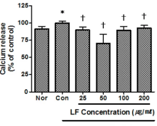
4. LPS로 유발된 세포내 Ca 생성의 변화
LF는 세포내 Ca 생성을 25 μg/ml 이
상의 모든 농도에서 유의하게 억제하였
다(Fig. 16).
Fig. 16. Effect of LF on Ca Release of RAW 264.7 Cell Treated with LPS.
LF : Ligustri Lucidi Fructus water extract Nor : Normal. Treated with media only.
Con : Control. Treated with LPS (1
μ
g/ml) only.LPS-induced Cells were incubated with LF at the concentration of 25, 50, 100, 200
μ
g/ml for 24 hrs.Results are represented as mean±SD of three independent experiments.
* : represents P<0.05 compared to the normal.
†: represents P<0.01 compared to the control.