38(4) : 339 348 (2007)
339
Genistein-4 ' -O- α -L-rhamnopyranosyl-(1-2)- β -D-glucopyranoside 의 RAW 264.7 세포에서 NF- κ B 불활성화를 통한 LPS 에 의해 유도되는
iNOS, COX-2 그리고 cytokine 들의 발현 저해효과
박승재1·김지연1·장영표1·조영욱2·안은미3·백남인4·이경태1*
1경희대학교약학대학
,
2경희대학교의과대학,
3대구한의과대학교한방식품 약리학과,
4경희대학교생명과학부Inhibition of LPS Induced iNOS, COX-2 and Cytokines Expression by Genistein-4 ' -O- α -L-Rhamnopyranosyl-(1-2)- β -D-Glucopyranoside through
the NF- κ B Inactivation in RAW 264.7 Cells
Seung Jae Park
1, Ji-Yeon Kim
1, Young Pyo Jang
1, Young-Wuk Cho
2Eun-Mi Ahn
3, Nam-In Baek
4and Kyung-Tae Lee
1*1
Department of pharmaceutical Biochemistry, College of Pharmacy, Kyung-Hee University, Seoul 130-701, South Korea
2
Department of Physiology, College of Medicin,e Kyung-Hee University, Seoul 130-701, South Korea
3
Department of Herbal Food Science, Daegu Haany University, Gyeongsan, 712-715, South Korea
4
Department of Life Science, Kyung-Hee University, Suwon 449-701, South Korea
Abstract −
This study were designed to evaluate the anti-inflammatory effects of genistein-4'-O-
α-L-rhamnopyranosyl-(1-2)-
β
-D-glucopyranoside (GRG) isolated from Sophora japonica (Leguminosae) on the lipopolysaccharide (LPS)-induced nitric oxide (NO) and prostaglandin (PGE
2) production by RAW 264.7 cell line. GRG significantly inhibited the LPS-induced NO and PGE
2production. Consistent with these observations, GRG reduced the LPS-induced expression of inducible nitric oxide synthase (iNOS) and cyclooxygenase-2 (COX-2) at the protein and mRNA levels in a concentration-dependent manner. In addi- tion, the release and the mRNA expression levels of tumor necrosis factor-
α(TNF-
α) and interleukin-6 (IL-6) were also reduced by GRG. Moreover, GRG attenuated the LPS-induced activation of nuclear factor-kappa B (NF-
κB), a transcription factor necessary for pro-inflammatory mediators, iNOS, COX-2, TNF-
αand IL-6 expression. These results suggest that the down regulation of iNOS, COX-2, TNF-
α, and IL-6 expression by GRG are achieved by the downregulation of NF-
κB activity, and that is also responsible for its anti-inflammatory effects.
Key words −
genistein-4'-O-
α-L-rhamnopyranosyl-(1-2)-
β-D-glucopyranoside, NF-
κB, LPS, Anti-inflammation, Sophora japonica
염증반응은생체나조직에물리적작용이나화학적물질
,
세균감염등의어떠한기질적변화를가져오는침습이가 해질때그손상부위를수복재생하려는기전이며
,
일단자극이가해지면국소적으로
histamine, serotonine, bradykinin, prostaglandins, hydroxyeicosatetraenoic acid (HETE)
및leukotriene
과같은혈관활성물질이유리되어혈관투과성이증대되면서염증을유발한다
.
그러나지속적인염증반응은오히려점막손상을촉진하고
,
그결과일부에서는암발생등의질환을유도한다
.
1)내독소로잘알려진
lipopolysaccaride (LPS)
는그람음성균의 세포외막에 존재하며
, RAW 264.7
세포와 같은macrophage
또는monocyte
에서tumor necrosis factor-alpha (TNF-
α), Interleukin-6 (IL-6), Interleukin-1
β(IL-1
β)
와 같은
proinflammatory cytokine
을증가시키는것으로알려져있다
.
2-6)이러한염증매개물질의형성은NO
생성 증가및phospholipase A
2의활성을촉진시켜prostaglandin (PG)
합성을유도한다
.
7-8)이중
NO
는체내방어기능,
신호전달기능,
신경독성,
혈*교신저자(E-mail):[email protected] (FAX):02-966-3885
관확장등의다양한생리기능을가지고있다
.
9)포유동물에서분리한
nitric oxide synthase (NOS)
는물리화학적성상에따라
Type I, II,
및III
등3
종류의동종효소로나누어진다
. Type I (neuronal NOS, nNOS)
과Type II (endotheli- al NOS, eNOS)
는세포속에계속적으로존재하기때문에구성
NOS(constitutive NOS)
로분류되며,
상대적으로일부세포에서
LPS
와cytokines
같은특수한자극제들에노출되는경우에만발현되는
Type II
인유도형NOS (iNOS)
로나누어진다
.
10)이들NOS
중iNOS
에의한NO
생성이절대적으로많으며이는병리적으로중요한작용을한다
.
일반적인NO
형성은박테리아를죽이거나종양을제거시키는중요한역할을하지만
,
병리적인원인에의한과도한NO
형성은염증을유발시키게되며조직의손상
,
유전자변이및신경손상등을유발한다
.
11-13)Cyclooxygenase (COX)
는arachidonic acid
를PGs
으로전환시키는효소로써
COX-1
과COX-2
로분류된다. COX-1
은체내에서혈소판의형성
,
위벽보호,
신장기능의유지등정상적 생체기능에 작용하지만
COX-2
는 염증매개물질인prostaglandins (PGs)
를형성시킨다.
14)TNF-
α는활성화된macrophage, fibroblast
및다른여러세포에서생성되는데이는종양세포에영향을미치는숙주 방어인자및염증매개물질로알려져있다
. IL-6
의생성은TNF-
α나IL-1
β같은요인외에도LPS
에의해유도된다.
15)IL-6
는proinflammatory cytokine
으로써endogenous pyrogen
으로작용을하며면역체계와조혈등에다양한영향을미친다
.
16)Nuclear transcription factor-kappa-B (NF-
κB)
는세포 분화
,
염증반응,
세포부착등에관련된여러유전자들의발현에가장중요한역할을하는전사인자이다
.
활성화된NF-
κ
B
는iNOS, COX-2, TNF-
α그리고IL-6
등여러염증매개물질의전사를촉진한다
.
17)회화나무
( Sophora japonica L.)
는콩과식물에속하는낙엽교목으로그높이는
25 m
에달하고가지가퍼지며우리나라
,
중국,
일본등지에날리분포한다.
18)잎은호생하고화기는
7-8
월로이시기의꽃을한방에서는괴화(
槐花)
라하고꽃봉오리는괴미
(
傀米),
그성숙한열매를괴각이라하여지혈
,
토혈,
변혈등에이용되며,
혈압강화효과와항염효과가있는것으로알려져있다
.
19-20)또한민간에서는잎을삶은물로치질부위를수세하고생
(
生)
초(
炒)
괴화를비(
鼻)
출혈
,
세균성치질의치유에,
고혈압환자가상복하여중풍의예방에사용하여왔다
.
21)이러한천연물로부터유효활성성분을분리하여이들의약리작용기전을규명함으로써다 양한약품의개발가능성을부여할수있다
.
본연구진은여러천연물의추출물및분리된단일화합물 들의항염증효과를검색하고그기전을규명함으로써새로 운항염증약물의개발을시도하고있다
.
본논문에서는이러한 연구중 괴각으로부터 분리한
genistein-4'-O-
α-L- rhamnopyranosyl-(1-2)-
β-D-glucopyranoside (GRG)
가LPS
에의해활성화된
RAW 264.7
세포에서항염효과를나타내는기전을 연구하였으며염증에 관련된다양한단백질
mRNA
그리고cytokine
의발현을측정하였다. 재료 및 방법
재료 −시료의추출과분획에사용한유기용매는대정 화학주식회사
(Gyonggi-do, Korea)
에서생산한1
급시약을사용하였다
. Column chromatography
용silica gel
은Kiesel gel 60 (Merck, Germany)
을사용하였다. TLC
는Kiesel gel 60 F
254와RP-18 F
254s를사용하였고, TLC
상의물질검출에는
UV lamp
와10% aq . H
2SO
4를사용하였다. NMR
스펙트럼은
Varian Inova AS 400 (Varian, USA)
으로, FABMS
는JMS-700 (JEOL, Japan)
로 측정하였다. Dulbecco’s modified Eagle’s minimum essential medium (DMEM), fetal bovine serum (FBS), penicillin, streptomy- cin
은life Technologies Inc. (Grand Island, NY)
에서구입하였다
. Genistein, 3-(4,5-dimethylthiazol-2-yl)-2,5-diphenyl- tetrazolium bromide (MTT), dimethyl sulfoxide (DMSO), sulfanilamide, aprotinin, leupeptin, phenylmethylsulfony- lfluride (PMSF), dithiothreitol (DTT), L-N
6-(1-iminoethyl) lysine (L-NIL), NS-398, Escherichia coli lipopolysaccha- ride (LPS)
는Sigma Chemical Co. (CA, U.S.A.)
에서 구입하였으며
, COX-2
와iNOS monoclonal antibodies
및peroxidase conjugated secondary antibody
는Santa Cruz Biotechnology (CA, U.S.A.)
에서구입하였다. iNOS, COX- 2, TNF-
α, IL-6
그리고 β-actin oligonucleotide primers
는Bioneer (Seoul, Korea)
에서구입하였다.
그리고TNF-
α, IL- 6, prostaglandin E
2측정을 위한kit
는R&D systems (MN, U.S.A.)
에서구입하였다.
시료의 추출및분리 −괴각
1.2 kg
에80% MeOH
용액(2 L
×3)
에24
시간실온에서3
회추출하다.
추출물을여과하고얻어진여액을모두합쳐감압농축하여
MeOH
추출물395 g
을얻었다. MeOH
추출물을증류수1
리터에현탁시킨후
EtOAc
및n -BuOH
로순차적으로분획하였다.
각각의분획물을 농축하여
EtOAc
분획물(9.42 g), n -BuOH
분획물(81.9 g)
및물분획물을얻었다. n -BuOH
분획물(81 g)
을CHCl
3-MeOH
의혼합용매를용출용매로사용하여기울기용리방식으로
silica gel column chromatography (c.c)
를실시하여총
11
개의소분획(B1~B11)
으로나누었다.
이중B10
번 분획을silica gel c.c.(CHCl
3-MeOH-H
2O = 65:35:10)
을 사용하여 노란분말상의genistein-4'-O-
α-L- rhamnopyranosyl-(1-2)-
β-D-glucopyranoside (528 mg)
을 분리하였다
.
이화합물의1H,
13C-NMR
및MS
의기존문헌과비교하여구조를확인동정하였다
.
22)Genistein-4'-O-
α-L-rhamnopyranosyl-(1-2)-
β-D- glucopyranoside (Fig.1)
: yellow amorphous powder, Negative FAB MS m/z : 577 ([M-H]
−),
1H-NMR (400 MHz, pyridine- d
5, δH): 8.06 (1H, s, H-2), 7.64 (2H, d, J=8.8, H2', 6'), 7.54 (2H, d, J = 8.8 Hz, H-3', 5'), 6.74 (1H, d, J = 2.0 Hz, H-6), 6.66 (1H, d, J=2.0 Hz, H-8), 6.43 (1H, br s,rha-1), 5.58 (1H, d, J=8.0 Hz, glc-1), 1.83 (3H, d, J=6.0 Hz, rha-6),
13C- NMR (100 MHz, pyridine- d
5, δC): 180.76 (C-4), 166.02 (C-7), 163.51 (C-5), 158.54 (C-9), 158.25 (C-4'), 153.59 (C-2), 131.92 (C-2', 6'), 125.23 (C-1'), 123.22 (C-3), 116.62 (C-3', 5'), 105.73 (C-10), 102.36 (rha-1'), 100.21 (C-6), 99.99 (glc-1), 94.68 (C-8), 79.40 (glc-5'), 78.76 (rha-4'), 77.75 (glc-3'), 74.18 (glc-2'), 72.76 (glc-4'), 72.53 (rha-2'), 71.35 (rha-3'), 69.94 (rha-5'), 62.16 (glc-6'), 18.98 (rha-6').
세포의 배양 −
RAW 264.7
세포는10% FBS
및penicillin (100
µg/ml), streptomycin (100 U/ml)
이 포함된DMEM
배지에서37
oC, 5% CO
2incubator
에서배양했다. RAW 264.7
세포에시료용액의여러농도(25, 50, 100, 200
µ
M)
또는양성대조군을1
시간전처리한후LPS (1
µg/mL)
를처리하고
24
시간배양하였다.
세포독성시험−
96 well plate
에1
×10
5cells/well
로세포를동일하게분주하고
24
시간동안배양한후여러농도의시료용액을두군으로나누어배지에희석하여첨가하였 다
. 1
시간후한군에만LPS (1
µg/ml)
를처리하였다. 24
시간이지난후
MTT
시약을넣고4
시간동안방치한후상등액을제거하고형성된
formazan
을DMSO 100
µl
를첨가하여녹였다
. 30
분후540 nm
에서흡광도를측정하였다. Nitrite
양의측정 −RAW 264.7
세포로부터생성된NO
의양은
Griess
시약을이용하여세포배양액중에존재하는
NO
2−의형태로서측정하였다.
즉세포배양상등액100
µ
l
와Griess
시약[1% (w/v) sulfanilamide in 5% (v/v)
phosphoric acid
와0.1% (w/v) naphtylethylenediamine- HCl] 100
µl
를혼합하여96 well plates
에서10
분동안반응시킨후
540 nm
에서흡광도를측정하였다.
Western blot
시험 −GRG
를처리한세포및대조군을PBS
로 씻어낸 후lysis buffer
인PRO-PREP (Intron Biotechnology)
으로단백질을추출한후원심분리하여상등액을취하였다
.
상등액을Bradford
시약을사용해단백질농도를정량하여
50
µg
의단백질을취했다.
추출된단백질은10%
의SDS-polyacrylamide gel
에 전기영동시킨 후nitro cellulose membrane
으로gel
의 단백질을blot
시켰다. 5%
skim milk
로하루밤동안blocking
한후1:500
의비율로iNOS
와COX-2 antibody
를4
시간동안상온에서방치한후TTBS
로15
분간격으로2
회세척하였다. 1:1000
의비율로희석한
secondary antibody
를1
시간동안상온에서방치시켰다
.
다시TTBS
로15
분간격으로3
회세척한후chemilu- minescence
로현상하였다.
PGE2, TNF-
α 및IL-6
양의측정 −세포배양액을취해각각
R&D kit
의지시에따라PGE
2, TNF-
α및IL-6
를정량하였다
.
RT-PCR
시험 −Easy Blue
Rkits (Intron Biotechnology)
를이용하여
Kit
의protocol
에따라전체cellular RNA
를추출하였다
.
각각의 시료에서MuLV reverse transcriptase, 1 mM dNTP
그리고oligo (dT
12-18) 0.5
µg/
µl
를이용하여1
Fig. 1.
Chemical structures of genistein and genistein-4'-O-
α-L- rhamnopyranosyl-(1-2)-
β-D-glucopyranoside (GRG).
Fig. 2.
The cytotoxicity of GRG or Genistein on RAW 264.7 cells. Cells were exposed to GRG and genistein (from 6.25
µ
M to 400
µM) with/without LPS (1
µg/ml). Cytotoxicity was
assessed by 3-(4, 5 dimethylthiazol-2-yl)-2, 5-diphenyltetrazo-
lium bromide (MTT) assay after 24 h incubation.
µ
g
의RNA
를역전사하여cDNA
를얻었다. cDNA
에Taq DNA polymerase 1 unit, 0.2 mM dNTP,
×10 reaction buffer
그리고5'
와3' primers 100 pmol
을포함한전체부피
25
µL
의 시료를thermal cycler (Perkin Elmer Cetus, Foster City, CA, USA)
를 이용하여PCR
분석을 하였다. PCR
반응은95
oC
에서2
분간initial denaturation
시킨 후iNOS (95
oC 1
분danaturation, 60
oC 1
분annealing
그리고72
oC 1.5
분extension), COX-2 (94
oC 1
분danaturaion, 60
oC 1
분annealing
그리고72
oC 1
분extension), TNF-
α(95
oC 1
분denaturation, 55
oC 1
분annealing
그리고72
oC 1
분
extension)
그리고IL-6 (94
oC 1
분denaturation, 56
oC 1
분
annealing
그리고72
oC 1
분extension)
를30
회amplifi-
cation
하였다.
이번 연구에서 아래의목록과 같은PCR primers
가사용되었으며Bioneer (Seoul, Korea)
에서구입하였다
. : sense strand iNOS, 5'-ATT GGC AAC ATC AGG- TCG GCC ATC ACT-3', anti-sense strand iNOS,5'- GCT GTG TGT CAC AGA AGT CTC GAA- CTC-3';
sense strand COX-2, 5'-GGA GAG ACT ATC AAG ATA GT-3' anti-sense strand COX-2, 5'-ATG GTC AGT AGA CTT TTA CA-3'; sense strand TNF-
α, 5'-ATG AGC ACA GAA AGC ATG- ATC-3', anti-sense strand TNF-
α, 5'- TAC AGG CTT GTC ACT CGA ATT-3'; sense strand IL-6, 5'-GAG GAT ACC ACT CCC AAC AGA CC-3', anti-sense strand IL-6, 5'-AAG TGC ATC ATC GTT GTT
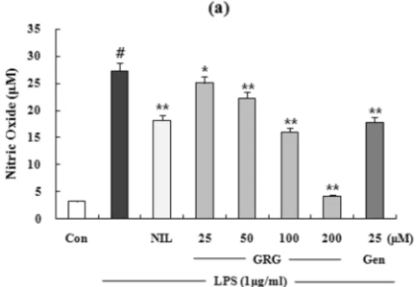
Fig. 3.
The effects of GRG on LPS-Induced NO production and iNOS protein and mRNA expressions in RAW 264.7 Cells. (a) Cells were treated with different concentrations of GRG or genistein (25
µM) for 1 h and then LPS (1
µg/ml) was added and the cells were incubated for 24 h. Control (Con) values were obtained in the absence of LPS or tested samples. L-N
6-(1-iminoethyl) lysine (L-NIL) was used as an assay positive control at a concentration of 10
µM. (b) Lysates were prepared from control or 24 h LPS (1
µg/ml)-stimulated cells alone or LPS plus with different concentration (25, 50, 100, 200
µM) of GRG or genistein (25
µM).
Total cellular proteins (40
µg) were resolved by SDS-PAGE, transferred to nitrocellulose membranes, and detected with specific
antibodies, as described in methods. A representative immunoblot of three separate experiments is shown. Total RNA was prepared
for the RT-PCR analysis of iNOS gene expression from RAW 264.7 macrophages stimulated with LPS (1
µg/ml) with/without
different concentration (25, 50, 100, 200
µM) of GRG or genistein (25
µM) for 4 h. iNOS-specific sequences (807 bp) was detected
by agarose gel electrophoresis, as described in methods. PCR of
β-actin was performed to verify that the initial cDNA contents of
the samples were similar. The experiment was repeated three times and similar results were obtained. The Western blot results have
some analogy with the RT-PCR results and these are also shown by relativc ratio graphs. The values are the mean
±S.D. of three
independent experiments.
#p <0.05 vs. the control group; * p <0.05, ** p <0.01 vs. the LPS-treated group; the significances of the
difference between the treated groups was evaluated using the Student’s t -test.
CAT ACA-3';5'-GTG CTG CCT- AAT GTC CCC TTG AAT C-3'; sense strand
β-actin, 5'-TCA TGA AGT GTG ACG TTG ACA- TCC GT-3', anti-sense strand
β-actin, 5’-CCT AGA AGC ATT TGC GGT GCA CGA TG-3'.
Amplification
후에PCR
반응시킨시료를2% agarose gel
에서전기영동하고
ethidim bromide
염색과UV
조사를통해확인하였다
.
NF-
κB Luciferase activity
측정 −RAW 264.7
세포를dish
에 각각2
×10
5cells/dish
농도로 분주한 후, superfect transfection reagent (Qiagen GmbH, Germany)
를이용하여NF-
κB luciferase reporter plasmid DNA
를 형질감염(transfection)
시켰다.
형질감염48
시간이경과한후3~4
×10
5cell/well
로12 well plate
에세포를분주하고GRG
를1
시간동안전처리한후
LPS (1
µg/ml)
를처리하였다. 24
시간후세포를수집하여
luciferase assay system (Promega, U.S.A.)
와
luminometer (Perkin Elmer Cetus, U.S.A)
를 이용하여luciferase
활성을측정하였다.
TLC
시험 −앞선실험과같은조건으로세포를배양하여
5
×10
5cells/dish
농도로분주하고시료(50
µM)
와LPS (1
µg/ml)
을처리하였다. LPS
처리24
시간후배지와세포를분리하여모았다
. Cold methanol
을이용하여단백질제거후질소가스로시료를농축하고
30
µl
의methanol
에녹였다. TLC plate
는silica gel aluminium sheets
를사용하였으며전개용매는
ethylacetate-formic acid-acetic acid-water
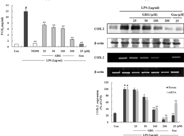
Fig. 4.
The effects of GRG on LPS-Induced PGE
2and COX-2 protein and mRNA expressions in RAW 264.7 Cells. (a) Effect of the GRG and genistein on PGE
2production by LPS-induced RAW 264.7 macrophage for 24 h. 10
µM of NS-398 was as a positive control in the assay. (b) Lysates were prepared from control or 24 h LPS (1
µg/ml)-stimulated cells alone or LPS plus with different concentration (25, 50, 100, 200
µM) of GRG or genistein (25
µM). Total cellular proteins (40
µg) were resolved by SDS-PAGE, transferred to nitrocellulose membranes, and detected with specific antibodies, as described in methods. A representative immunoblot of three separate experiments is shown. Total RNA was prepared for the RT-PCR analysis of COX-2 gene expression from RAW 264.7 macrophages stimulated with LPS (1
µg/ml) with/without different concentration (25, 50, 100, 200
µM) of GRG or genistein (25
µM) for 4 h. COX-2-specific sequences (721 bp) was detected by agarose gel electrophoresis, as described in methods. PCR of
β
-actin was performed to verify that the initial cDNA contents of the samples were similar. The experiment was repeated three times and similar results were obtained. The Western blot results have some analogy with the RT-PCR results and these are also shown by relativc ratio graphs. The values are the mean
±S.D. of three independent experiments.
#p <0.05 vs. the control group;
* p <0.05, ** p <0.01 vs. the LPS-treated group; the significances of the difference between the treated groups was evaluated using
the Student’s t -test.
(30:1.2:1.2:1.5)
로전개한다음말리고AlCl
3를분무한후UV (254 nm)
로확인하였다.
통계학적분석 −실험치의값은
mean
±S.D.
로나타냈으며분석은
Student’s t -test
로그유의성을나타내었다.
결 과
GRG
의세포독성에대한효과 −GRG
와GRG
의agly- cone
인genistein
의RAW 264.7
세포에대한세포독성을측정하기위해
MTT assay
를수행하였다. GRG
와genistein
모두농도의존적으로
RAW 264.7
세포의생존능력을감소시켰으며
(Fig. 2a) LPS (1
µg/ml)
는세포의viability
에영향이없음을확인하였다
. GRG
의IC
50는284.25
µM
로확인되었으며
genistein (IC
50:79.69
µM)
에비해RAW 264.7
세포에서세포독성이낮음을확인하였다
(Fig. 2b).
GRG
의Nitrite
형성및iNOS
단백질과mRNA
발현저해 −
LPS
에의해활성화된RAW 264.7
세포의배양액중에생성된
nitrite
의양을Griess
시약을사용하여GRG
의NO
생성저해효과를조사하였다. GRG
는농도의존적으로NO
생성을저해하였으며(Fig.3a) 100
µM
에서NO
생성을46.9%
저해하였다. genistein
은25
µM
에서NO
생성을39.4%
저해하였으며양성대조군으로는L-arginene
과의기질경쟁에의하여
iNOS
저해제로알려진L-NIL (10
µM)
을사용하였다
. GRG
에의한염증인자(NO)
의 생성억제와iNOS
발현의상관성을알아보기 위하여Western blot
과RT-PCR
로iNOS
단백질과mRNA
발현을조사하였다. LPS
에의해
iNOS
단백질이뚜렷하게증가하였으며, GRG
에의해
iNOS
단백질의발현이농도의존적으로저해되었다.
β- actin
의band density
비율에따라iNOS
단백질의발현 정도를보정하였을때
GRG
의aglycone
인genistein
은25
µM
에서
iNOS
단백질발현을68.7%
저해하였으며GRG
는50
µM
농도에서iNOS
단백질발현을68.9%
저해함을확인하였다
. GRG
에의한iNOS mRNA
발현저해는농도의존적이며단백질발현저해와상관성있게나타났다
(Fig.3b).
GRG
의PGE2
형성및COX-2
단백질과mRNA
발현저해효과 −
GRG
가RAW 264.7
세포에서LPS
처리에의한
PGE
2의생성을농도의존적으로유의성있게감소시키는 것을 확인할수있었으며(Fig. 4a) IC
50는105.97
µM
로확인되었다
.
양성대조군으로사용한NS398 (5
µM)
과GRG
의
aglycone
인genistein (25
µM)
에서PGE
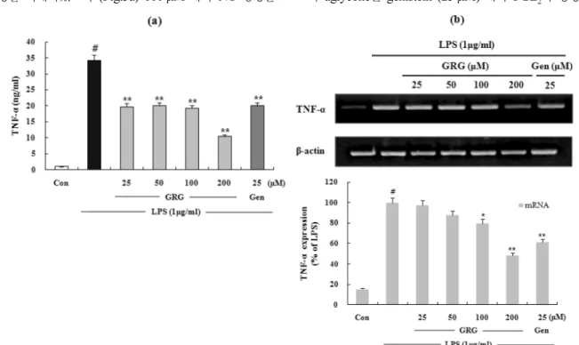
2의생성을뚜렷Fig. 5.
The effects of GRG and genistein on LPS-induced TNF-
αrelease and mRNA expression in RAW 264.7 cells. Cells were
treated with different concentrations of GRG or genistein (25
µM) for 1 h and then LPS (1
µg/ml) was added and the cells were
incubated for 24 h. Control (Con) values were obtained in the absence of LPS or tested samples. Total RNA was prepared for the
RT-PCR analysis of TNF-
αgene expression from RAW 264.7 macrophages stimulated with LPS (1
µg/ml) with/without different
concentration (25, 50, 100, 200
µM) of GRG or genistein (25
µM) for 4 h. TNF-
α-specific sequences (351 bp) was detected by
agarose gel electrophoresis, as described in methods. PCR of
β-actin was performed to verify that the initial cDNA contents of the
samples were similar. TNF-
αrelease results have some analogy with the RT-PCR results and these are also shown by relativc ratio
graphs. The values are the mean
±S.D. of three independent experiments.
#p <0.05 vs. the control group; * p <0.05, ** p <0.01 vs. the
LPS-treated group; the significances of the difference between the treated groups was evaluated using the Student’s t -test.
하게저해하는것을확인하였다
. GRG
에의한PGE
2생성 저해와COX-2
발현의상관성을알아보기위하여Western blot
과RT-PCR
로COX-2
단백질과mRNA
발현을조사하였다
. GRG
는COX-2
단백질과mRNA
의발현을농도의존적으로저해하였으며
GRG
의aglycone
인genistein (25
µM)
에서도
COX-2
단백질과mRNA
의발현이뚜렷하게저해되는것을확인하였다
(Fig. 4b).
GRG
의TNF-
α와IL-6
의형성및mRNA
발현저해효과 −
GRG
가RAW 264.7
세포에서LPS
에 의한pro- inflammatory cytokine
의형성을억제하는지알아보기위해ELISA
와RT-PCR
을이용하여TNF-
α와IL-6
의생성 및mRNA
발현을측정하였다. LPS
처리에의한TNF-
α와IL- 6
의생성이25, 50, 100
그리고200
µM GRG
에서모두유의성감소되는것을확인하였다
. GRG
는25
µM
에서TNF-
α의생성을
42.3%
저해하여같은농도의genistein (41.2%
저해
)
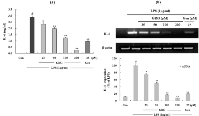
과비슷한효과를나타내었다(Fig. 5a). LPS
에의한IL-6
생성은GRG
의100
µM
농도에서56.7%
저해되었으며GRG
의aglycone
인genistein (25
µM)
에서는66.2%
가저해됨을확인하였다
(Fig. 6a).
또한GRG
는LPS
에의한TNF-
α와
IL-6 mRNA
의발현을유의성있게저해하며TNF-
α와
IL-6
생성저해효과와상관성이있음을확인하였다(Figs.
5b and 6b).
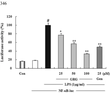
GRG
의NF-
κB
활성저해효과−LPS
에의해유도되는iNOS, COX-2, TNF-
α,
그리고IL-6
의발현에NF-
κB
의활성이 중요한역할을한다고보고되어있으므로23)
GRG
가LPS
에의한NF-
κB
의활성화를억제하는지알아보기위해luciferase assay
를수행하였다. RAW 264.7
세포에일시적으로
pNF-
κB-luc plasmid
를transfection
시키고GRG
또는genistein
을처리한군과처리하지않은대조군에LPS (1
µ
g/ml)
로자극을가한다. GRG
가LPS
에의해유도된NF-
κ
B
의존적인luciferase
효소의발현을농도의존적으로유의성있게감소시키는것을확인하였다
(Fig. 7).
TLC
에의한세포와배지내시료의측정 −In vitro
상에서
GRG
의항염증효과가당이떨어진형태인genistein
에의한것인지확인하기위해세포와배양액중의
GRG
와그aglycone
인genistein
을각각확인하였다. genistein
을처리한경우에는세포와배양액에서모두
genistein
표준물질과같은위치에서
spot
이확인되었다.
그러나GRG
를처리한세포와배양액에서는
genistein
이존재하지않음을확인하였으며배지에서는
GRG
표준물질과같은위치에서spot
이확Fig. 6.