The Role of Phosphofructokinase-2/Fructose-2,6-bisphosphatase 2 (PFKFB2) in Wnt-induced Epithelial-mesenchymal Transition
Su Yeon Lee1, Min Kyung Ju1, Hyun Min Jeon1, Cho Hee Kim1,2, Hye Gyeong Park3 and Ho Sung Kang1*
1Department of Molecular Biology, College of Natural Sciences, Pusan National University, Busan 609-735, Korea
2DNA Identification Center, National Forensic Service, Seoul 158-707, Korea
3Nanobiotechnology Center, Pusan National University, Busan 609-735, Korea
Received August 17, 2017 /Revised September 26, 2017 /Accepted September 27, 2017
Most cancer cells produce ATP predominantly through glycolysis instead of through mitochondrial oxidative phosphorylation, even in the presence of oxygen. The phenomenon is termed the Warburg effect, or the glycolytic switch, and it is thought to increase the availability of biosynthetic precursors for cell proliferation. EMTs have critical roles in the initiation of the invasion and metastasis of cancer cells. The glycolytic switch and EMT are important for tumor development and progression; however, their correlation with tumor progression is largely unknown. The Snail transcription factor is a major factor involved in EMT. The Snail expression is regulated by distal-less homeobox 2 (Dlx-2), a homeo- domain transcription factor that is involved in embryonic and tumor development. The Dlx-2/Snail cascade is involved in Wnt-induced EMTs and the glycolytic switch. This study showed that in re- sponse to Wnt signaling, the Dlx-2/Snail cascade induces the expression of PFKFB2, which is a glyco- lytic enzyme that synthesizes and degrades fructose 2, 6-bisphosphate (F2,6BP). It also showed that PFKFB2 shRNA prevents Wnt-induced EMTs in the breast-tumor cell line MCF-7. The prevention in- dicated that glycolysis is linked to Wnt-induced EMT. Additionally, this study showed PFKFB2 shRNA suppresses in vivo tumor metastasis and growth. Finally, it showed the PFKFB2 expression is higher in breast, colon and ovarian cancer tissues than in matched normal tissues regardless of the cancers' stages. The results demonstrated that PFKFB2 is an important regulator of EMTs and meta- stases induced by the Wnt, Dlx-2 and Snail factors.
Key words : Dlx-2, epithelial-mesenchymal transition (EMT), glycolytic switch, PFKFB2, Snail
*Corresponding author
*Tel : +82-51-510-2275, Fax : +82-51-513-9258
*E-mail : [email protected]
This is an Open-Access article distributed under the terms of the Creative Commons Attribution Non-Commercial License (http://creativecommons.org/licenses/by-nc/3.0) which permits unrestricted non-commercial use, distribution, and reproduction in any medium, provided the original work is properly cited.
Journal of Life Science 2017 Vol. 27. No. 11. 1245~1255 DOI : https://doi.org/10.5352/JLS.2017.27.11.1245
서 론
암세포는 세포죽음 억제, 무한 증식 기작 획득, 유전체 불안 정성과 돌연변이, invasion과 metastasis, oncogenic metabo- lism 등의 다양한 특징들을 나타낸다[14, 15]. 암세포는 암세포 의 생존 및 증식을 위해 oncogenic metabolism을 나타내는 것으로 알려져 있다[5-7, 10, 13, 17, 23, 52, 58]. Oncogenic me- tabolism의 대표적인 현상으로 glycolytic switch (aerobic gly- colysis, Warburg effect)가 보고되어 있다. Glycolytic switch 는 암세포가 정상세포와는 달리 산소가 충분한 상태에서도 미토콘드리아에 의존하지 않고 glycolysis를 통해 대부분의 ATP 에너지를 생성하는 현상으로, 악성종양세포(malignant cell)에서 보편적으로 나타나는 특징이다[17, 23, 58, 61]. 암세 포가 oxidative phosphorylation 보다 비효율적인 glycolysis
에 의해 ATP를 얻는 방식을 채택하는 이유는 암세포 증식에 필요한 대사산물(metabolite)의 원활한 공급을 위해서인 것으 로 알려져 있다[5-7, 10, 13, 17, 23, 52, 58]. 실제로 많은 human cancer에서 glycolysis가 활성화되어져 있는 것으로 보고되어 있다[5-7, 10, 13, 17, 23, 52, 58, 61]. Glycolytic switch는 glu- cose 소비량 증가로 인해 과량의 lactic acid를 생성하며, mi- croenvironmental acidosis를 일으키며, 또한 chemotherapy 또는 radiotherapy에 대한 저항성을 부여한다[4, 16, 18, 24, 34, 44, 51, 68, 69].
암세포는 glycolytic switch 뿐만 아니라 invasion 및 meta- stasis 능력을 획득하기 위해 epithelial-mesenchymal tran- sition (EMT)를 나타낸다. EMT는 종양 세포가 epithelial mor- phology를 잃어버리고, motile, invasive fibroblast-mesen- chymal phenotype 획득을 나타내는 것으로 cell adhesion molecule인 E-cadherin 발현 감소를 유도한다[9, 25, 56, 57].
EMT는 항암제에 대한 감수성을 감소시킬 뿐만 아니라, 암발 생과 밀접한 연관성을 지니고 있는 cancer stem cells (CSC)의 발생을 유도하는 것으로 알려져 있다[8, 32, 48, 53, 57, 59].
EMT를 유도하는 대표적인 단백질인 Snail은 전사인자로서 E-cadherin 등과 같은 다양한 epithelial markers 유전자의 발 현을 억제하고, fibronectin, vitronectin, ɑSMA, fibroblast-
Table 1. shRNA target sequences used in this study
Genes Target sequence 5’ to 3’
Con shRNA Dlx-2 shRNA Snail shRNA PFKFB2 shRNA
AATTCTCCGAACGTGTCACGT TTCGGATAGTGAACGGGAA GCGAGCTGCAGGACTCTAA
TCAGGAAGGCTCCTTACCCTGTAGCAG specific protein 1 (FSP1) 등과 같은 mesenchymal markers
유전자의 발현을 활성화함으로써 EMT를 유도하는 것으로 알 려져 있다[1, 43, 46, 60].
암성장을 조절하는 Wnt는 cancer cell migration, invasion 및 metastasis를 유도하며, 악성유방암(TNBC) 발생과도 깊은 연관성을 지니고 있는 것으로 밝혀져 있다[1, 19-21, 47, 49, 60]. Wnt는 Snail 전달계를 통하여 EMT를 유도하고, 그 결과 invasion 및 metastasis를 조절한다[9, 43, 49, 56, 60]. 또한 Wnt 에 의해 활성화된 Snail은 EMT 뿐만 아니라 glycolytic switch 를 유도하며, 미토콘드리아 산소호흡 및 전자전달계의 com- plex IV (cytochrome c oxidase) 활성을 억제하는 것으로 알려 져 있다[28]. 또한 Snail은 fructose-1,6-biphosphatase (FBP1) 의 발현을 감소시킨다[12]. 그리고 발생 유전자의 발현을 조절 하는 것으로 알려져 있는 Dlx-2 [33, 42]가 tumor growth와 metastasis 등을 통해 tumor malignancy를 유도하며[29, 55, 64, 66], Snail 발현을 조절하여 Wnt에 의한 EMT 및 glycolytic switch를 조절하는 것으로 최근 밝혀져 있다[27].
종양에서 관찰되는 glycolytic switch와 EMT는 tumor growth 및 invasion과 연관된 매우 중요한 과정임에도 불구하 고, 그 조절 기작 및 상호작용에 대한 연구는 아직 밝혀져 있지 않다. 이에 본 연구에서는 glycolysis가 Wnt에 의한 EMT에 관여할 가능성을 조사하였다. 그 과정에서 glycolytic switch target 유전자로 PFKFB2를 발굴하였다. 6-phosphofructo-2- kinase/fructose-2,6-biphosphatase (PFKFB)는 PFKFB1-4 등 4 가지 isozymes이 존재하며, fructose-2,6-bisphosphate (F2,6BP) 의 합성 및 분해에 관여하는 효소이다. PFKFB 대사산물인 F2,6BP는 glycolytic pathway에서 rate-limiting enzyme으로 작용하는 6-phosphofructo-1-kinase (PFK-1)을 활성화시켜, glycolysis를 촉진한다[39, 40, 50, 63]. 본 연구에서는 Wnt-Dlx- 2/Snail cascade에 의한 PFKFB2의 발현 조절 기작을 분석하 고, xenograft mouse model에서 tumor growth 및 metastasis 에 미치는 PFKFB2 shRNA 영향을 연구하였다. 아울러 PFKFB2 가 Wnt에 의한 EMT에 미치는 영향에 대한 연구를 통해 gly- colytic switch와 EMT의 상호작용 가능성을 분석하고, 그 기작 을 조사하고자 하였다.
재료 및 방법
세포배양 및 Wnt3a conditioned medium (CM) 처리 MCF-7, MDA-MB231, L cells은 American Type Culture Collection (ATCC, USA)으로부터 구입하여 사용하였으며, 실 험실에서 확립한 방법에 의해 배양하였다[26]. Wnt3a-secret- ing L cells은 Dr. Min DS (Pusan National University, Pusan, Korea)로부터 제공받았다. 각각의 세포들은 10% fetal bovine serum (FBS) 및 1% penicillin/streptomycin이 포함된 Eagle's Minimum Essential Medium (EMEM), RPMI, Dulbecco’s
modified Eagle’s medium (DMEM)을 사용하여 37℃, 5% CO2
조건 하에서 배양하였다. Wnt3a conditioned medium (CM) 은 Wnt3a-secreting L cell을 100mm dish에 배양하여 72시간 뒤에 medium을 모아 획득하였다. 원심분리를 통해 medium 내의 cell debris를 제거한 후, 배양 중인 세포에 처리하였다.
Transfection 및 short hairpin RNA (shRNA) interfer- ence
MCF-7에 jetPEI (Polyplus transfection, SA, USA)를 이용하 여 expression vectors pCAGGS-Dlx-2 (Dr. John L.R. (Ruben- stein, University of California at San Francisco)로부터 제공 받음)와 pCR3.1-Snail-Flg (Dr. Yook J.I. (Yonsei University, Korea)로부터 제공받음)을 transfection하였다. Control 및 Dlx-2, Snail, PFKFB2에 특이적으로 반응하는 shRNA/pSUPER vectors를 실험실에서 확립한 방법에 의해 제작하여 trans- fection 하였다[26]. shRNA target sequences는 Table 1에 정리 되어 있다.
Western blotting 및 real time qRT-PCR
Western blotting 및 real time qRT-PCR은 실험실에서 확립 한 방법으로 수행하였다[26]. Western blotting에 사용한 anti- bodies는 PFKFB2 (Abgent, San Diego, CA, USA); α-tubulin (Biogenex, CA, USA)이다. TRIzol (Invitrogen, Carlsbad, CA, USA)을 이용하여 total RNA를 cell에서 분리하였다. Tran- scription levels은 real time qRT-PCR로 측정하였다. 사용한 primer sequences는 Table 2에 정리되어 있다. 값은 β-actin으 로 normalization하였다.
Chromatin immunoprecipitation (ChIP)
ChIP assay는 ChIP assay kit (Millipore)를 이용하여 수행 하였다. Isotype control IgG와 anti-Snail (Santa Cruz, CA, USA)를 이용하여 DNA-containing complexes와 immun- oprecipitate하였다. PFKFB2 promoter에 존재하는 것으로 추 정되는 Snail binding sites를 포함한 specific primers를 이용 하여 PCR하여 분석하였다. PFKFB2 promoter region의 Snail binding sites (Table 3)와 primer sequence (Table 2)는 표에 정리되어 있다.
동물 실험
실험동물은 6주령의 암컷 BALB/c nude mice를 오리엔트
Table 2. Primer sequences used in this study
Gene Sequence 5’ to 3’ Annealing °C
Real-time qRT-PCR
β-actin NM_001101.3 sense
antisense
ACTCTTCCAGCCTTCCTTCC TGTTGGCGTACAGGTCTTTG
PFKFB2 NM_001018053.1 sense
antisense
GCGTCCCTCATGTTGCCTTGCT TCCAGGCCTCGTTTTGGTGGC
62
Snail NM_005985 sense
antisense
ATCGGAAGCCTAACTACAGC CAGAGTCCCAGATGAGCATT
55
E-cadherin NM_004360 sense
antisense
GATTTTGAGGCCAAGCAGCA AGATGGGGGCTTCATTCACA
55
ERα NM_000125.3 sense
antisense
CCAGGGAAGCTACTGTTTGC ACACAAACTCCTCTCCCTGC
62
FOXA1 NM_004496.3 sense
antisense
GGGGGTTTGTCTGGCATAGC GCACTGGGGGAAAGGTTGTG
62
EGFR NM_005228.3 sense
antisense
AGGCACGAGTAACAAGCTCAC ATGAGGACATAACCAGCCACC
62
SPARC NM_001309443.1 sense
antisense
GTGCAGAGGAAACCGAAGAG TCATTGCTGCACACCTTCTC
62
Caveolin-1 NM_001172895.1 sense
antisense
TCTCTACACCGTTCCCATCC TCCCTTCTGGTTCTGCAATC
62
ChIP assay
PFKFB2 #2 (for E2) sense
antisense
CCGAGCCAGTTAGGCGATT GCAAATCATGTGAGCCCGTC
Table 3. Putative Snail binding sites in promoter region
Gene Snail binding sites
Positions form transcription start site
PFKFB2
E1 E2 E3
-911 CACCTG -906
-105 CAGGTG -100
+65 CAGCTG +70
(Seoul, Korea)로부터 구입하여, light/dark 주기 12시간을 유 지하며, 부산대학교 규정에 따라 사육하였다. Tumor 성장을 측정하기 위해, nude mice 대퇴부에 6×10⁶ MDA-MB231 sta- ble cells (control shRNA, PFKFB2 shRNA)을 subcutaneously (SC) injection 하였다 (n=3). Tumor volume은 caliper를 이용 하여 일주일에 2~3번 측정하였다(tumor volume(v)=(length
× width2)/2). Tumor weight은 마지막에 측정하였다. Tumor 는 절제하여 10% formalin solution (Sigma)에 고정시켰다.
Lung metastasis를 조사하기 위해, nude mice에 1×10⁶ MDA-MB231 stable cells (control shRNA, PFKFB2 shRNA) 을 lateral tail vein에 injection 하였다(n=3). Injection 후 mouse의 몸무게 및 사료 섭취량 감소, 움직임 둔화 시 해부하 여 lung은 10% formalin solution (Sigma)에 고정시켰다. 각각 의 lung에 있는 surface metastatic nodule을 현미경으로 관찰 하며 counting 하였다. 고정시킨 조직들은 조직 section (5 um) 하여 haemotoxylin and eoxin (H&E) 염색하였다. 모든 동물 실험은 부산대학교 동물실험윤리위원회의 승인(승인번호: PNU-
2016-1135)을 받은 후 진행하였다.
Human 암조직 연구
모든 human 조직은 기관생명윤리위원회(IRB)의 심의(IRB no. 2011-20)를 받은 후 한국인체자원은행 네트워크에 소속된 부산대학교 병원 인체자원은행(National Biobank of Korea, PNUH)에서 제공받았다. 유방암(breast cancer)은 환자 #70331,
#70648 (infiltrating ductal carcinoma)과 #69965 (invasive ductal carcinoma), #69941 (metaplastic carcinoma), #70168 (pleomorphic lobular carcinoma)이고, 대장암(colon cancer) 은 환자 #71335, #71593, #70852, #71304 (adenocarcinoma)과
#70825 (mucinous adenocarcinoma)이다. 그리고 난소암 (ovarian cancer)은 환자 #1963, #1844 (clear cell carcinoma)과
#1903 (mucinous cystadenocarcinoma), #2281, #2297 (serous adenocarcinoma)이다. 각각의 암조직은 매치된 정상 조직과 대조하여 분석하였다. 조직 50-100 mg 당 TRIzol (Invitrogen, Carlsbad, CA, USA) 1 ml을 사용하여, tissuelyser (QIAGEN) 로 30 Hz에서 2-3분 homogenization한 후, total RNA를 분리 하였다. Real time qRT-PCR을 실시하여 human 암 조직에서 의 유전자 발현을 조사하였다.
통계학적 분석
Real time qRT-PCR은 최소 3번 수행하였으며, 모든 실험은 두 번 이상 반복하였다. 분석된 실험 데이터의 통계적 유의성
A B C
D E
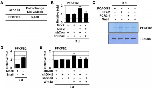
Fig. 1. Wnt induces PFKFB2 expression by activating Dlx-2 and Snail. (A) MCF-7 cells were transfected with Dlx-2. Changes in cellular gene transcription were detected by microarray analysis. Fold increase in expression was compared with Mock.
(B) MCF-7 cells co-transfected with Dlx-2 and shSnail were analyzed by real-time qRT-PCR for PFKFB2 expression. **p<0.01 versus Mock, #p<0.05 versus shCon. (C) MCF-7 cells transfected with Dlx-2 or Snail were analyzed by immunoblotting using the indicated antibodies. (D) MCF-7 cells transfected with Snail were analyzed by real-time qRT-PCR for PFKFB2 expression. **p<0.01 versus Mock. (E) MCF-7 cells were transfected with shDlx-2 or shSnail and then treated with Wnt3a CM were analyzed by real-time qRT-PCR for PFKFB2 expression. **p<0.01 versus untreated, #p<0.05; ##p<0.01 versus shCon.
All error bars represent SE.
은 Student’s t test를 통하여 검증하였다. 모든 결과는 mean
± SE로 나타내었으며, p<0.05는 통계학적으로 유의하다고 간 주하였다.
결과 및 고찰
Wnt-Dlx-2/Snail 신호전달계에 의한 glycolytic switch target 유전자 PFKFB2 발굴
Wnt 신호전달계는 EMT 및 glycolytic switch를 유도하는 것으로 알려져 있다[1, 9, 26-28, 49, 56]. EMT와 glycolytic switch 모두 암세포 성장 및 invasion에 중요한 과정이므로, 본 연구에서는 glycolytic switch가 Wnt에 의한 EMT를 조절 할 가능성을 조사하였다. Dlx-2가 Snail 발현을 증가시켜 Wnt 에 의한 EMT 및 glycolytic switch를 조절하므로[27], glyco- lytic switch 관련 Dlx-2 target gene(s)을 발굴하기 위해 cDNA microarray (Agilent Human Genome 8x60K array, Agilent technologies, CA)를 이용하여 분석하였다. 분석한 42,400여종 유전자 중 대사와 관련된 많은 효소들 (PFKFB2 (5.430 fold 증가), GLS (2.233), H6PD (2.660), acetyl-CoA carboxylase (2.525), HK3 (2.362), SUCLG2 (2.406), 17β-HSD (3.029), DGKI (4.538), HIF-1α (4.186))의 발현이 Dlx-2에 의해 증가하였다 (microarray dataset GSE61009) [26]. Glycolysis 관련 효소 중
6-phosphofructo-2-kinase/fructose-2,6-biphosphatase 2 (PFKFB2) 의 발현이 Dlx-2에 의해 5배 이상 증가됨을 관찰하였다(Fig.
1A).
대부분의 암세포는 암세포의 생존 및 성장을 위해 glyco- lytic switch를 나타낸다[5-7, 10, 13, 17, 23, 52, 58]. PFKFB2는 fructose-2,6-bisphosphate (F2,6BP)의 합성 및 분해에 관여하 는 enzyme으로 glycolysis pathway에서 중요하게 작용한다.
F2,6BP는 glycolytic pathway에서 rate-limiting enzyme으로 작용하는 6-phosphofructo-1-kinase (PFK-1)을 활성화시켜, glycolysis를 촉진한다[39, 40, 50, 63]. Human lung cancers에 서 PFKFB 유전자의 발현이 증가되어 있고, hypoxia에 의해 PFKFB2, PFKFB3, PFKFB4의 발현이 증가됨이 보고되어 있다 [37]. 또한 PFKFB는 HIF-1α, c-Myc, ras에 의해 활성화되고, p53에 의해 불활성화 되는 것으로 알려져 있다[3, 22, 35, 36, 41].
Wnt-Dlx-2/Snail cascade에 의한 glycolytic switch 조절 기 작을 분석하기 위해 Dlx-2, Snail 및 Wnt에 의한 PFKFB2 발현 변화를 real time qRT-PCR 및 Western blotting을 이용하여 조사하였다. MCF-7 세포에서 Dlx-2와 Snail 및 Wnt에 의해 PFKFB2 발현이 증가됨을 관찰하였다(Fig. 1B-Fig. 1E). 또한 Dlx-2에 의해 증가한 PFKFB2는 Snail shRNA에 의해 억제되 는 것으로 나타났다(Fig. 1B). 뿐만 아니라 Wnt에 의한
A
C B
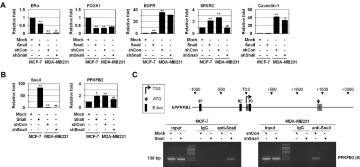
Fig. 2. Snail induces PFKFB2 expression. MCF-7 cells were transfected with Snail. MDA-MB231 cells were transfected with shSnail.
(A, B) The cells were analyzed by real-time qRT-PCR using the indicated primers. *p<0.05; **p<0.01 versus Mock, #p<0.05;
##p<0.01 versus shCon. (C) A schematic diagram of the human PFKFB2 proximal promoter region is shown in upper panel.
ChIP assays were performed using IgG or anti-Snail antibodies and ChIP-enriched DNA was analyzed by PCR using primers complementary to the Snail binding region (lower panel). All error bars represent SE.
PFKFB2 증가는 Dlx-2 shRNA 또는 Snail shRNA에 의해 억제 됨을 관찰하였다(Fig. 1E). 이러한 결과를 바탕으로 Wnt-Dlx-2/Snail cascade에 의해 PFKFB2 발현이 조절됨을 알 수 있었다.
PFKFB2가 Wnt에 의한 EMT에 미치는 영향 분석 Wnt 및 Snail에 의한 EMT는 암 발생과 밀접한 연관성을 지니고 있는 악성유방암(TNBC) 발생을 유도하는 것으로 알려 져 있다[11, 19, 20, 47]. 유방암 세포는 발현 유전자에 따라 luminal A, luminal B, HER-2/neu+, basal-like (TNBC) 등 4가 지 subtype으로 분류된다. 임상에서 luminal subtype은 예후 가 좋지만, TNBC는 estrogen, progesterone 및 HER-2 (ERBB2) receptor의 발현 감소를 나타내며, EMT markers 발현 및 CSC- like characteristics을 보이며, aggressive하고 전이 및 재발 위 험이 높은 것으로 알려져 있다. TNBC는 다른 breast cancer subtype 보다 예후가 나쁘고, 환자에 대한 효과적인 targeted therapy가 없는 것으로 보고되어 있다[2, 38, 45, 54, 65].
MCF-7 세포는 ER (estrogen receptor) 및 PR (progesterone receptor)에 positive하고, non-invasive한 luminal A subtype breast cancer cell lines으로 Snail의 발현이 낮다. 반면, MDA- MB231 세포는 TNBC subtype으로 autocrine Wnt signaling을 가지고 있으며 Snail 발현이 높아 invasive한 성질을 가진다 [11, 19, 20, 47]. MCF-7과 MDA-MB231 세포의 특성 및 PFKFB2 의 발현 변화를 분석하기 위해 Snail을 과발현시킨 MCF-7 세 포와 Snail를 knockdown시킨 MDA-MB231 세포를 이용하여
조사하였다. 이를 위해 먼저 luminal 및 basal markers의 발현 을 조사하였다. MCF-7에 Snail을 과발현시킨 세포에서 lumi- nal markers (ER-a and FoxA1)의 발현이 감소되는 반면, triple negative TNBC의 basal markers (EGFR, SPARC, and Caveo- lin-1)의 발현이 증가됨을 관찰하였다(Fig. 2A). 이러한 결과를 바탕으로 Snail은 MCF-7 세포의 invasiveness를 높이고 CSC- like characteristics를 부여하며 luminal phenotype에서 임상 적으로 예후가 나쁜 TNBC로 변화시키고, tumorigenicity를 증 가 시키는 것으로 생각된다. 반면, MDA-MB231 세포는 MCF- 7 세포에 비해 luminal markers (ER-a and FoxA1) 발현이 낮 고 basal markers (EGFR, SPARC, and Caveolin-1)의 발현이 증가됨을 관찰하였다. 또한 MDA-MB231 세포에서 SPARC, Caveolin-1 발현이 Snail shRNA에 의해 감소되는 것으로 나 타났다(Fig. 2A).
또한 MCF-7과 MDA-MB231 세포에서 Snail에 의한 PFKFB2 발현을 조사하였다. 그 결과, MCF-7 세포에 Snail을 과발현시 킨 경우 Snail에 의해 PFKFB2 발현이 증가되는 것으로 나타났 다(Fig. 2B). 반면, MDA-MB231 세포는 MCF-7 세포에 비해 PFKFB2의 basal level이 높고 Snail shRNA에 의해 PFKFB2의 발현이 감소됨을 관찰하였다(Fig. 2B). 이러한 결과를 바탕으 로 Snail이 PFKFB2 발현에 밀접하게 연관되어 있음을 알 수 있었다.
Snail이 직접 PFKFB2 유전자 발현을 조절하는지를 ChIP analysis를 이용하여 분석하였다. PFKFB2의 promoter를 조사 한 결과, PFKFB2 유전자의 transcription start site (TSS)로부
A B
Fig. 3. Inhibition of PFKFB2 prevents Wnt-induced EMT. MCF-7 cells were transfected with shPFKFB2 and then treated with Wnt3a CM. The cells were analyzed by phase-contrast microscopy for cell morphology (A) and by real-time qRT-PCR (B) using the indicated primers. **p<0.01 versus untreated, ##p<0.01 versus shCon. All error bars represent SE.
터 –1,000과 +1,000 사이에 Snail binding site가 3개 존재하였 다(Fig. 2C). PFKFB2 promoter region의 Snail biding site 3개 에 대한 primer는 #1, #2, #3로 명시하였다(Fig. 2C). 이를 대상 으로 ChIP analysis를 수행한 결과, MCF-7에 Snail을 과발현 시킨 세포에서 PFKFB2의 Snail binding site에 Snail이 직접 binding함을 관찰하였다(Fig. 2C). 또한 MDA-MB231 세포에 서 PFKFB2의 Snail binding site에 Snail이 직접 binding하고 Snail shRNA에 의해서 억제됨을 관찰하였다(Fig. 2C). Snail이 직접 PFKFB2의 promoter region에 결합하여 발현을 조절함 을 알 수 있었다.
나아가, Snail이 Wnt에 의한 EMT를 조절하는 것으로 알려 져 있으므로[9, 43, 49, 56, 60], Wnt에 의한 EMT에 미치는 PFKFB2의 영향을 조사하였다. 그 결과, Wnt에 의해 inter- cellular adhesion 감소, spindle-like cell shape 및 pseudopo- dia 형성 등의 morphological changes를 통하여 전형적인 mesenchymal cells로의 전환이 유도됨을 관찰하였고(Fig. 3A), PFKFB2 shRNA가 Wnt에 의한 EMT 유도(Fig. 3A) 및 E-cad- herin 발현 감소(Fig. 3B)를 상당히 억제하는 것으로 나타났다.
이러한 결과를 바탕으로 PFKFB2가 Wnt에 의한 EMT에 관여 함을 알 수 있었다. 이는 glycolysis pathway가 Wnt에 의한 EMT에 밀접하게 연관되어져 있음을 제시한다.
또한 Panc1 세포에서 TGF-β에 의해 PFKFB3 발현이 증가 되고, PFKFB3 발현을 억제하면 TGF-β에 의한 Snail 발현 증가 및 invasion이 억제됨이 보고되어 있다[62]. 뿐만 아니라 colon cancer에서 glycolysis에 관여하는 enzyme인 GAPDH를 knockdown하면 Snail 발현 감소를 통해 EMT 및 cell migra- tion을 억제하는 것으로 나타났다[31]. 이를 통해 glycolysis가 EMT에 관여함을 알 수 있었다.
PFKFB2 불활성화에 의한 xenograft mouse의 암 성장 및 전이 조절
PFKFB2가 xenograft mouse model에서 tumor growth 및
metastasis에 미치는 영향을 조사하였다. 먼저, xenograft mouse에서 PFKFB2가 tumor growth에 미치는 영향을 조사하 였다. PFKFB2를 knockdown 시킨 MDA-MB231 stable cell lines (6×10⁶ cells)을 nude mouse에 subcutaneous injection하 여 tumor 형성을 관찰하였다. 3일 간격으로 tumor size를 측정 한 결과, PFKFB2 shRNA에 의해 tumor growth가 감소함을 관찰하였다. 뿐만 아니라 PFKFB2 shRNA에 의해 tumor vol- ume 및 weight가 감소되는 것으로 나타났다(Fig. 4A-C).
또한 metastasis에 미치는 영향을 조사하기 위해 PFKFB2를 knockdown 시킨 MDA-MB231 stable cell line (1×10⁶ cells)을 tail vein injection 하였다. Injection 137일 이후, Con shRNA group에서 mouse 움직임이 둔해지기 시작하여 몸무게가 20%
이상 감소함을 관찰하였으며, lung metastasis가 일어남을 metastatic nodules을 통해 관찰하였다. 반면, PFKFB2 shRNA group은 metastatic nodules이 감소됨을 관찰하였다. 또한 PFKFB2 shRNA에 의해 metastatic lesion이 감소됨을 H&E staining를 통해 관찰하였다(Fig. 4D-Fig. 4F). 동물 모델 실험 을 통해 PFKFB2가 xenograft tumor 성장 및 invasion, meta- stasis에 관여함을 알 수 있었다.
Human 암조직에서 PFKFB2의 발현 분석
Cancer와의 연관성을 조사하기 위해 실제 human 암조직을 대상으로 PFKFB2의 발현을 조사하였다. 그 결과 breast, colon 및 ovarian cancer tissue의 다양한 stage에서 PFKFB2 발현이 증가됨을 관찰하였다(Fig. 5).
뿐만 아니라, ovarian cancer의 metastatic lesion에서 PFKFB2의 발현이 높음이 보고되어 있다. Metastasis를 동반한 ovarian cancer 환자의 경우, cancer-associated fibroblasts (CAF)에서 chemokine CXCL14의 발현이 높으며, CXCL14가 높게 발현된 부위에서 long noncoding RNA LINC00092 발현 도 높게 나타나는데, LINC00092가 PFKFB2와 결합하여, PFKFB2의 발현 또한 높게 관찰되는 것으로 밝혀져 있다[67].
A B C
D E F
Fig. 4. shPFKFB2 inhibits tumor growth and metastasis. MDA-MB231 cells stably transfected with shCon or shPFKFB2 were injected subcutaneously into the dorsal flank of nude mice (n=3). Tumor growth curves (A) and photographs of representative mice, tumors (B) and tumor weight (C) are shown. *p<0.05; **p<0.01 versus shCon. 1×10⁶ MDA-MB231 cells stably transfected with shCon or shPFKFB2 were injected into the tail vein of nude mice (n=3). Photographs of representative lungs (D) and H&E staining of lung sections are shown (E). The number of metastatic nodules (F). *p<0.05 versus shCon. All error bars represent SE. All scale bars represent 100 μm.
Fig. 5. The expression of PFKFB2 in human tumors. Real-time qRT-PCR data showing expression of PFKFB2 mRNA in normal (N) and tumor (T) tissues from the indicated tumor types and histological stages (TNM classification) of breast, colon and ovarian cancer. **p<0.01 versus matched normal (N) tissues. All error bars represent SE.
이러한 결과들은 암성장 및 암전이에 있어 PFKFB2가 중요하 게 작용함을 제시한다.
암세포는 glycolytic switch를 포함한 oncogenic metabo- lism의 활성화를 통해 암세포의 성질을 획득하여 tumor mi- croenvironment에서 암세포의 생존 및 증식을 조절하는 것으 로 알려져 있다[5-7, 10, 13, 17, 23, 52, 58]. 본 연구에서는 Wnt 신호전달계에 의한 glycolytic switch가 암세포의 성장 및 증식 에 관여할 뿐만 아니라 EMT 및 metastasis, tumor progression 에도 관여함을 알 수 있었다.
정상세포는 Wnt/TGF-β/EGF 등의 자극에 의해 oncogene 활성화 및 tumor suppressor 불활성화를 통해 암세포 특성을 획득하는 것으로 알려져 있다[30]. 본 연구진은 oncogene 활성 화/tumor suppressor 불활성화를 통해 가장 먼저 변화하는 것이 metabolism이라고 생각하며(oncogenic metabolism), oncogenic metabolism은 tumor growth 및 proliferation 뿐만 아니라, cancer의 다른 특징을 나타내는데 기본 바탕(basis)이 될 것으로 생각하고 있다. 즉 Wnt-Dlx-2/Snail cascade는 PFKFB2를 통해 oncogenic metabolism을 일으킴으로써 EMT 를 유도하고 tumor metastasis를 촉진할 것으로 보인다. 이러 한 결과를 바탕으로 PFKFB2가 metastasis 및 tumor pro- gression을 예방하기 위한 새로운 항암 치료법 개발의 중요한 target이 될 것임을 시사한다.
감사의 글
이 논문은 부산대학교 기본연구지원사업(2년)에 의하여 연 구되었음.
References
1. Anastas, J. N. and Moon, R. T. 2013. WNT signalling path- ways as therapeutic targets in cancer. Nat. Rev. Cancer 13, 11-26.
2. Barnard, M. E., Boeke, C. E. and Tamimi, R. M. 2015.
Established breast cancer risk factors and risk of intrinsic tumor subtypes. Biochim. Biophys. Acta 1856, 73-85.
3. Bensaad, K., Tsuruta, A., Selak, M. A., Vidal, M. N., Nakano, K., Bartrons, R., Gottlieb, E. and Vousden, K. H. 2006.
TIGAR, a p53-inducible regulator of glycolysis and apopto- sis. Cell 126, 107-120.
4. Bhatt, A. N., Chauhan, A., Khanna, S., Rai, Y., Singh, S., Soni, R., Kalra, N. and Dwarakanath, B. S. 2015. Transient elevation of glycolysis confers radio-resistance by facilitat- ing DNA repair in cells. BMC Cancer 15, 335.
5. Boroughs, L. K. and DeBerardinis, R. J. 2015. Metabolic pathways promoting cancer cell survival and growth. Nat.
Cell Biol. 17, 351-359.
6. Cairns, R. A., Harris, I. S. and Mak, T. W. 2011. Regulation of cancer cell metabolism. Nat. Rev. Cancer 11, 85-95.
7. Dang, C. V. 2012. Links between metabolism and cancer.
Genes Dev. 26, 877-890.
8. Dave, B., Mittal, V., Tan, N. M. and Chang, J. C. 2012.
Epithelial-mesenchymal transition, cancer stem cells and treatment resistance. Breast Cancer Res. 14, 202.
9. De Craene, B. and Berx, G. 2013. Regulatory networks defin- ing EMT during cancer initiation and progression. Nat. Rev.
Cancer 13, 97-110.
10. DeBerardinis, R. J., Lum, J. J., Hatzivassiliou, G. and Thompson, C. B. 2008. The biology of cancer: metabolic re- programming fuels cell growth and proliferation. Cell Metab.
7, 11-20.
11. Dhasarathy, A., Phadke, D., Mav, D., Shah, R. R. and Wade, P. A. 2011. The transcription factors Snail and Slug activate the transforming growth factor-beta signaling pathway in breast cancer. PLoS ONE 6, e26514.
12. Dong, C., Yuan, T., Wu, Y., Wang, Y., Fan, T. W., Miriyala, S., Lin, Y., Yao, J., Shi, J., Kang, T., Lorkiewicz, P., St Clair, D., Hung, M. C., Evers, B. M. and Zhou, B. P. 2013. Loss of FBP1 by Snail-mediated repression provides metabolic advantages in basal-like breast cancer. Cancer Cell 23, 316- 331.
13. Finley, L. W., Zhang, J., Ye, J., Ward, P. S. and Thompson, C. B. 2013. SnapShot: cancer metabolism pathways. Cell Metab. 17, 466-466 e462.
14. Hanahan, D. and Weinberg, R. A. 2000. The hallmarks of cancer. Cell 100, 57-70.
15. Hanahan, D. and Weinberg, R. A. 2011. Hallmarks of cancer:
the next generation. Cell 144, 646-674.
16. Hirschey, M. D., DeBerardinis, R. J., Diehl, A. M., Drew, J. E., Frezza, C., Green, M. F., Jones, L. W., Ko, Y. H., Le, A., Lea, M. A., Locasale, J. W., Longo, V. D., Lyssiotis, C.
A., McDonnell, E., Mehrmohamadi, M., Michelotti, G., Muralidhar, V., Murphy, M. P., Pedersen, P. L., Poore, B., Raffaghello, L., Rathmell, J. C., Sivanand, S., Vander Heiden, M. G., Wellen, K. E. and Target Validation, T. 2015. Dysre- gulated metabolism contributes to oncogenesis. Semin.
Cancer Biol. 35 Suppl, S129-150.
17. Hsu, P. P. and Sabatini, D. M. 2008. Cancer cell metabolism:
Warburg and beyond. Cell 134, 703-707.
18. Hua, G., Liu, Y., Li, X., Xu, P. and Luo, Y. 2014. Targeting glucose metabolism in chondrosarcoma cells enhances the sensitivity to doxorubicin through the inhibition of lactate dehydrogenase-A. Oncol. Rep. 31, 2727-2734.
19. Iida, J., Dorchak, J., Lehman, J. R., Clancy, R., Luo, C., Chen, Y., Somiari, S., Ellsworth, R. E., Hu, H., Mural, R. J. and Shriver, C. D. 2012. FH535 inhibited migration and growth of breast cancer cells. PLoS ONE 7, e44418.
20. King, T. D., Suto, M. J. and Li, Y. 2012. The Wnt/beta-cat- enin signaling pathway: a potential therapeutic target in the treatment of triple negative breast cancer. J. Cell. Biochem.
113, 13-18.
21. Klaus, A. and Birchmeier, W. 2008. Wnt signalling and its impact on development and cancer. Nat. Rev. Cancer 8, 387- 398.
22. Kole, H. K., Resnick, R. J., Van Doren, M. and Racker, E.
1991. Regulation of 6-phosphofructo-1-kinase activity in
ras-transformed rat-1 fibroblasts. Arch. Biochem. Biophys. 286, 586-590.
23. Koppenol, W. H., Bounds, P. L. and Dang, C. V. 2011. Otto Warburg's contributions to current concepts of cancer metabolism. Nat. Rev. Cancer 11, 325-337.
24. Kwon, T., Youn, H., Son, B., Kim, D., Seong, K. M., Park, S., Kim, W. and Youn, B. 2016. DANGER is involved in high glucose-induced radioresistance through inhibiting DAPK-mediated anoikis in non-small cell lung cancer.
Oncotarget 7, 7193-7206.
25. Lamouille, S., Xu, J. and Derynck, R. 2014. Molecular mecha- nisms of epithelial-mesenchymal transition. Nat. Rev. Mol.
Cell Biol. 15, 178-196.
26. Lee, S. Y., Jeon, H. M., Ju, M. K., Jeong, E. K., Kim, C. H., Park, H. G., Han, S. I. and Kang, H. S. 2016. Dlx-2 and gluta- minase upregulate epithelial-mesenchymal transition and glycolytic switch. Oncotarget 7, 7925-7939.
27. Lee, S. Y., Jeon, H. M., Ju, M. K., Jeong, E. K., Kim, C. H., Yoo, M. A., Park, H. G., Han, S. I. and Kang, H. S. 2015.
Dlx-2 is implicated in TGF-beta- and Wnt-induced epi- thelial-mesenchymal, glycolytic switch, and mitochondrial repression by Snail activation. Int. J. Oncol. 46, 1768-1780.
28. Lee, S. Y., Jeon, H. M., Ju, M. K., Kim, C. H., Yoon, G., Han, S. I., Park, H. G. and Kang, H. S. 2012. Wnt/Snail signaling regulates cytochrome C oxidase and glucose metabolism.
Cancer Res. 72, 3607-3617.
29. Lee, S. Y., Jeon, H. M., Kim, C. H., Ju, M. K., Bae, H. S., Park, H. G., Lim, S. C., Han, S. I. and Kang, H. S. 2011.
Homeobox gene Dlx-2 is implicated in metabolic stress-in- duced necrosis. Mol. Cancer 10, 113.
30. Lee, S. Y., Jeong, E. K., Ju, M. K., Jeon, H. M., Kim, M.
Y., Kim, C. H., Park, H. G., Han, S. I. and Kang, H. S. 2017.
Induction of metastasis, cancer stem cell phenotype, and on- cogenic metabolism in cancer cells by ionizing radiation.
Mol. Cancer 16, 10.
31. Liu, K., Tang, Z., Huang, A., Chen, P., Liu, P., Yang, J., Lu, W., Liao, J., Sun, Y., Wen, S., Hu, Y. and Huang, P. 2017.
Glyceraldehyde-3-phosphate dehydrogenase promotes can- cer growth and metastasis through upregulation of SNAIL expression. Int. J. Oncol. 50, 252-262.
32. Mani, S. A., Guo, W., Liao, M. J., Eaton, E. N., Ayyanan, A., Zhou, A. Y., Brooks, M., Reinhard, F., Zhang, C. C., Shipitsin, M., Campbell, L. L., Polyak, K., Brisken, C., Yang, J. and Weinberg, R. A. 2008. The epithelial-mesenchymal transition generates cells with properties of stem cells. Cell 133, 704-715.
33. Merlo, G. R., Zerega, B., Paleari, L., Trombino, S., Mantero, S. and Levi, G. 2000. Multiple functions of Dlx genes. Int.
J. Dev. Biol. 44, 619-626.
34. Mims, J., Bansal, N., Bharadwaj, M. S., Chen, X., Molina, A. J., Tsang, A. W. and Furdui, C. M. 2015. Energy metabo- lism in a matched model of radiation resistance for head and neck squamous cell cancer. Radiat. Res. 183, 291-304.
35. Minchenko, A., Leshchinsky, I., Opentanova, I., Sang, N., Srinivas, V., Armstead, V. and Caro, J. 2002. Hypoxia-in- ducible factor-1-mediated expression of the 6-phospho-
fructo-2-kinase/fructose-2,6-bisphosphatase-3 (PFKFB3) gene.
Its possible role in the Warburg effect. J. Biol. Chem. 277, 6183-6187.
36. Minchenko, O., Opentanova, I. and Caro, J. 2003. Hypoxic regulation of the 6-phosphofructo-2-kinase/fructose-2,6-bi- sphosphatase gene family (PFKFB-1-4) expression in vivo.
FEBS Lett. 554, 264-270.
37. Minchenko, O. H., Ogura, T., Opentanova, I. L., Minchenko, D. O., Ochiai, A., Caro, J., Komisarenko, S. V. and Esumi, H. 2005. 6-Phosphofructo-2-kinase/fructose-2,6-bisphospha- tase gene family overexpression in human lung tumor. Ukr.
Biokhim. Zh. (1999) 77, 46-50.
38. Neve, R. M., Chin, K., Fridlyand, J., Yeh, J., Baehner, F. L., Fevr, T., Clark, L., Bayani, N., Coppe, J. P., Tong, F., Speed, T., Spellman, P. T., DeVries, S., Lapuk, A., Wang, N. J., Kuo, W. L., Stilwell, J. L., Pinkel, D., Albertson, D. G., Waldman, F. M., McCormick, F., Dickson, R. B., Johnson, M. D., Lippman, M., Ethier, S., Gazdar, A. and Gray, J. W. 2006.
A collection of breast cancer cell lines for the study of func- tionally distinct cancer subtypes. Cancer Cell 10, 515-527.
39. Okar, D. A., Manzano, A., Navarro-Sabate, A., Riera, L., Bartrons, R. and Lange, A. J. 2001. PFK-2/FBPase-2: maker and breaker of the essential biofactor fructose-2,6-bi- sphosphate. Trends Biochem. Sci. 26, 30-35.
40. Okar, D. A., Wu, C. and Lange, A. J. 2004. Regulation of the regulatory enzyme, 6-phosphofructo-2-kinase/fructose- 2,6-bisphosphatase. Adv. Enzyme Regul. 44, 123-154.
41. Osthus, R. C., Shim, H., Kim, S., Li, Q., Reddy, R., Mukherjee, M., Xu, Y., Wonsey, D., Lee, L. A. and Dang, C. V. 2000.
Deregulation of glucose transporter 1 and glycolytic gene expression by c-Myc. J. Biol. Chem. 275, 21797-21800.
42. Panganiban, G. and Rubenstein, J. L. 2002. Developmental functions of the Distal-less/Dlx homeobox genes. Develop- ment 129, 4371-4386.
43. Peinado, H., Olmeda, D. and Cano, A. 2007. Snail, Zeb and bHLH factors in tumour progression: an alliance against the epithelial phenotype? Nat. Rev. Cancer 7, 415-428.
44. Peng, X., Gong, F., Chen, Y., Jiang, Y., Liu, J., Yu, M., Zhang, S., Wang, M., Xiao, G. and Liao, H. 2014. Autophagy pro- motes paclitaxel resistance of cervical cancer cells: involve- ment of Warburg effect activated hypoxia-induced factor 1-alpha-mediated signaling. Cell Death Dis. 5, e1367.
45. Perou, C. M., Sorlie, T., Eisen, M. B., van de Rijn, M., Jeffrey, S. S., Rees, C. A., Pollack, J. R., Ross, D. T., Johnsen, H., Akslen, L. A., Fluge, O., Pergamenschikov, A., Williams, C., Zhu, S. X., Lonning, P. E., Borresen-Dale, A. L., Brown, P.
O. and Botstein, D. 2000. Molecular portraits of human breast tumours. Nature 406, 747-752.
46. Pickup, M., Novitskiy, S. and Moses, H. L. 2013. The roles of TGFbeta in the tumour microenvironment. Nat. Rev.
Cancer 13, 788-799.
47. Pohl, S. G., Brook, N., Agostino, M., Arfuso, F., Kumar, A.
P. and Dharmarajan, A. 2017. Wnt signaling in triple-neg- ative breast cancer. Oncogenesis 6, e310.
48. Polyak, K. and Weinberg, R. A. 2009. Transitions between epithelial and mesenchymal states: acquisition of malignant
and stem cell traits. Nat. Rev. Cancer 9, 265-273.
49. Puisieux, A., Brabletz, T. and Caramel, J. 2014. Oncogenic roles of EMT-inducing transcription factors. Nat. Cell Biol.
16, 488-494.
50. Ros, S. and Schulze, A. 2013. Balancing glycolytic flux: the role of 6-phosphofructo-2-kinase/fructose 2,6-bisphosphatases in cancer metabolism. Cancer Metab. 1, 8.
51. Sattler, U. G., Meyer, S. S., Quennet, V., Hoerner, C., Knoerzer, H., Fabian, C., Yaromina, A., Zips, D., Walenta, S., Baumann, M. and Mueller-Klieser, W. 2010. Glycolytic metabolism and tumour response to fractionated irradiation.
Radiother. Oncol. 94, 102-109.
52. Schulze, A. and Harris, A. L. 2012. How cancer metabolism is tuned for proliferation and vulnerable to disruption.
Nature 491, 364-373.
53. Singh, A. and Settleman, J. 2010. EMT, cancer stem cells and drug resistance: an emerging axis of evil in the war on cancer. Oncogene 29, 4741-4751.
54. Sorlie, T., Perou, C. M., Tibshirani, R., Aas, T., Geisler, S., Johnsen, H., Hastie, T., Eisen, M. B., van de Rijn, M., Jeffrey, S. S., Thorsen, T., Quist, H., Matese, J. C., Brown, P. O., Botstein, D., Lonning, P. E. and Borresen-Dale, A. L. 2001.
Gene expression patterns of breast carcinomas distinguish tumor subclasses with clinical implications. Proc. Natl. Acad.
Sci. USA 98, 10869-10874.
55. Tang, P., Huang, H., Chang, J., Zhao, G. F., Lu, M. L. and Wang, Y. 2013. Increased expression of DLX2 correlates with advanced stage of gastric adenocarcinoma. World J. Gas- troenterol. 19, 2697-2703.
56. Thiery, J. P. and Sleeman, J. P. 2006. Complex networks or- chestrate epithelial-mesenchymal transitions. Nat. Rev. Mol.
Cell Biol. 7, 131-142.
57. Tsai, J. H. and Yang, J. 2013. Epithelial-mesenchymal plasti- city in carcinoma metastasis. Genes Dev. 27, 2192-2206.
58. Vander Heiden, M. G., Cantley, L. C. and Thompson, C.
B. 2009. Understanding the Warburg effect: the metabolic requirements of cell proliferation. Science 324, 1029-1033.
59. Wang, S. S., Jiang, J., Liang, X. H. and Tang, Y. L. 2015.
Links between cancer stem cells and epithelial-mesenchymal transition. Onco Targets Ther. 8, 2973-2980.
60. Wang, Y., Shi, J., Chai, K., Ying, X. and Zhou, B. P. 2013.
The role of snail in EMT and tumorigenesis. Curr. Cancer Drug Targets 13, 963-972.
61. Warburg, O. 1956. On respiratory impairment in cancer cells. Science 124, 269-270.
62. Yalcin, A., Solakoglu, T. H., Ozcan, S. C., Guzel, S., Peker, S., Celikler, S., Balaban, B. D., Sevinc, E., Gurpinar, Y. and Chesney, J. A. 2017. 6-phosphofructo-2-kinase/fructose 2,6- bisphosphatase-3 is required for transforming growth factor beta1-enhanced invasion of Panc1 cells in vitro. Biochem.
Biophys. Res. Commun. 484, 687-693.
63. Yalcin, A., Telang, S., Clem, B. and Chesney, J. 2009. Regula- tion of glucose metabolism by 6-phosphofructo-2-kinase/
fructose-2,6-bisphosphatases in cancer. Exp. Mol. Pathol. 86, 174-179.
64. Yan, Z. H., Bao, Z. S., Yan, W., Liu, Y. W., Zhang, C. B., Wang, H. J., Feng, Y., Wang, Y. Z., Zhang, W., You, G., Zhang, Q. G. and Jiang, T. 2013. Upregulation of DLX2 con- fers a poor prognosis in glioblastoma patients by inducing a proliferative phenotype. Curr. Mol. Med. 13, 438-445.
65. Yao, H., He, G., Yan, S., Chen, C., Song, L., Rosol, T. J. and Deng, X. 2016. Triple-negative breast cancer: is there a treat- ment on the horizon? Oncotarget 8, 1913-1924.
66. Yilmaz, M., Maass, D., Tiwari, N., Waldmeier, L., Schmidt, P., Lehembre, F. and Christofori, G. 2011. Transcription fac- tor Dlx2 protects from TGFbeta-induced cell-cycle arrest and apoptosis. EMBO J. 30, 4489-4499.
67. Zhao, L., Ji, G., Le, X., Wang, C., Xu, L., Feng, M., Zhang, Y., Yang, H., Xuan, Y., Yang, Y., Lei, L., Yang, Q., Lau, W.
B., Lau, B., Chen, Y., Deng, X., Yao, S., Yi, T., Zhao, X., Wei, Y. and Zhou, S. 2017. Long noncoding RNA LINC00092 acts in cancer-associated fibroblasts to drive glycolysis and pro- gression of ovarian cancer. Cancer Res. 77, 1369-1382.
68. Zhao, Y., Butler, E. B. and Tan, M. 2013. Targeting cellular metabolism to improve cancer therapeutics. Cell Death Dis.
4, e532.
69. Zhou, M., Zhao, Y., Ding, Y., Liu, H., Liu, Z., Fodstad, O., Riker, A. I., Kamarajugadda, S., Lu, J., Owen, L. B., Ledoux, S. P. and Tan, M. 2010. Warburg effect in chemosensitivity:
targeting lactate dehydrogenase-A re-sensitizes taxol-re- sistant cancer cells to taxol. Mol. Cancer 9, 33.
초록:Wnt에 의한 epithelial-to-mesenchymal transition에서 PFKFB2의 역할
이수연1․주민경1․전현민1․김초희1․박혜경2․강호성1*
(1부산대학교 자연과학대학 분자생물학과, 2한국나노바이오테크놀러지센터)
암세포는 정상세포와는 다른 metabolism 특히 glycolytic switch를 나타낸다. Glycolytic switch는 암세포가 정 상세포와 달리 산소가 충분한 상태에서도 미토콘드리아에 의존하지 않고 glycolysis를 통해 대부분의 ATP 에너지 를 생성하는 현상이다. 또한 암세포는 invasion 및 metastasis 능력을 획득하기 위해 epithelial-mesenchymal tran- sition (EMT)를 나타낸다. EMT와 glycolytic switch는 암세포의 생존 및 증식에 관여하는 중요한 현상이지만, 이들 상호작용 및 그 기작에 대한 연구는 아직 밝혀져 있지 않다. Snail은 EMT를 유도하는 주요한 전사인자이다. 본 연구진은 이전 연구에서 Snail이 발생 및 암성장에 관여하는 전사인자인 Dlx-2에 의해 조절됨을 밝혔다. 또한 Wnt 가 Dlx-2/Snail cascade을 통하여 EMT 및 glycolytic switch을 유도함을 밝혔다. 본 연구에서는 glycolytic switch 가 Wnt에 의한 EMT에 미치는 영향을 규명하고자 하였다. Dlx-2/Snail의 glycolytic switch target 유전자로 phos- phofructokinase-2/fructose-2,6-bisphosphatase 2 (PFKFB2)를 발굴하였다. PFKFB2는 fructose-2,6-bisphosphate (F2,6BP)의 합성 및 분해에 관여하는 효소로서 glycolysis에서 중요하게 작용한다. Wnt에 의해 PFKFB2 발현이 Dlx-2/Snail 의존적으로 증가함을 관찰하였다. 또한 PFKFB2를 knockdown한 결과 Wnt에 의한 EMT가 억제되므 로 glycolytic switch가 Wnt에 의한 EMT에 관여할 가능성이 높을 것으로 보인다. 뿐만 아니라 PFKFB2 shRNA가 xenograft mouse model에서 tumor 성장 및 metastasis를 억제하는 것으로 나타났다. 또한 Human 암조직에서 정상조직에 비해 PFKFB2의 발현이 높음을 관찰하였다. 따라서 PFKFB2가 Wnt-Dlx-2/Snail-induced EMT 및 metastasis에서 중요한 역할을 할 것으로 예상된다.