증례 보고
평소에 별 증상이 없이 잘 지내던 27세 된 남자가 오토바이 를 타고 가다가 승용차에 부딪혀서 다발성 사지 골절과 복부 둔상을 입었다. 수상 직후에 시행한 복부 전산화단층 촬영에서 혈복강과 우 하복부의 장간막에 혈종과 희미한 증가음영을 보 여 주었다 (Fig. 1A). 간, 췌장, 비장, 그리고 신장은 특이 소 견이 없었다. 복강 내에 유리 공기음영이나, 정맥으로 주입한 조영제의 유출은 없었다. 보존적인 치료로 증상이 호전되고 1 주일 후에 시행한 초음파 검사에서 복강내의 혈복강이 사라진 것이 확인이 되었다. 입원치료 중 수상 5주 후 우 하복부 통 증, 구토, 그리고 복부 팽창을 호소 했다. 추적 복부 초음파 검 사에서 10 cm 길이의 장벽 비후 소견이 보이고, 그 주위의 장 간막은 에코가 광범위하게 증가되었다 (Fig. 1B). 복부 전산화 단층 촬영에서는 우 하복부의 회장에 분절성 장벽 비후의 소 견이 보이고 (Fig. 1C), 협착부위 상부의 소장은 확장이 되었 다. 소장 조영술에서 회장에 10 cm 길이의 협착이 있고 궤양 이 동반되어 있으며 협착부위의 상부소장은 확장이 되어 있었 다 (Figs. 1D, E). 수술은 협착된 부위를 절제하고 유착된 부 위를 박리하였다. 수술 소견에서 회장이 협착되어 있었고, 그 주위의 장간막은 단단하게 섬유화된 지방괴사의 소견이 있고 유착이 심했으며, 장간막근(mesenteric root)은 짧아져 있었 다. 병리조직 검사에서는 궤양이 있고 그 하부로 점막근층과 점막하조직까지 육아조직이 침범하고 있었다 (Fig. 1F). 점막 하조직에는 섬유화를 보였지만 장벽에 육아종의 소견은 없었 다. 환자는 퇴원하여 4년 6개월이 지난 현재까지 특별한 문제
가 없이 잘 지내고 있다.
고 찰
복부의 외상은 장 허혈의 원인 중의 하나로 알려져 왔다 (1).
외상 후에 생긴 허혈성 장 협착은 장간막에 가해진 열상이나 타박상으로 올 수 있고 (2), 안전 벨트에 의한 장의 손상 후에 도 올 수 있는 것으로 알려져 왔다 (3). 외상 후에 생긴 허혈 성 장 협착은 장벽의 점막층의 궤양이나 점막하층의 염증과 섬 유화를 동반한 부분적인 허혈을 가져오거나, 전 층의 섬유화를 동반한 허혈을 가져 올 수 있다. 이런 흉터화는 협착형성과 장 폐색을 가져온다 (2).
장간막의 손상과 일치하는 전산화 단층촬영소견은 장간막의 지방 내에 줄무늬가 있는 연부조직 침윤, 장간막 주름사이의 액체 저류, 그리고 국소적인 장간막 내의 혈종이다. 그리고 고 형의 기관의 손상이 없는 다량의 복강내의 액체저류는 장간막 이나 장손상의 중요한 증후일수도 있다 (4). 전산화 단층촬영 에서 장간막에 희미하게 증가된 음영 또한 장간막 타박상을 의 미한다. 이러한 장간막 손상의 소견이 보일 때는 후에 허혈성 장 협착이 올 가능성을 고려 해야 하겠다.
문헌의 보고에 의하면 외상 후의 장 협착은 증상이 지연되 어서 나타나는 특징을 가진다. 그러나 수상으로부터 증상의 시 작까지의 기간은 1주에서 18주까지로 다양하다 (3, 5). 방사 선학적 소견은 관 모양의 협착이 긴 길이의 분절을 포함하며 환상의 궤양을 가진다. 협착은 직접적인, 기계적인 압박에 의 해서 생기는 것 같다. 협착의 길이도 1 cm에서 28 cm로 다양 하며 대개 한 부위에 국한되나 다발성인 경우도 있다 (3). 주 로 소장의 협착을 가져오나 대장의 협착을 가져올 수 있다 (5).
초음파 검사에서의 소장 벽의 비후와 층의 소실은 특이한 소 견은 아니지만 장의 허혈에서도 볼 수 있는 소견이다 (6). 장 간막의 균질하게 증가된 에코는 장간막의 손상으로 인한 출혈 대한방사선의학회지 2002;47:213-215
─ 213 ─
외상 후 생긴 소장 협착: 1예 보고1,2
권 중 혁1,2・김 갑 철3
외상 후에 발생하는 장 협착은 복부 둔상(blunt abdominal trauma)의 드문 합병증으로 장간 막이나 장벽의 손상으로 인해 상당기간 후 국소적인 허혈성 협착을 가져오는 질환이다. 저자들 은 복부 둔상을 받은 후 5주 후부터 불완전한 소장 폐색증상을 보인 젊은 환자가 방사선학적 검사, 수술소견, 및 병리소견상에서 외상 후 발생한 허혈성 소장 협착으로 진단된 1예를 경험 하였기에 그 소견을 보고하고자 한다.
1계명대학교 의과대학 진단방사선과학교실
2동강병원 진단방사선과
3울산보람병원 방사선과
이 논문은 2001년 12월 20일 접수하여 2002년 4월 29일에 채택되었음.
에 의한 지방괴사의 소견이다. 이러한 장간막의 변화는 손상이 장벽에 가해졌다기 보다는 장간막에 가해진 것을 암시한다 (7).
전산화 단층촬영에서는 다양한 길이의 동심성의 두꺼워진 장 벽과 이 부위의 상부에 위치한 장의 확장과 하부에 위치한 장 의 내경의 감소의 소견을 보여준다 (8). 소장 바륨조영술에서 는 다양한 길이의 동심성의 좁아진 장의 내경과 점막층의 궤 양을 보여준다 (2). 그리고 상부에 위치한 장의 확장과 하부에 위치한 장의 허탈을 보여준다. 혈관촬영술에서는 상장간막동맥
의 몇 개의 분지가 막혀 있는 것을 보여줄 수 있고 허혈성 장 벽의 조영을 보여줄 수도 있다 (2).
진단의 기준은 복부 둔상의 명백한 병력, 수상 전에 병이 없 고, 수상 후에 장의 증상의 발발, 방사선학적 검사에서 장 협 착의 확인, 그리고 수술로 절제를 한 부위에서 염증성이나 종 양성의 변화가 없다는 점이다 (5). 방사선학적 검사에서 크론 병, 장결핵, 방사선 장염, 그리고 소장의 암 등과 감별이 필요 할 수도 있다 (5). 특히 크론병 (9)이나 장결핵과 감별하기가 권중혁 외: 외상 후 생긴 소장 협착
─ 214 ─
A B C
D E F
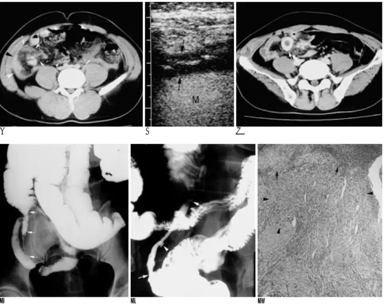
Fig. 1. A 27-year-old man with posttraumatic intestinal stenosis.
A. Postcontrast CT scan performed on the day of the traffic accident shows mesenteric hematoma and mesenteric haziness (ar- rows) and hemoperitoneum (arrowheads) in the right lower abdomen.
B. Transverse US image of the right lower abdomen performed 5 weeks later, shows a thickened small bowel loop (arrows) with loss of wall stratification, surrounded by a mesentery (M) with homogeneously increased echogenicity.
C. Postcontrast CT scan shows a diffuse, uniform, mural thickening of small bowel loop (arrows) and dilatation of small bowel loops proximal to this stenotic segment (not shown).
D. Small bowel follow-through study shows a long stenotic segment (approximately 10 cm in length) within the ileum. An irregular contour (arrows) is demonstrated throughout the stenotic segment. Small bowel loops proximal to the stenotic segment are moder- ately dilated.
E. Close detail of this segment shows a discrete ulcer (arrowhead) within the stenotic segment (arrows).
F. Photomicrograph of resected small bowel shows ulceration of mucosa covered with granulation tissue (arrows), and granulation tissue (arrowheads) extending into the muscularis mucosa and submucosa (H & E stain, ×40).
어렵다. 이 질환에 특별한 방사선학적 소견을 규명하기는 어려 워도 복부 둔상의 병력이 확인될 때 진단의 어려움은 없다. 드 물지라도 두꺼워진 장이 보일 시는 이 질환이 감별진단에 고 려되어야만 한다. 복부의 둔상을 받은 환자가 장간막에 혈종이 나 타박상을 가질 경우, 장의 손상의 소견이 없고 혈복강이 다 량이 아니거나 증가하지 않을 때는 대부분의 경우에 보존적으 로 치료한다. 이런 환자가 몇 주 후에 장폐색의 증후와 증상을 가져오면 외상 후에 생긴 장협착을 고려해야 한다.
참 고 문 헌
1. Rha SE, Ha HK, Lee SH, et al. CT and MR imaging findings of bowel ischemia from various primary causes. RadioGraphics 2000;
20:29-42
2. De Backer AI, De Schepper AMA, Vaneerdeweg W, Pelckmans P.
Intestinal stenosis from mesenteric injury after blunt abdominal trauma. Eur Radiol 1999;9:1429-1431
3. Lynch JM, Albanese CT, Meza MP, Wiener ES. Intestinal stricture
following seat belt injury in children. J Pediatr Surg 1996;31:1354- 1357
4. Breen DJ, Janzen DL, Zwirewich CV, Nagy AG. Blunt bowel and mesenteric injury: diagnostic performance of CT signs. J Comput Assist Tomogr 1997;21:706-712
5. Hirota C, Iida M, Aoyagi K, Matsumoto T, Yao T, Fujishima M.
Posttraumatic intestinal stenosis: clinical and radiographic features in four patients. Radiology 1995;194:813-815
6. Teefey SA, Roarke MC, Brink JA, Middleton WD, et al. Bowel wall thickening: differentiation of inflammation from ischemia with color Doppler and duplex Doppler US. Radiology 1996;198:547-551 7. Loberant N, Szvalb S, Herskovits M, Cohen I, Salamon V.
Posttraumatic intestinal stenosis: radiographic and sonographic ap- pearance. Eur Radiol 1997;7:524-526
8. Tsushima Y, Yamada S, Aoki J, Endo K. Ischaemic ileal stenosis following blunt abdominal trauma and demonstrated by CT. Br J Radiol 2001;74:277-279
9. Fanelli C, Bassotti G, Giansanti M, Bartoli A, Bolli GB.
Posttraumatic ileal stenosis mimicking Crohn’s disease. J Clin Gastroenterol 1995;20:338-340
대한방사선의학회지 2002;47:213-215
─ 215 ─
J Korean Radiol Soc 2002;47:213-215
Address reprint requests to : Jung Hyeok Kwon, M.D., Department of Diagnostic Radiology, Dongsan Medical Center, Keimyung University College of Medicine, 194 Dongsan-dong, Jung-gu, Taegu 700-712, Korea.
Tel. 82-53-250-7767 Fax. 82-53-250-7766 E-mail: [email protected],kr
Posttraumatic Intestinal Stenosis: A Case Report1,2
Jung Hyeok Kwon, M.D.1,2, Gab Chul Kim, M.D.3
1Department of Diagnostic Radiology, Dongsan Medical Center, Keimyung University College of Medicine
2Department of Diagnostic Radiology, Dongkang General Hospital
3Department of Diagnostic Radiology, Ulsan Boram Hospital
Post-traumatic intestinal stenosis (PIS) is an uncommon sequela of blunt abdominal trauma, in which injury to the mesentery or bowel wall leads to focal ischemic stricture of that segment. We present a case of PIS of the ileum diagnosed on the basis of radiological studies and surgical and pathologic findings in a patient with par- tial small bowel obstruction occurring five weeks after blunt abdominal trauma.
Index words :Intestines, injuries Intestines, ischemia
Intestines, stenosis or obstruction Trauma