CASE REPORT
진행성 총담관암에서 고식적 치료법으로써 내시경적 고주파열제거술 후 인공담관 삽입술과 담낭 내 에탄올 치료
이용우1,2, 김현정1,2, 이상엽3, 허준1,2, 정민규1,2
경북대학교 의과대학 내과학교실1, 경북대학교병원 소화기내과2, 경북대학교 의과대학 영상의학교실3
Palliative Measures with Ethanol Gallbladder Ablation and Endobiliary Radiofrequency Ablation Followed by Endoscopic Biliary Stent Placement in an Advanced Case of Common Bile Duct Cancer: A Case Report
Yong-woo Lee1,2, Hyun Jeong Kim1,2, Sang Yub Lee3, Jun Heo1,2 and Min Kyu Jung1,2
Department of Internal Medicine, School of Medicine, Kyungpook National University1; Division of Gastroenterology and Hepatology, Department of Internal Medicine, Kyungpook National University Hospital2; Department of Radiology, School of Medicine, Kyungpook National University3, Daegu, Korea
Endobiliary radiofrequency ablation (RFA) is a procedure performed widely to induce locoregional tumor control by the transfer of thermal energy to the lesion and subsequent tumor necrosis. A 72-year-old male with a prior history of acute calculous cholangitis and perforated cholecystitis was admitted to the Kyungpook National University Hospital complaining of fever and nausea. He had an indwelling percutaneous transhepatic gallbladder drainage (PTGBD) catheter from the previous episode of perforated cholecystitis. An abdominal CT scan showed marked dilation of both the intrahepatic and extrahepatic bile ducts. Common bile duct cancer was confirmed histologically after an endobiliary biopsy. A surgical resection was considered to be the initial treat- ment option. During open surgery, multiple metastatic nodules were present in the small bowel mesentery and anterior abdomi- nal wall. Resection of the tumor was not feasible, so endobiliary RFA was performed prior to biliary stenting. Cholecystectomy was required for the removal of the PTGBD catheter, but the surgical procedure could not be performed due to a cystic ductal invasion of the tumor. Instead, chemical ablation of the gallbladder (GB) with pure ethanol was performed to breakdown the GB mucosa.
Palliative treatment for a biliary obstruction was achieved successfully using these procedures. In addition, a PTGBD catheter was removed successfully without significant side effects. As a result, an improvement in the patient’s quality of life was accomplished. (Korean J Gastroenterol 2020;75:50-55)
Key Words: Gallbladder; Radiofrequency ablation; Cholangiocarcinoma; Cholangiopancreatography, endoscopic retrograde
Received August 4, 2019. Revised August 27, 2019. Accepted October 2, 2019.
CC This is an open access article distributed under the terms of the Creative Commons Attribution Non-Commercial License (http://creativecommons.org/licenses/
by-nc/4.0) which permits unrestricted non-commercial use, distribution, and reproduction in any medium, provided the original work is properly cited.
Copyright © 2020. Korean Society of Gastroenterology.
교신저자: 정민규, 41944, 대구시 중구 동덕로 130, 경북대학교병원 소화기내과
Correspondence to: Min Kyu Jung, Division of Gastroenterology and Hepatology, Department of Internal Medicine, Kyungpook National University Hospital, 130 Dongdeok-ro, Jung-gu, Daegu 41944, Korea. Tel: +82-53-200-5514, Fax: +82-53-426-8773, E-mail: minky1973@hanmail.net, ORCID: https://orcid.org/0000-0001-8749-408X Financial support: None. Conflict of interest: None.
INTRODUCTION
Endobiliary radiofrequency ablation (RFA) is performed wide- ly to induce locoregional tumor control by inducing tumor ne- crosis through the transfer of thermal energy.1 Endobiliary RFA
was performed prior to biliary stenting in a patient with an inoperable common bile duct (CBD) cancer. Cholecystectomy was required to remove the percutaneous transhepatic gall- bladder drainage (PTGBD) catheter, but the surgical procedure could not be performed due to cystic ductal invasion of the
Fig. 1. Abdominal computed tomography scan showed a perforated gallbladder and abscesses at lesser sac and ligament teres, which is suspicious for acute perforated cholecystitis, diffusely thickened common bile duct and intrahepatic duct dilatation with a stricture.
Fig. 2. Endobiliary radiofrequency ablation was performed under seven watts using the 22 mm RFA catheter (ELRAⓇ RF catheter, STARmed, Goyang, Korea) for 2 minutes. The arrows indicate the RFA catheter electrode. The partially covered biliary stent was placed through the narrowed common bile duct after performing the endobiliary radiofrequency ablation.
tumor. Although gallbladder (GB) ablation is not a standard treatment, it could be an alternative option for cholecystectomy such as in inoperable circumstances.2 Pure ethanol was used to breakdown the GB mucosa. This paper reports a case, in which the procedures mentioned above were performed suc- cessfully without significant side effects for 3 months.
CASE REPORT
A 69-year-old male was admitted for 1 week owing to the complaints of right upper quadrant pain and chills. The pa-
tient had hypertension and type 2 diabetes. His initial labo- ratory findings were as follows: white blood cell count, 7,130/mm3; hemoglobin level, 8.6 g/dL; platelet count, 164,000/mm3; erythrocyte sedimentation rate, 43 mm/h;
CRP, 14.25 mg/dL; AST, 91 U/L; ALT, 78 U/L; total bilirubin, 4.14 mg/dL; and direct bilirubin, 3.55 mg/dL. Abdominal ultrasound showed that his GB was distended. Abdominal CT scans revealed high-density nodules in the distal CBD, which indicated a CBD stone with a distended GB and dilated pe- ripheral intrahepatic duct. ERCP, performed on the day of ad- mission, revealed some filling defects and the presence of
A B
C D
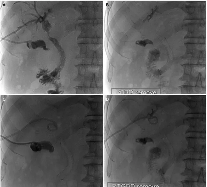
Fig. 3. (A-D) PTBD was removed successfully after endobiliary radiofrequency ablation followed by biliary stent placement. PTGBD was also removed after chemical ablation of the gallbladder using ethanol. PTBD, percutaneous transhepatic biliary drainage; PTGBD, percutaneous transhepatic gallbladder drainage.
several black-pigmented stones. The stones were removed through endoscopic sphincterotomy and balloon sweeping.
Cholecystectomy was not considered at this time owing to a lack of evidence of combined cholecystitis and the presence of a GB stone.
Three years after the last discharge, the patient was admit- ted again because of complaints of right upper quadrant pain and chills. His laboratory test results were as follows: white blood cell count, 13,640/mm3; hemoglobin level, 13.2 g/dL;
platelet count, 246,000/mm3; CRP level, 23.10 mg/dL; AST value, 35 U/L; ALT value, 51 U/L; total bilirubin level, 1.62 mg/dL; direct bilirubin level, 1.28 mg/dL; and CA 19-9,
187.64. The abdominal CT scans showed a perforated GB and abscesses in the lesser sac and ligament teres, which indicated acute perforated cholecystitis and also showed a diffusely thickened CBD and intrahepatic duct dilatation with a stricture (Fig. 1). The ERCP cholangiogram showed that the upstream bile duct was dilated with focal segmental narrow- ing at the proximal CBD. A plastic biliary stent (double pigtail typed C-flex, 7 Fr×10 cm) was placed into the upstream duct and PTGBD catheter. An endobiliary biopsy revealed chronic inflammation. The endoscopic retrograde biliary drainage could be removed after several days of antibiotic treatment, but the PTGBD catheter could not be removed.
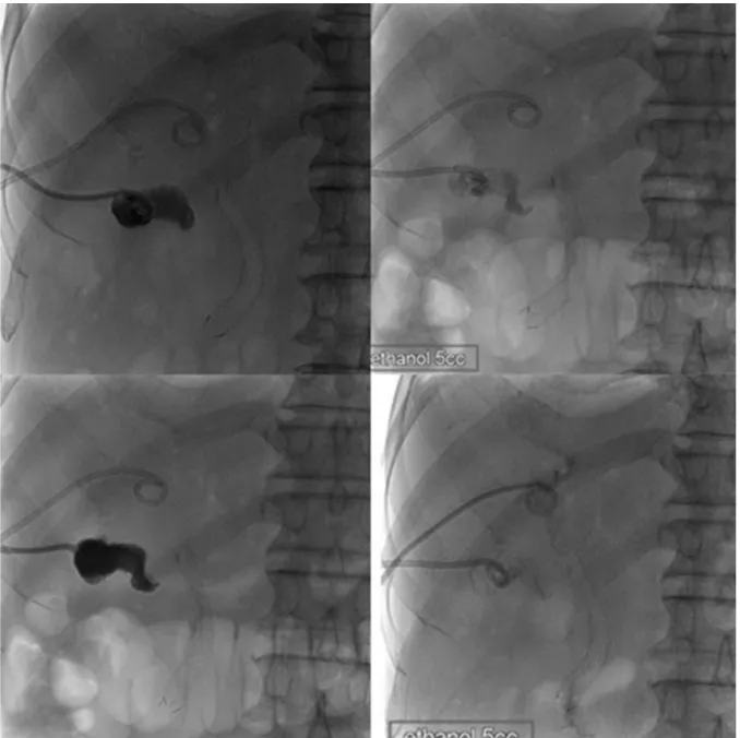
Fig. 4. Two cycles of chemical ablation of the gallbladder using 5 mL of pure ethanol at one-week intervals were performed to remove percutaneous transhepatic gallbladder drainage catheter.
Two weeks after the last discharge, he was admitted to the hospital for the third time owing to fever and nausea.
His laboratory results revealed the following: white blood cell count, 10,390/mm3; hemoglobin, 11.3/mm3; platelet count, 202,000/mm3; erythrocyte sedimentation rate, 59 mm/h;
CRP level, 1.05 mg/dL; AST value, 599 U/L; ALT value, 323 U/L; ALP value, 541 U/L; GGT value, 1249 U/L; total bilirubin level, 2.81 mg/dL; and direct bilirubin level, 2.46 mg/dL. An abdominal CT scan showed that the intrahepatic and extra- hepatic ducts were markedly dilated. Percutaneous trans- hepatic biliary drainage (PTBD) was performed at the distal
CBD, which had been occluded completely. An endoscopic ultrasound scan showed that the biliary duct was dilated and that there was an internal echogenic lesion in the distal CBD.
The PTBD cholangiogram showed that the biliary duct was dilated with a focal segmental narrowing at the proximal CBD.
A histology examination after the endobiliary biopsy confirmed the presence of atypical epithelial cells, which were indicative of adenocarcinoma.
Tumor removal was attempted but was unsuccessful be- cause it was challenging to dissect the tissue owing to its adhesion beginning at the liver hilum and extending up to
the GB. In addition, multiple metastatic nodules were already present in the small bowel mesentery, anterior abdominal wall, and pelvic cavity. These abdominal wall and small bowel mesentery nodules were diagnosed as adenocarcinoma after analyzing the frozen biopsy tissue samples. Consequently, a decision was made to perform endobiliary RFA for such an unresectable CBD cancer. RFA was performed under seven watts using an 22 mm RFA catheter (ELRAⓇ RF catheter, STARmed, Goyang, Korea) for 2 minutes. The partially cov- ered biliary stent was placed against the narrowed CBD after performing endobiliary RFA (Fig. 2). Subsequently, the PTBD catheter was removed (Fig. 3A, B). After performing two cy- cles of chemical ablation of the GB using 5 mL of pure ethanol at one-week intervals (Fig. 4), the PTGBD catheter was also removed (Fig. 3C, D). Since then, he did not have any compli- cations, such as jaundice or cholecystitis, for 3 months.
DISCUSSION
In Western countries, the most common cause of biliary stenosis is malignant diseases, such as pancreatic head can- cer, extrahepatic cholangiocarcinoma, GB cancer, malignant hilar lymphadenopathy, and hepatocellular carcinoma.3 Because many of these cancers are often inoperable at the time of diagnosis, palliative care, particularly with regard to the resolution of a biliary obstruction, is of primary importance.
Continual biliary decompression is achieved through biliary stenting; for this purpose, maintaining the patency of the stent becomes a key task. Recent studies suggested that performing RFA prior to biliary stenting yields better results in terms of stent patency. Endobiliary RFA is believed to induce locore- gional tumor control by inducing tumor necrosis and trans- ferring the thermal energy through the active electrode of a probe tip.
According to a recently published randomized trial by Yang et al.4 involving 19 patients with a Klatskin tumor and 46 with a distal cholangiocarcinoma, 32 patients were treated with RFA combined with plastic stent placement and 33 pa- tients were treated only with plastic stent placement. The oc- currence of side effects were similar between the two groups, whereas stent patency was significantly higher in the RFA and subsequent biliary stent inserted group compared to the only biliary stent inserted group; the results were 6.8 months and 3.4 months, respectively (p=0.02). Moreover, the mean over-
all survival period was also significantly longer in the RFA group (13.2 vs. 8.3 months, p<0.001). Sharaiha et al.5 re- ported favorable results regarding the bile duct diameter and survival rate in the RFA combined with self-expandable metal stent group compared to the results of the self-expandable metal stent only group. The study included 66 patients with a biliary obstruction complicated by cholangiocarcinoma and pancreatic cancer, and showed that there was no significant difference in adverse effects. According to a meta-analysis of reviewing nine investigations, RFA showed substantial pro- longation of the stent patency and survival among patients with a biliary stricture caused by a malignancy. Regarding the adverse events, the frequency of abdominal pain was sig- nificantly higher in the RFA group; the frequency of other ad- verse events did not show notable differences between the two groups.6
The side effects after biliary stent placement include chol- angitis, cholecystitis, and pancreatitis as well as stent obstruction. Recently, the frequency of such side effects has also increased because there has been an increase in the use of stents during biliary obstruction. Isayama et al.7 re- ported that cholecystitis occurred at a rate of 5.3% (13 out of 246 patients) after the insertion of a covered or an un- covered metallic stent. In addition, as in the case of this study, an obstruction of the cystic duct owing to a malignancy is one of the risk factors for cholecystitis occurring after biliary stent placement. Suk et al.8 reported nine cystic duct malig- nant involvements among a total of 15 patients with chol- ecystitis, who underwent the placement of a metal stent ow- ing to a malignant biliary obstruction. In this case, the patient had no scope for undergoing cholecystectomy because of the cystic duct invasion of cancer. Cholecystostomy is also a safe option for cholecystectomy. On the other hand, GB ablation was performed because the PTGBD could not be removed in the present case. Chemical ablation of the GB could be an alternative option for patients in whom cholecystectomy cannot be performed. Many studies have been conducted on the selection of a sclerosant. Lee et al.9 reported successful treatment with GB sclerosis using pure ethanol in a case of a patient with a suspicious cystic ductal invasion of cholangiocarcinoma. Xu et al.10 performed the chemical abla- tion of GB in 34 patients who underwent minicholecystectomy for the treatment of cholelithiasis and/or acute cholecystitis.
As a side effect of the chemical ablation of GB, five out of
34 patients developed a mucocele 2 to 5 years after the procedure.
RFA can be beneficial to the stent patency and survival and does not cause a significant increase in side effects in cases of biliary obstruction caused by malignancy. Accordingly, endoscopic RFA was performed before placing the biliary stent in the present case of an inoperable CBD cancer complicated by a biliary obstruction. Therefore, chemical ablation of GB was performed using pure ethanol for removing PTBD and because the patient had a prior history of acute cholecystitis with a GB rupture. This paper reports this case, where palliative treatment for biliary obstruction was achieved successfully.
The PTGBD catheter was removed successfully through ethanol ablation of the GB with no significant side effects for 3 months.
Consequently, PTGBD removal was performed, which brought about an improvement in the patient’s quality of life.
REFERENCES
1. Cho JH, Lee KH, Kim JM, Kim YS, Lee DH, Jeong S. Safety and ef- fectiveness of endobiliary radiofrequency ablation according to the different power and target temperature in a swine model. J Gastroenterol Hepatol 2017;32:521-526.
2. Majeed AW, Reed MW, Stephenson TJ, Johnson AG. Chemical ablation of the gallbladder. Br J Surg 1997;84:638-641.
3. Dawwas MF, Oppong KW, Webster GJ. Endoscopic assessment and management of biliary strictures. Frontline Gastroenterol 2016;7:170-175.
4. Yang J, Wang J, Zhou H, et al. Efficacy and safety of endoscopic radiofrequency ablation for unresectable extrahepatic chol- angiocarcinoma: a randomized trial. Endoscopy 2018;50:
751-760.
5. Sharaiha RZ, Sethi A, Weaver KR, et al. Impact of radiofrequency ablation on malignant biliary strictures: results of a collaborative registry. Dig Dis Sci 2015;60:2164-2169.
6. Sofi AA, Khan MA, Das A, et al. Radiofrequency ablation com- bined with biliary stent placement versus stent placement alone for malignant biliary strictures: a systematic review and meta-analysis. Gastrointest Endosc 2018;87:944-951.e1.
7. Isayama H, Kawabe T, Nakai Y, et al. Cholecystitis after metallic stent placement in patients with malignant distal biliary obstruction. Clin Gastroenterol Hepatol 2006;4:1148-1153.
8. Suk KT, Kim HS, Kim JW, et al. Risk factors for cholecystitis after metal stent placement in malignant biliary obstruction.
Gastrointest Endosc 2006;64:522-529.
9. Lee TH, Park SH, Kim SP, et al. Chemical ablation of the gall- bladder using alcohol in cholecystitis after palliative biliary stenting. World J Gastroenterol 2009;15:2041-2043.
10. Xu Z, Wang L, Zhang N, Ling X, Hou C, Zhou X. Chemical ablation of the gallbladder: clinical application and long-term observations.
Surg Endosc 2005;19:693-696.