Results of Endovascular Coil Embolization Treatment for Small (≤ 5 mm) Unruptured Intracranial Aneurysms
Siwoo Lee, Tae-Sik Gong, Yong-Woo Lee, Hyo-Joon Kim, Chang-young Kweon Department of Neurosurgery, Presbyterian Medical Center, Jeonju, Korea
Objective : Researchers and clinicians have been unable to fully elucidate the natural course of and proper treatment for unruptured intracranial aneurysms (UIAs) smaller than or equal to 5 mm, particularly with regard to whether close observation or surgery is more appropriate. In this retro- spective study, we evaluated the safety and efficacy of endovascular coil embolization of small (≤ 5 mm) asymptomatic UIAs by analyzing out- comes and complications associated with the procedure.
Materials and Methods : We analyzed data from 150 patients with small asymptomatic UIAs (≤ 5 mm) treated with coil embolization between January 2011 and December 2015. Three-dimensional angiography was used to measure aneurysm size. We evaluated procedure-related morbidity and mortality, immediate post-operative angiographic results, brain com- puted thomography follow-up results on post-operative day one, and clin- ical progress.
Results : UIAs occurred primarily in the anterior circulation area (142 cases, 94.67%), though eight patients exhibited UIAs of the posterior circulation.
Following coil embolization, aneurysms with complete occlusion were ob- served in 137 cases (91.3%). Partial occlusion occurred in five cases (3.33%), while the procedure had failed in eight cases (5.33%). Procedure-related morbidity and mortality were five cases (3.33%) and zero cases, respectively.
Conclusion : The endovascular treatment of small asymptomatic UIAs is as- sociated with good short-term outcomes without permanent neurologic complications as well as low overall complication and morbidity rates. Thus, the procedure should be considered for patients with smaller asympto- matic UIAs.
J Cerebrovasc Endovasc Neurosurg.
2016 September;18(3):229-233 Received : 30 April 2016
Revised : 5 September 2016 Accepted : 15 September 2016 Correspondence to Tae-Sik Gong
Department of Neurosurgery, Presbyterian Medical Center, 365 Seowonro, Wansan-gu, Jeonju 54987, Korea
Tel : 82-63-230-1420 Fax : 82-63-285-9573 E-mail : [email protected]
ORCID : http://orcid.org/0000-0002-7147-7805
This is an Open Access article distributed under the terms of the Creative Commons Attribution Non- Commercial License (http://creativecommons.org/li- censes/by-nc/3.0) which permits unrestricted non- commercial use, distribution, and reproduction in any medium, provided the original work is properly cited.
Keywords Embolization, Endovascular procedures, Intracranial aneurysm, Morbidity
INTRODUCTION
Due to advancements in the field of radiology and increased public interest in neurological health, the number of patients in whom incidental unruptured in- tracranial aneurysms (UIA) have been identified con- tinues to increase. However, clinicians remain divided with regard to whether close observation or surgery is
most appropriate in cases of small UIA. As rupture of UIAs is associated with high morbidity and mortality, prophylactic surgery is often performed, though the risks associated with surgery should be considered.
The natural course of small UIAs remains unclear.
Though the rupture rate is low, it is not zero, and there is no standard size of UIA at which treatment must begin. Juvela et al. stated that UIAs require sur-
A B C D
Fig. 1. Transfemoral cerebral angiograms in a 71-year-old male patient. The 3.6 mm × 3.3 mm unruptured intracranial aneurysm (UIA) is visible on the left posterior communicating artery. (A, B) Frontal and lateral views showing pre-embolization UIA. (C, D) Frontal and lateral views showing a totally occluded aneurysmal sac after coil embolization.
gical treatment regardless of their size or risk factors for rupture as long as surgery is technically possible and not contraindicated.6) In contrast, Sonobe et al. as- serted that patients with single UIAs ≤ 5 mm in size should undergo careful observation due to their very low annual rupture rate. However, patients with multi- ple UIAs larger than 4 mm in size, patients under the age of 50, and those with a history of hypertension and/or smoking require surgical treatment due to the increased risk of rupture.5)10)12)
Evidence suggests that endovascular treatment is asso- ciated with lower morbidity than surgical treatment.3)4) Therefore, we evaluated the safety and efficacy of en- dovascular coil embolization of small (≤ 5 mm) UIA by analyzing procedure-related complications and outcomes.
MATERIALS AND METHODS
We analyzed data from patients treated at our hos- pital between January 2011 and December 2015. Over these 5 years, endovascular coil embolization was per- formed on a total of 324 patients. Small intracranial aneurysms (≤ 5 mm) were observed in 162 patients, 12 of whom had ruptured intracranial aneurysms. In the present retrospective study, we evaluated data from 150 patients with small asymptomatic UIAs.
Patients typically underwent computed thomography (CT) or MR angiography for evaluation of persistent symptoms such as headache or dizziness. Transfemoral
cerebral angiography was then used to confirm diag- noses of suspected UIA. All embolization procedures were performed through femoral access under general anesthesia by a single skilled neurosurgeon. For com- plete packing, stent assisted coiling was performed in wide-neck aneurysms. Aneurysm occlusion was as- sessed using frontal, lateral, and working projection views. Patients underwent close observation in the in- tensive care unit for one day after endovascular coil embolization. Non-enhanced brain CT was performed to check for hemorrhagic or thromboembolic injury.
We categorized the results of coil embolization as complete occlusion, partial occlusion, or failed occlu- sion as follows. Complete occlusion was defined as occlusion of the aneurysm and aneurysm sac (Fig. 1), while partial occlusion was defined as occlusion with a space at the neck of aneurysm sac (Fig. 2).
RESULTS
Embolization procedures were performed in 37 men (24.67%) and 113 women (75.33%). The average age of patients was 62.23 years (range: 30-83 years) (Table 1).
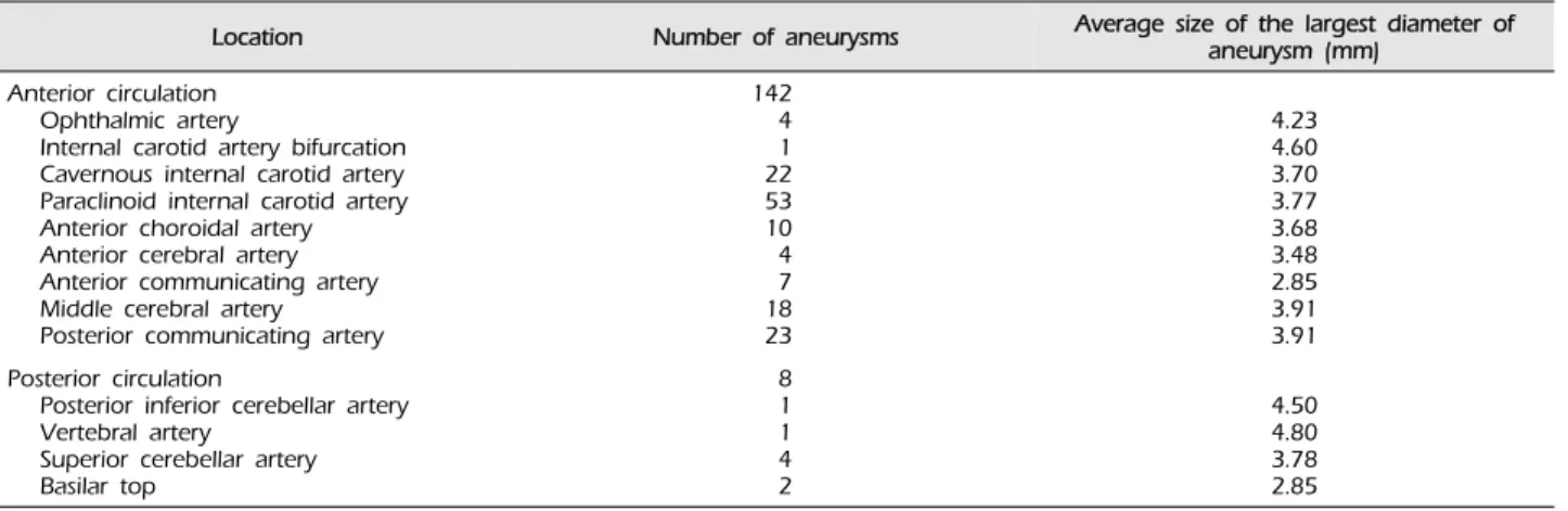
UIAs were located primarily in the anterior circu- lation area (142 cases, 94.67%). The origins of aneur- ysms were as follows: ophthalmic artery, 4; internal carotid artery (ICA) bifurcation, 1; cavernous ICA, 22;
paraclinoid ICA, 53; anterior choroidal artery, 10; an- terior cerebral artery (ACA), 4; anterior communicat-
A B C D
Fig. 2. Transfemoral cerebral angiograms in a 61-year-old female patient. The 4.9 mm × 2.7 mm UIA is visible on the right oph- thalmic artery. (A, B) Frontal and lateral views showing pre-embolization UIA. (C, D) Frontal and lateral views showing a partially oc- cluded aneurysmal sac after coil embolization.
Characteristics Number of patients Age (years)
< 40 40-60
> 60
3 (2%)
63 (42%)
84 (56%) Sex
Female
Male 113 (75.33%)
37 (24.67%) Table 1. Patient demographics
ing artery, 7; middle cerebral artery (MCA), 18; poste- rior communicating artery, 23; posterior inferior cer- ebellar artery, 1; vertebral artery, 1; superior cerebellar artery, 4; and basilar top, 2 (Table 2).
The average aneurysm sac size was 3.76 mm (range:
2.2 mm to 5 mm). UIA size was < 3 mm in 25 cases, 3-4 mm in 65 cases, and > 4 mm in 60 cases. Complete occlusion was observed in 137 cases (91.33%), while partial occlusion was observed in five cases (3.33%).
The procedure had failed in eight cases (5.33%).
Among these, there were three cases of coil herniation in spite of assistive stent and five cases in which mi- crocatheter access was impaired due to severe angula- tion and tortuous parent vessels.
No patients experienced new-onset neurological symptoms following coil embolization. Neither hem- orrhage nor cerebral infarction was observed on brain CT at 1 day post-operation. Two patients experienced bleeding and swelling at the femoral artery puncture site. In these cases, sand bags were applied at the puncture site, and patients were restricted to absolute
bed rest. Ultrasonography revealed a femoral arterio- venous fistula associated with pain and mild flow sounds at the access site. Two patients experienced systemic allergic responses including rash and itching, though these symptoms resolved following steroid treatment and hydration (Table 3).
DISCUSSION
Recent advances in the sensitivity of neuroimaging techniques have increased the ability for clinicians to identify small asymptomatic UIAs. Though the rup- ture risk of small UIAs is quite low, rupture can be associated with devastating results such as death or brain damage from subarachnoid hemorrhage (SAH).
Accordingly, preventive treatment of UIA prior to rupture has been considered necessary, though the risks of surgery must also be considered. As patients with UIAs at low risk for rupture may experience se- vere complications following surgery, much debate has centered around whether surgical treatment or close observation is more appropriate.
Zaccarello reported that mortality and morbidity re- lated to surgical treatment were higher than rupture risk, with a 0.05% chance of SAH incidence for UIAs under 10 mm in patients without previous SAH history.13) In a study by Wiebers et al., 5-year cumu- lative rupture rates of UIAs (7 mm) in areas of the anterior circulation such as the ICA, anterior commu-
Location Number of aneurysms Average size of the largest diameter of aneurysm (mm)
Anterior circulation Ophthalmic artery
Internal carotid artery bifurcation Cavernous internal carotid artery Paraclinoid internal carotid artery Anterior choroidal artery Anterior cerebral artery Anterior communicating artery Middle cerebral artery Posterior communicating artery
142 41 22 5310 47 18 23
4.234.60 3.70 3.773.68 3.482.85 3.91 3.91 Posterior circulation
Posterior inferior cerebellar artery Vertebral artery
Superior cerebellar artery Basilar top
8 11 42
4.504.80 3.782.85 Table 2. Location and mean size of aneurysms
Complications Number of cases
Total
Thromboembolism or hemorrhage on brain CT 1 day post-operation, n Contrast-related allergic response, n
Femoral access site bleeding & swelling, n Femoral arteriovenous fistula, n
5 (3.33%) 02 (1.33%) 2 (1.33%) 1 (0.67%) CT = computed thomography
Table 3. Procedure-related complications
nicating artery, ACA, and MCA were 0%. However, a 2.5-fold increase in such rates was associated with UIAs in areas of the posterior circulation such as the posterior communicating artery.11)
Recently, Byoun et al.1) retrospectively evaluated the natural course of UIA progression in 1,006 patients from 2000 to 2008. Nine-year follow-up results re- vealed a rupture risk of 1.00%. UIAs smaller than 7 mm in patients without previous SAH history were associated with an even lower risk of rupture (0.79%).
Moreover, even untreated small UIAs were often as- sociated with a benign natural course and very low rupture risk. Nevertheless, this low risk of rupture does not definitely indicate that conservative treat- ment is optimal small UIAs.11) Furthermore, the retro- spective nature, short follow-up period and recruit- ment bias of the aforementioned studies may have in- fluenced the respective results of these studies. Indeed, may authors recommend active treatment for incidental asymptomatic UIAs due to the risk of rupture.7)
Skilled neurointerventionists can often provide suc-
cessful preventive treatment for small UIAs using an- tiplatelet and anticoagulation therapies, which have significantly diminished morbidity rate in this patient population.8) Although a number of technically chal- lenging procedures occurred after coil embolization in patients of the present study, procedural morbidity (five cases, 3.33%) and mortality (zero cases) were low.
Thromboembolic complications may also produce vague or mild cognitive or neurological abnormalities.2) However, several studies have revealed that endovas- cular coil embolization of UIAs is associated with a lower risk of such complications than surgical clipping.
In a study of 130 patients with UIA, Johnston et al.
reported that changes in modified Rankin Scale (mRS) scores were 25% for surgical clipping and 8% for en- dovascular coil embolization.3)4) Recent studies have also revealed that patients who have undergone endo- vascular coil embolization exhibit prognosis better than or similar to those who have undergone surgical clipping, although these studies did not consider dif- ferences in procedural morbidity and characteristics of
UIA among patients.
Ogilvy and Carter reported that UIAs located in areas of the anterior circulation were associated with very low morbidity after clipping, and that long-term effi- cacy and durability of endovascular treatment for UIA was not yet certain.9) Therefore, further studies should investigate the ability of embolization treatment to lower rupture risk using longer clinical and neuro- imaging follow-up periods and examine differences among patients according to various characteristics.
Treatment for UIAs smaller than 3 mm remains challenging, regardless of whether endovascular treat- ment or surgical clipping is performed. The risk of rupture for such small UIAs is high during endovas- cular treatment, though no such cases were observed in the present study. Some authors recommend that patients under 50 years old, those with hypertension, and those with multiple UIAs larger than 4 mm should be treated due to the high risk of rupture.5)10)12)
CONCLUSION
Much debate persists regarding the potential risk of rupture and benefits of preventive treatment for small asymptomatic UIAs. In the present study, we ob- served satisfactory short-term outcomes and no per- manent neurologic complications following endovas- cular treatment of UIA. Therefore, endovascular treat- ment should be considered for the treatment of small asymptomatic UIAs.
Disclosure
The authors report no conflict of interest concerning the materials or methods used in this study or the findings specified in this paper.
REFERENCES
1. Byoun HS, Huh W, Oh CW, Bang JS, Hwang G, Kwon OK. Natural history of unruptured intracranial aneurysms:
a retrospective single center analysis. J Korean Neurosurg Soc. 2016 Jan;59(1):11-6.
2. Im SH, Han MH, Kwon OK, Kwon BJ, Kim SH, Kim JE, et al. Endovascular coil embolization of 435 small asymp- tomatic unruptured intracranial aneurysms: procedural mor- bidity and patient outcome. AJNR Am J Neuroradiol. 2009 Jan;30(1):79-84.
3. Johnston SC, Wilson CB, Halbach VV, Higashida RT, Dowd CF, McDermott MW, et al. Endovascular and surgical treat- ment of unruptured cerebral aneurysms: comparison of risks. Ann Neurol. 2000 Jul;48(1):11-9.
4. Johnston SC, Zhao S, Dudley RA, Berman MF, Gress DR.
Treatment of unruptured cerebral aneurysms in California.
Stroke. 2001 Mar;32(3):597-605.
5. Juvela S, Porras M, Poussa K. Natural history of unruptured intracranial aneurysms: probability and risk factors for aneurysm rupture. Neurosurg Focus. 2000;8(5): Preview 1.
6. Juvela S, Porras M, Poussa K. Natural history of unruptured intracranial aneurysms: probability of and risk factors for aneurysm rupture. J Neurosurg. 2000 Sep;93(3):379-87.
7. Juvela S, Poussa K, Lehto H, Porras M. Natural history of unruptured intracranial aneurysms: a long-term follow-up study. Stroke. 2013 Sep;44(9):2414-21.
8. Lanterna LA, Tredici G, Dimitrov BD, Biroli F. Treatment of unruptured cerebral aneurysms by embolization with gu- glielmi detachable coils: case-fatality, morbidity, and effec- tiveness in preventing bleeding--a systematic review of the literature. Neurosurgery. 2004 Oct;55(4):767-75; discussion 775-8.
9. Ogilvy CS, Carter BS. Stratification of outcome for surgically treated unruptured intracranial aneurysms. Neurosurgery.
2003 Jan;52(1):82-7; discussion 87-8.
10. Sonobe M, Yamazaki T, Yonekura M, Kikuchi H. Small unruptured intracranial aneurysm verification study: SUAVe study, Japan. Stroke. 2010 Sep;41(9):1969-77.
11. Wiebers DO, Whisnant JP, Huston J 3rd, Meissner I, Brown RD Jr, Piepgras DG, et al. Unruptured intracranial aneur- ysms: natural history, clinical outcome, and risks of surgical and endovascular treatment. Lancet. 2003 Jul;362(9378):103-10.
12. Yonekura M. Small unruptured aneurysm verification (SUAVe Study, Japan)--interim report. Neurol Med Chir (Tokyo).
2004 Apr;44(4):213-4.
13. Zuccarello M. Treatment strategy for patients with un- ruptured intracranial aneurysms. Neurol Med Chir (Tokyo).
2001 Dec;41(12):571-5.