Assessment of Capsular Insertion Type and of Capsular Elongation in Patients with Anterior Shoulder Instability and It’s Correlation with Surgical Outcome: A Quantitative Assessment
with Computed Tomography Arthrography
Do Hoon Kim, Do Yeon Kim, Hye Yeon Choi1, Ji Soon Park2, Ye Hyun Lee3, Joo Han Oh
Department of Orthopedic Surgery, Seoul National University Bundang Hospital, Seoul National University College of Medicine, Seongnam, 1Department of Orthopedic Surgery, Seoul Medical Center, 2Department of Orthopedic Surgery, Korea University Anam Hospital, 3Department of Orthopedic Surgery, National Police Hospital, Seoul, Korea
Background: The study aimed to determine the type of capsular insertion and the extent of capsular elongation in anterior shoulder in- stability by quantitatively evaluating their computed tomography arthrographic (CTA) findings, and to investigate the correlation of these parameters with surgical outcomes.
Methods: We retrospectively reviewed 71 patients who underwent CTA and arthroscopic capsulolabral reconstruction for anterior shoulder instability between April 2004 and August 2008. The control group comprised 72 patients diagnosed as isolated type II superior labrum anterior to posterior (SLAP) lesion during the period. Among the 143 patients, 71 were examined with follow-up CTA at an aver- age 13.8 months after surgery. It was measured the capsular length and cross-sectional area at two distinct capsular regions: the 4 and 5 o’clock position of the capsule.
Results: With regards to the incidence of the type of anterior capsular insertion, type I was more common in the control group, whereas type III more common than in the instability group. Anterior capsular length and cross-sectional area were significantly greater in the in- stability group than in the control group. Among patients of the instability group, the number of dislocations and the presence of anterior labroligamentous periosteal sleeve avulsion lesion were significantly associated with anterior capsular redundancy. Postoperatively, recur- rence was found in 3 patients (4.2%) and their postoperative capsular length and cross-sectional area were greater than those of patients without recurrence.
Conclusions: Capsular insertion type and capsular redundancy derived through CTA may serve as important parameters for the man- agement of anterior shoulder instability.
(Clin Shoulder Elbow 2016;19(3):155-162)
Key Words: Capsular elongation; Computed tomography; Arthrography; Joint instability; Shoulder
Clinics in Shoulder and Elbow
Copyright © 2016 Korean Shoulder and Elbow Society. All Rights Reserved. pISSN 2383-8337
Clinics in Shoulder and Elbow Vol. 19, No. 3, September, 2016 http://dx.doi.org/10.5397/cise.2016.19.3.155
Received December 29, 2015. Revised May 25, 2016. Accepted May 27, 2016.
Correspondence to: Joo Han Oh
Department of Orthopedic Surgery, Seoul National University Bundang Hospital, Seoul National University College of Medicine, 82 Gumi-ro 173beon-gil, Bundang-gu, Seongnam 13620, Korea
Tel: +82-31-787-7197, Fax: +82-31-787-4056, E-mail: [email protected] IRB approval (No. B-1608-360-103).
Financial support: None. Conflict of interests: None.
Introduction
Dislocations most commonly occur in the glenohumeral joint.
And the incidence of shoulder dislocations in individuals aged between 18 and 70 years is around 2%; of whom, reports have shown that 75% develop shoulder instability.1-3) The methods of
diagnosis and of treatment for shoulder instability are still evolv- ing. And the incidence of recurrence despite reparative surgery has been shown to be around 10%.4,5) Shoulder instability has been shown to result by means of a large spectrum of patho- logical conditions such as labral deficiency, capsular elongation, patulousness of the shoulder capsule, ligament injury, bony defi-
ciencies, and etc.6-8)
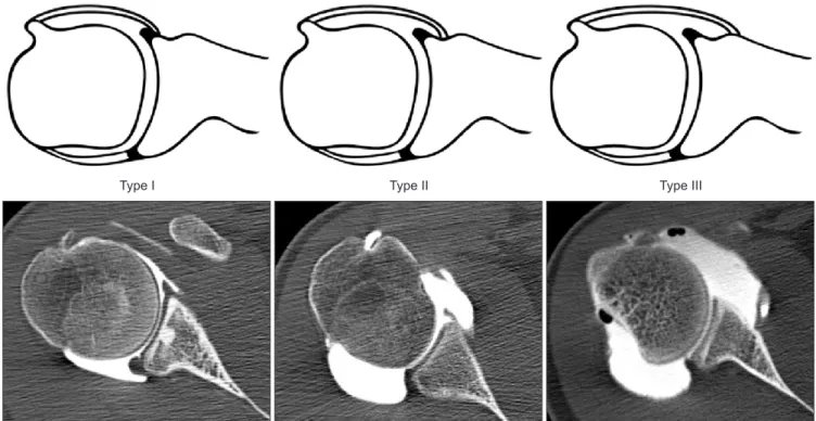
In 1962 Moseley9) introduced a classification system based on the mode of capsular insertion for categorizing anatomical varia- tions in the capsule that surrounds the glenohumeral joint. The three types of capsular insertion were as follows: type I, capsular insertion into the labral base; type II, capsular insertion into the glenoid fossa; and type III, capsular insertion into a more medial position following the scapular neck (Fig. 1)9). They reported a significant association of type III insertions with anterior shoulder instability.9-11) Additionally, Massengill et al.10) reported that a large anterior pouch or a repaired anterior capsular tear required differentiation from a type III capsular insertion as its appearance was likened to those of the former two.
Previous reports have implicated numerous factors for gleno- humeral dislocations. For instance, some reports have suggested that capsular stretching after instances of repeated glenohumeral dislocations and a lax capsule resultant from permanent plastic deformation, which can be determined through either bio- mechanical evaluation or arthroscopic inspection, were more prominent causative factors for glenohumeral dislocation than Bankart lesions.12,13) In another study, functional deficiency of the inferior glenohumeral ligament (IGHL), usually found at the site of the glenoid attachment (as in a Bankart lesion) but rarely at the humeral side or at the midsubstance, was shown to be caus- ative of recurrent anterior shoulder dislocation.14)
Substantial deformation in anatomy prevails during repeti- tive dislocations before paving way to ruptures of the ligament
and of the capsule.14,15) McMahon et al.16) reported that with shoulder dislocations, the IGHL not only sustains a Bankart le- sion but also permanent stretching of an average 2.3 mm and capsular elongation, which can be clinically observed by joint angiography or intraoperatively.17,18) Yet, when we reviewed the literature, we could not find reports that quantitatively analyzed capsular elongation in patients with anterior shoulder instability.
Therefore, this study aimed to undertake the following goals: (1) use computed tomography arthrography (CTA) to assess the cor- relation between the type of capsular insertion an individual has and anterior shoulder instability; (2) to quantitatively evaluate capsular elongation in patients with anterior shoulder instability by pre- and postoperatively measuring length and cross-section- al area of the anterior glenoid capsule; and (3) to investigate the relationship between the type of capsular insertion or capsular elongation and clinical factors of the patients with anterior shoul- der instability.
Methods
Patients
The following inclusion criteria were used to recruit patients who were admitted between April 2004 and August 2008 into the instability group: 1) those whose condition was diagnosed as an anterior shoulder instability; 2) recipients of preoperative CTA; 3) recipients of arthroscopic capsulolabral reconstruction;
and 4) those able to participate in at least a year of postoperative
Type I Type II Type III
Fig. 1. An illustration of the anatomical variations of anterior capsular attachment and their corresponding computed tomography images from patients with an- terior shoulder instability. Revised from the article of Massengill et al. (Radiographics. 1994;14:1211-23)10) with original copyright holder’s permission.
follow-up for CTA. A total of 71 patients were enrolled into the instability group in our retrospective study. Between the same period, we recruited individuals who fulfilled the following in- clusion criteria into the control group: 1) those whose condition were diagnosed as an isolated SLAP type II lesion; 2) recipients of preoperative CTA; 3) recipients of an arthroscopic superior labrum anterior to posterior (SLAP) repair; and 4) those able to participate in at least a year of postoperative follow-up for CTA.
A total of 72 participants were enrolled for the comparative analysis. All operations included Bankart operations, which com- prised an arthroscopic labral repair with concomitant plication of the anterior capsular including the IGHL were performed by a single surgeon.
Clinical Variables
We analyzed clinical characteristics (age at the time of op- eration, sex, hand dominance, age at first dislocation, and total number of dislocation events) of the patients and performed a detailed diagnosis for classic Bankart, bony Bankart, and anterior labroligamentous periosteal sleeve avulsion (ALPSA) lesion in patients in the instability group. We classified the mechanism of injury concerning the first dislocation event into six types: a spontaneous dislocation, a lateral contusion, an abduction-ex- ternal rotation injury, a traction injury, a fall onto an outstretched
hand, and a hyperextension injury. The severity of these injures was classified into three levels: injury without contact, injury with contact, and high energy injury.
Imaging Technique
Under fluoroscopic guidance, we injected contrast medium into the patient’s glenohumeral joint for shoulder CTA. A maxi- mum 20 ml of contrast medium was intra-articularly injected until the patient felt pain (12 ml Omnipaque 300+8 ml normal saline). Then, CTA was taken within 15 minutes (with a maxi- mum time lapse of 30 minutes). Throughout the imaging the pa- tient was positioned into a in a neutral position with the upper extremity in anatomical position.6) Basic axial computed tomog- raphy (CT) imaging was taken in 16-sections or in 64-sections of either 2 or 3 mm thickness with a multi-detector CT.
Capsular Measurements
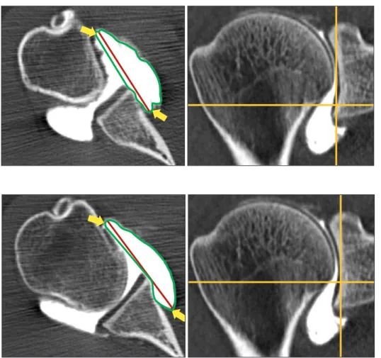
Following the classification system proposed by Moseley,9) we evaluated the type of anterior capsular insertion by examining the glenoid morphology at the mid-glenoid level. We quanti- tatively measured capsular length and capsular cross-sectional area by employing the picture archiving and communication system (PACS) as our image analyzing software. We defined an- terior capsular space as the area of joint highlighted just anterior
Fig. 2. Capsule at the 5 o’clock position. Axial computed tomography slices at the level that is 6 mm superior to the inferior margin of the glenoid. White arrows mean anterior and posterior margin of anterior joint capsule.
Black line expresses anterior capsular length and gray line indicates anterior capsular space.
Fig. 3. Capsule at the 4 o’clock position. Axial computed tomography slices at the level that is 12 mm superior to inferior margin of the glenoid. White arrows mean anterior and posterior margin of anterior joint capsule.
Black line expresses anterior capsular length and gray line indicates anterior capsular space.
to the point where the anterior glenoid tip and the capsule insert into the humerus; then, taking this definition of anterior capsular space we took the longest axial length as the capsular length. Af- ter the anterior capsular space margin was drawn out, we used the PACS systems to calculate the capsular cross-sectional area (Fig. 2, 3). Using the axial plane derived from CTA imaging, we measured the capsular length and the capsular cross-sectional area at two distinct capsular regions on PACS: the 5 o’clock po- sition of the capsule 6 mm superior to the inferior margin of the glenoid rim (Fig. 2) and the 4 o’clock position 12 mm superior to the inferior margin of the glenoid rim (Fig. 3). Two orthopedic surgeons made the measurements independently of each other and in two repeats, where each measurement was taken after a week interval. To normalize the measured capsular length and capsular cross-sectional area, we divided the measured data by the ratio of the measured and the average humeral head diam- eter.7)
Statistical Analysis
All statistical analyses were performed with SPSS ver. 12.0 software (SPSS Inc., Chicago, IL, USA). Statistical significance was set to a p-value of less than 0.05. To analyze in terms of vari- ables, we used the independent t-test, the paired t-test, the chi- square test, the Fisher’s exact test, and the intra-class correlation coefficient analysis.
Results
We summarized the patients’ clinical data in Table 1. The av- erage age of the patients in the instability group was 22.4 years and in the control group, 39.5 years. The ratio of gender in the instability group was 65 men to 6 women (n=71) and in the control group, 62 men to 10 women (n=72). Among the insta- bility group, three patients showed postoperative complications after an average 13.8 months: two dislocations and one sublux- ation. The recurrent capsular insertion was a type II and a type
III in the two patients with dislocation and a type II insertion in the patient with subluxation. Although the patient with recurrent dislocation and a type II capsular insertion exhibited a lax cap- sule, the patient showed labral healing. The other two patients presented with a labral tear and a lax capsule. We performed reoperations on all three patients.
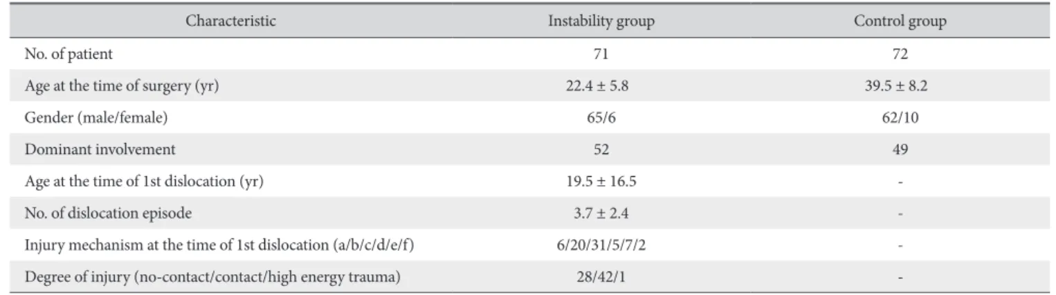
We assessed the type of capsular insertion at the mid-anterior glenoid level. We found that ten patients had a type I capsular insertion in the control group (13.9%) and 15 patients had a type III capsular insertion in the instability group (21.1%), thereby showing a statistically significant difference (Table 2). The inter- observer and intra-observer correlation coefficients were 0.89 and 0.93, respectively, for the measurements of capsular length and capsular cross-sectional area, denoting an excellent reli- ability of our measurements. We found that the capsular length measured at the 5 o’clock position was 17.1% longer in the instability group than in the control group and that the capsular cross-sectional area was 46.0% significantly larger. Likewise, the same measurements made at the 4 o’clock position showed the corresponding increases, 19.2% and 69.5%, respectively (Table 3). The incidence of lesions among the patients in the instability group was as follows: 52 patients had classic Bankart lesion; 5 had bony Bankart lesion; and 14, ALPSA lesion. We found that compared to those with classic Bankart lesion or with bony Bankart lesion, those with ALPSA lesion showed a statisti- Table 1. Clinical Characteristics
Characteristic Instability group Control group
No. of patient 71 72
Age at the time of surgery (yr) 22.4 ± 5.8 39.5 ± 8.2
Gender (male/female) 65/6 62/10
Dominant involvement 52 49
Age at the time of 1st dislocation (yr) 19.5 ± 16.5 -
No. of dislocation episode 3.7 ± 2.4 -
Injury mechanism at the time of 1st dislocation (a/b/c/d/e/f) 6/20/31/5/7/2 -
Degree of injury (no-contact/contact/high energy trauma) 28/42/1 -
Values are presented as number only or mean ± standard deviation.
a: spontaneous dislocation, b: lateral contusion, c: abduction-external rotation injury, d: traction injury, e: fall onto outstretched hand, f: hyper extension injury.
Table 2. Type of Capsular Insertion in Both Groups
Type Instability group Control group p-value
Type I 3 (4.2) 10 (13.9) 0.038
Type II 53 (74.6) 55 (76.4) 0.813
Type III 15 (21.1) 7 (9.7) 0.049
Values are presented as number (%).
Type I: capsular insertion into the labral base, Type II: capsular insertion into the glenoid fossa, Type III: capsular insertion into a more medial position fol- lowing the scapular neck.
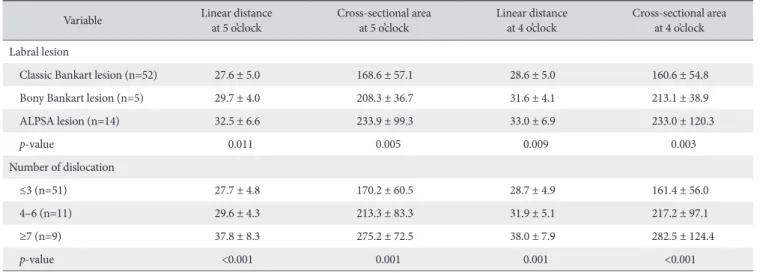
cally significant elongation of capsule length and increase in cross-sectional area. And we also found that those with ALPSA lesion associated with more frequent preoperative dislocation episodes had a significantly greater increase in capsular length and cross-sectional area than those with less frequent episodes (Table 4). However, we did not observe a significant correlation between anterior capsular elongation and clinical factors such as
injury mechanism, severity of injury, hand dominance, gender, and age at first dislocation.
A comparative analysis of the pre- and postoperative CTA findings of the instability group revealed that the postoperative capsular length and capsular cross-sectional area were significantly lower than their corresponding preoperative values (Table 5). We found that the postoperative capsular length and cross-sectional area of the instability group were comparable to those of the preoperative values of the control group (Table 6). Further, the postoperative CT arthrograms of the three patients (4.2% of the instability group) with recurrent dislocation showed that the an- terior capsular length and cross-sectional area were significantly greater in these patients than those of the patients without recur- rence.
Discussion
We found that type I capsular insertion was significantly prev- alent in the control group whereas type III capsular insertion was prevalent in the instability group. This association of capsular insertion type in patients with anterior shoulder instability had already been suggested by previous studies19,20)—our study con- Table 3. Linear Distance and Cross-sectional Area of the Capsule at the 5
o’clock Position* and at the 4 o’clock Position†
Position Instability group Control group p-value 5 o’clock position
Linear distance 28.7 ± 5.6 (17.1) 24.5 ± 5.6 <0.001 Cross-sectional area 184.3 ± 70.7 (46.0) 126.2 ± 53.1 <0.001 4 o’clock position
Linear distance 29.8 ± 5.7 (19.2) 25.0 ± 6.4 <0.001 Cross-sectional area 178.5 ± 76.7 (69.5) 105.5 ± 48.7 <0.001 Values are presented as mean ± standard deviation (% enlargement) or mean
± standard deviation only.
*Axial slices at 6 mm superior to the inferior margin of the glenoid. †Axial slices at 12 mm superior to the inferior margin of the glenoid.
Table 4. Correlation of Capsular Redundancy with Labral Lesions and with the Number of Dislocation Episodes
Variable Linear distance
at 5 o’clock Cross-sectional area
at 5 o’clock Linear distance
at 4 o’clock Cross-sectional area at 4 o’clock Labral lesion
Classic Bankart lesion (n=52) 27.6 ± 5.0 168.6 ± 57.1 28.6 ± 5.0 160.6 ± 54.8
Bony Bankart lesion (n=5) 29.7 ± 4.0 208.3 ± 36.7 31.6 ± 4.1 213.1 ± 38.9
ALPSA lesion (n=14) 32.5 ± 6.6 233.9 ± 99.3 33.0 ± 6.9 233.0 ± 120.3
p-value 0.011 0.005 0.009 0.003
Number of dislocation
≤3 (n=51) 27.7 ± 4.8 170.2 ± 60.5 28.7 ± 4.9 161.4 ± 56.0
4–6 (n=11) 29.6 ± 4.3 213.3 ± 83.3 31.9 ± 5.1 217.2 ± 97.1
≥7 (n=9) 37.8 ± 8.3 275.2 ± 72.5 38.0 ± 7.9 282.5 ± 124.4
p-value <0.001 0.001 0.001 <0.001
Values are presented as mean ± standard deviation.
ALPSA: anterior labroligamentous periosteal sleeve avulsion.
Table 5. Comparison between the Preop. and Postop. Capsular Parameters in the Instability Group
Variable Instability group
Preop. CTA Postop. CTA p-value
Linear distance at 5 o’clock 28.5 ± 5.8 22.4 ± 6.5 <0.001
Cross-sectional area at 5 o’clock 181.0 ± 63.7 119.8 ± 56.7 <0.001
Linear distance at 4 o’clock 29.2 ± 6.3 23.6 ± 5.4 <0.001
Cross-sectional area at 4 o’clock 167.4 ± 61.9 96.6 ± 36.1 <0.001
Values are presented as mean ± standard deviation.
Preop.: preoperative, Postop.: postoperative, CTA: computed tomography arthrography.
firms the results of other studies. For example, in their retrospec- tive analysis of CTA findings in 54 patients with recurrent dislo- cation, Singson et al.19) reported that anterior shoulder instability is associated with a type III capsular attachment. However, Ng et al.21) suggested that the type of capsular insertion has no clear relationship with anterior shoulder instability. But their study differs from ours in that their classification of capsular insertion type was based on findings of magnetic resonance imaging (MRI) results and that their average number of patients in any given group, into which the patients were allocated in terms of their total number of dislocation events, was much smaller than that of our study.
Reports that evaluate the quantitative assessment of capsular redundancy in anterior shoulder instability are limited. The study by Urayama et al.22) was the first of its kind to describe the meth- od of quantifying capsular redundancy. Although their study is limited in that only the capsular length of 12 patients with anteri- or shoulder instability was measured, they found that an average of 19% of capsular elongation was seen in these patients. In a separate, recent study, MRIs taken at neutral position and at the abduction-external rotation position were used to assess changes in joint volume to quantitatively measure the extent of capsular laxity.21) But since this would require MRIs to be taken at two different positions, its applicability is realistically low. In current study, we used CTA, which is widely used in the clinical context, to measure the length and the surface area of the capsule. We found that the increase in capsular size was significantly greater in patients with anterior shoulder instability and in those who sustained a more severe injury than in those without anterior shoulder instability. We found that their capsular size returned to the pre-injury, norm level after a successful operation showing that our findings were closely reflective of the clinical situation.
Past studies have implied ALPSA lesion as being more of a chronic lesion than Bankart lesion is.23,24) For instance, Haber- meyer et al.23) suggested that the ALPSA lesion is a progressed state of Bankart lesion that has undergone repeated dislocations.
Ozbaydar et al.24) reported in their study comparing ALPSA lesions to Bankart lesions that the patients with the former condition had a significantly greater number of preoperative dislocations than the latter. In this study, we found that capsular elongation was significantly greater in patients with ALPSA lesion
than in those without—a finding that is in agreement with previ- ous reports that the ALPSA lesion is a more chronic condition than Bankart lesion.
Known factors other than ones concerning anatomy to influ- ence anterior shoulder instability include age at first dislocation, gender, hyperlaxity, fracture of the greater tuberosity, and etc.25-27) In a recent systematic review, which analyzed the results of six retrospective cohort studies, it was reported that the risk factors implicated in inducing the transition from a dislocation episode to anterior shoulder instability were age of less than 14 years, male sex, and etc.28) But contrastingly we could not find a signifi- cant association between capsular elongation with either gender or age. The apparent discrepancy may be explained for a couple of reasons: our sample of patients may not have been an appro- priate one to investigate and to compare the difference between genders since the gender ratio was uneven; we evaluated the current capsular volume of patients in the instability group in a retrospective manner, which meant that we could not recruit a heterogeneous age group—none of the patients was aged 15 years or younger; and lastly, the systematic review included only studies comprising a retrospective cohort study of level III and below.
Since our findings show that, compared to those without re- currence, patients with recurrent shoulder dislocations showed a greater postoperative increase in capsular length and in capsular cross-sectional area, it suggests that the postoperative evalua- tion of these two parameters may be useful in several ways. For example, the changes in these parameters provide a meaningful predictive value for estimating prognosis and grounds to develop a more rigorous rehabilitative approach of conservative manage- ment of anterior shoulder instability29,30) than those of current lines of management like muscle strengthening exercises of the anterior and the posterior shoulder.
Distinctive from previous attempts, our study deserves merit in that it attempted to quantitatively measure and evaluate cap- sular redundancy and to assess its correlation with clinical factors and with surgical outcomes. However, limitations of this study still exist. First, as a retrospective study, ours is limited in that the control group and the patient group showed a significant difference in term of average age and that the control group composed of patients who had received SLAP repair, thus not Table 6. Comparison of Capsular Parameters between the Postop. CTA Findings of the Instability Group and the Preop. CTA Findings of the Control Group
Variable Instability group (postop. CTA) Control group (preop. CTA) p-value
Linear distance at 5 o’clock 22.4 ± 6.5 24.5 ± 5.6 0.094
Cross-sectional area at 5 o’clock 119.8 ± 56.7 126.2 ± 53.1 0.575
Linear distance at 4 o’clock 23.6 ± 5.4 25.0 ± 6.4 0.296
Cross-sectional area at 4 o’clock 96.6 ± 36.1 105.5 ± 48.7 0.351
Values are presented as mean ± standard deviation.
Postop.: postoperative, CTA: computed tomography arthrography, Preop.: preoperative.
being a completely “healthy” counterpart. Yet we can justify the use of our “controls” with SLAP but without lesions of the anterior capsule as an alternative sample because taking CTA of healthy individuals is an ethically difficult task. And since during consultations, by standard, we perform the same pre- and post- operative CTA in patients who receive a SLAP repair as in pa- tients with anterior shoulder instability, we considered that they are compatible controls in terms of the nature of data in our comparative study. Second, we could not differentiate whether the capsule was elongated or reattached with displacement after the tear in preoperative CTA, for these two cannot be discrimi- nated until the pre-injury condition can be known. As afore- mentioned, some authors proposed that these two lesions are either the same condition or part of a continuum of the same condition.10,14-16) Regardless, even if the etiology may differ the extent of capsular augmentation is an important consideration point for operation. Third, the stretching of the capsule may have been influenced by the volume of intra-articular injection of the contrast material. And lastly, because our parameters rely on manual measurements they are susceptible to variations re- sulting from human error; but by showing that our approach has high inter-, intra-observer correlation coefficients, and thereby, a high reproducibility we were able to reject this possibility.
Conclusion
In sum, we found that a type III capsular attachment was significantly associated with anterior shoulder instability. And we found that compared to those of patients with type I and type II attachments the capsular length and capsular cross-sectional area, measured on CT arthrogram, significantly increased in pa- tients with type III capsular attachment. Additionally, we found that a medical history of preoperative dislocation episodes or presence of a concomitant ALPSA lesion was significantly associ- ated with the augmentation of the capsular length and of the capsular cross-sectional area in patients with anterior shoulder instability. We found that after these patients received operation with a successful outcome their capsular length and capsular cross-sectional area did not significantly differ to the correspond- ing preoperative values of the control group. In conclusion, the identification of capsular insertion type and the measurement of capsular elongation through CTA provide a meaningful reference for diagnosis and prognosis for patients with anterior shoulder instability.
References
1. Hovelius L. Incidence of shoulder dislocation in Sweden. Clin Orthop Relat Res. 1982;(166):127-31.
2. Bottoni CR, Wilckens JH, DeBerardino TM, et al. A prospec- tive, randomized evaluation of arthroscopic stabilization versus
nonoperative treatment in patients with acute, traumatic, first- time shoulder dislocations. Am J Sports Med. 2002;30(4):576- 80.
3. Wheeler JH, Ryan JB, Arciero RA, Molinari RN. Arthroscopic versus nonoperative treatment of acute shoulder dislocations in young athletes. Arthroscopy. 1989;5(3):213-7.
4. Kim YS. Ok JH. Arthroscopic reconstruction of bony defect in shoulder instability. Clin Shoulder Elbow. 2011;14(1):117-24.
5. Kim YK. Traumatic anterior instability: failed repairs and com- plicated problems. J Korean Shoulder Elbow Soc. 2007;10(1):
1-9.
6. Dewing CB, McCormick F, Bell SJ, et al. An analysis of capsular area in patients with anterior, posterior, and multidirectional shoulder instability. Am J Sports Med. 2008;36(3):515-22.
7. Bigliani LU, Kelkar R, Flatow EL, Pollock RG, Mow VC. Gle- nohumeral stability. Biomechanical properties of passive and active stabilizers. Clin Orthop Relat Res. 1996;(330):13-30.
8. O’Brien SJ, Neves MC, Arnoczky SP, et al. The anatomy and histology of the inferior glenohumeral ligament complex of the shoulder. Am J Sports Med. 1990;18(5):449-56.
9. Moseley HF. Recurrent dislocation of the shoulder. Postgrad Med. 1962;31:23-9.
10. Massengill AD, Seeger LL, Yao L, et al. Labrocapsular ligamen- tous complex of the shoulder: normal anatomy, anatomic variation, and pitfalls of MR imaging and MR arthrography.
Radiographics. 1994;14(6):1211-23.
11. Iannotti JP, Zlatkin MB, Esterhai JL, Kressel HY, Dalinka MK, Spindler KP. Magnetic resonance imaging of the shoulder. Sen- sitivity, specificity, and predictive value. J Bone Joint Surg Am.
1991;73(1):17-29.
12. Speer KP, Deng X, Borrero S, Torzilli PA, Altchek DA, Warren RF. Biomechanical evaluation of a simulated Bankart lesion. J Bone Joint Surg Am. 1994;76(12):1819-26.
13. Turkel SJ, Panio MW, Marshall JL, Girgis FG. Stabilizing mecha- nisms preventing anterior dislocation of the glenohumeral joint. J Bone Joint Surg Am. 1981;63(8):1208-17.
14. Bokor DJ, Conboy VB, Olson C. Anterior instability of the gleno- humeral joint with humeral avulsion of the glenohumeral liga- ment. A review of 41 cases. J Bone Joint Surg Br. 1999;81(1):93- 6.
15. Wolf EM, Cheng JC, Dickson K. Humeral avulsion of gleno- humeral ligaments as a cause of anterior shoulder instability.
Arthroscopy. 1995;11(5):600-7.
16. McMahon PJ, Dettling J, Sandusky MD, Tibone JE, Lee TQ.
The anterior band of the inferior glenohumeral ligament. As- sessment of its permanent deformation and the anatomy of its glenoid attachment. J Bone Joint Surg Br. 1999;81(3):406-13.
17. Hara H, Ito N, Iwasaki K. Strength of the glenoid labrum and adjacent shoulder capsule. J Shoulder Elbow Surg. 1996;5(4):
263-8.
18. Tijmes J, Loyd HM, Tullos HS. Arthrography in acute shoulder
dislocations. South Med J. 1979;72(5):564-7.
19. Singson RD, Feldman F, Bigliani L. CT arthrographic patterns in recurrent glenohumeral instability. AJR Am J Roentgenol.
1987;149(4):749-53.
20. Chandnani VP, Yeager TD, DeBerardino T, et al. Glenoid labral tears: prospective evaluation with MRI imaging, MR arthrography, and CT arthrography. AJR Am J Roentgenol.
1993;161(6):1229-35.
21. Ng AW, Chu CM, Lo WN, Lai YM, Kam CK. Assessment of capsular laxity in patients with recurrent anterior shoulder dis- location using MRI. AJR Am J Roentgenol. 2009;192(6):1690-5.
22. Urayama M, Itoi E, Sashi R, Minagawa H, Sato K. Capsular elongation in shoulders with recurrent anterior dislocation.
Quantitative assessment with magnetic resonance arthrogra- phy. Am J Sports Med. 2003;31(1):64-7.
23. Habermeyer P, Gleyze P, Rickert M. Evolution of lesions of the labrum-ligament complex in posttraumatic anterior shoulder instability: a prospective study. J Shoulder Elbow Surg. 1999;
8(1):66-74.
24. Ozbaydar M, Elhassan B, Diller D, Massimini D, Higgins LD, Warner JJ. Results of arthroscopic capsulolabral repair: Bankart lesion versus anterior labroligamentous periosteal sleeve avul- sion lesion. Arthroscopy. 2008;24(11):1277-83.
25. Robinson CM, Howes J, Murdoch H, Will E, Graham C. Func- tional outcome and risk of recurrent instability after primary traumatic anterior shoulder dislocation in young patients. J Bone Joint Surg Am. 2006;88(11):2326-36.
26. Hoelen MA, Burgers AM, Rozing PM. Prognosis of primary anterior shoulder dislocation in young adults. Arch Orthop Trauma Surg. 1990;110(1):51-4.
27. Vermeiren J, Handelberg F, Casteleyn PP, Opdecam P. The rate of recurrence of traumatic anterior dislocation of the shoulder.
A study of 154 cases and a review of the literature. Int Orthop.
1993;17(6):337-41.
28. Olds M, Donaldson K, Ellis R, Kersten P. In children 18 years and under, what promotes recurrent shoulder instability after traumatic anterior shoulder dislocation? A systematic review and meta-analysis of risk factors. Br J Sports Med. 2015. doi:
10.1136/bjsports-2015-095149.
29. Gibson K, Growse A, Korda L, Wray E, MacDermid JC. The effectiveness of rehabilitation for nonoperative management of shoulder instability: a systematic review. J Hand Ther.
2004;17(2):229-42.
30. Dumont GD, Russell RD, Robertson WJ. Anterior shoulder instability: a review of pathoanatomy, diagnosis and treatment.
Curr Rev Musculoskelet Med. 2011;4(4):200-7.