Patch Augmentation for Massive Rotator Cuff Tears
Jong Pil Yoon
Department of Orthopedic Surgery, Kyungpook National University School of Medicine, Daegu, Korea
Rotator cuff tears commonly affect the shoulder joints. Despite developments in surgical techniques and instrumentation, the treatment of massive rotator cuff tears remains challenging. The problems associated with rotator cuff repairs, such as inferior mechanical properties and high retear rates are yet to be solved. Recently, patch augmentation has been suggested as an alternative treatment because it can reinforce mechanical properties at the initial stage of healing and reduce gap formation. The purpose of this article was to comprehen- sively summarize the concepts and the consensus surrounding patch augmentation and evaluate the clinical and anatomical outcomes after patch augmentation for massive rotator cuff tears.
(Clin Shoulder Elbow 2017;20(2):105-112) Key Words: Rotator cuff; Tear; Tendon; Shoulder
Clinics in Shoulder and Elbow
Copyright © 2017 Korean Shoulder and Elbow Society. All Rights Reserved. pISSN 2383-8337
Clinics in Shoulder and Elbow Vol. 20, No. 2, June, 2017 https://doi.org/10.5397/cise.2017.20.2.105
Received May 12, 2017. Accepted May 15, 2017.
Correspondence to: Jong Pil Yoon
Department of Orthopedic Surgery, Kyungpook National University Hospital, Kyungpook National University School of Medicine, 130 Dongdeok-ro, Jung-gu, Daegu 41944, Korea
Tel: +82-53-420-5628, Fax: +82-53-422-6605, E-mail: [email protected] Review article does not need an IRB approval.
Financial support: This research was supported by the Basic Science Research Program of the National Research Foundation of Korea and funded by the Ministry of Science, ICT, and Future Planning (2015R1C1A1A02036478).
Conflict of interests: None.
Introduction
A rotator cuff tear is a common disease of the shoulder. These tears are often classified by their size as small, medium, large, or massive. The most severe, massive tears are defined as a com- plete separation of two or more tendons. And, although litera- ture reports vary in terms of its prevalence, massive rotator cuff tears have been shown to account for around 10% to 30% of all cuff tears.1) The first-line of treatment for rotator cuff tears should ideally be conservative, and failed conservative management or large tears should be treated surgically. Among the surgical treat- ments, the primary rotator cuff repair has been shown to give a functionally complete recovery and is also the recommended choice of treatment. However, for massive rotator cuff repairs, which often include the repair of the anatomical footprint, only a subset of studies has shown good outcomes.2,3) This is thought to be because the stiffened and shortened cuff tendons in pa- tients with massive cuff tears causes sufficient footprint coverage.
Despite developments in surgical techniques and instrumenta-
tion, repairs of massive cuff tears remain a challenge to many orthopedic surgeons.
Furthermore, outcomes of massive rotator cuff repairs have often been shown to be either unsatisfactory or unpredictable.
And the early-stage excessive tension that the cuff complex has to endure has been associated with the high failure rates of this procedure even when a successful restoration is initially ob- served.4,5) The reported values of this failure rate differ depending on tear size, patient age, muscle atrophy, fatty degeneration, and chronicity, even reaching a rate of 94%.6-10) Improving the suc- cess rates of rotator cuff repairs, thereby the clinical outcomes, involves two essential requirements: 1) the rotator cuff complex must be able to endure early tension during the early repara- tive stage (between the repair of mechanical properties and the formation of tendon) and 2) biological ability that maximizes the healing capacity of the bone-tendon junction must be obtained.
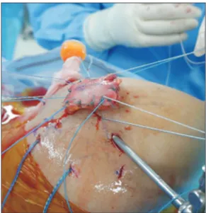
As a solution to these milestones, an augmentation graft made up of patch material was introduced for large-to-massive rotator cuff tears, for irreparable tears, or for poor quality tendons (Fig. 1).
Classification of Patch Grafts
Patch grafts can be classified broadly into xenografts, synthetic grafts, allografts, and autografts. The common harvest sources for xenografts are the porcine dermis and the porcine small intesti- nal submucosa; for synthetic grafts, the polyglycolic acid sheet, polypropylenes, the mersilene mesh, the gore-tex patch, and etc.; for allografts, the human dermal matrix and freeze-dried cadaveric rotator cuffs; and for autografts, the biceps tendon, the fascia lata, and the patellar tendon (Table 1).11-13) In the past, xenografts had been the most widespread form of patch grafts, but they fell out of popularity because, compared to other grafts, they were associated with less favorable structural healing rates.
Its superiority to primary repairs was never demonstrated and there were several reports of immunogenic complications that disfavored their use.14) Further, compared to groups that had not use synthetic grafts or to groups that had used allografts, groups that had used synthetic grafts were associated with bet- ter structural and functional results. Theoretically, these synthetic composite materials can be engineered in a way that key biome- chanical properties, such as strength are optimally manipulated.
But the biological consequences of these synthetic materials, such as foreign body reaction, poor integration, and synovitis, upon insertion into the body may be inevitable. Well designed
controlled studies that investigate the effectiveness of these syn- thetic grafts are still lacking.
Findings from a recent systemic review of animal and clini- cal experiments suggest that the effectiveness of xenografts are questionable, whereas those for synthetic grafts and allografts indicated they were associated with good clinical outcomes.15) A study that estimated re-tear rates according to patch graft type reported that xenografts showed the worst re-tear rate (xenografts [44%] vs. synthetic graft [15%] vs. allografts [23%]).16) These negative findings of xenografts have been succeeded with a corresponding decrease in the use of xenografts in the clinic. As an alternative to xenografts, patch grafts—a type of allografts consisting of human dermal matrix—are increasingly coming under light because of their low incidence of complica- tions, such as disease transmission and graft rejection, and their relatively safety.11,17) Patch grafts are de-cellularized freeze-dried human cadaveric material consisting of collagen type I, II, IV, and VII, elastin, chondroitin sulfate, proteoglycans, and fibroblast growth factors and has in it preserved basement membrane and vascular channels, two constituents which have been shown to help host incorporation.18,19) However, well-designed and long- term studies that investigate the mechanism of patch grafts and their effects are limited. And the possibility of remnant allogenic proteins within the scaffolds means that inflammatory responses may occur and cause tissue degeneration. Furthermore, bony in-growth formation, which is fundamental for functional resto- ration of the tendon, is nonexistent in allografts, restricting their use.12,20-22)
Common harvest sources of autografts include the tensor fas- cia lata, the biceps long head tendon, the hamstring tendon, the patellar tendon, and the Achilles tendon. Because these grafts originate from the person’s own body, they are regarded as the gold-standard choice of graft. Studies on autografts have shown favorable clinical outcomes,23-25) and the successful outcomes have been shown to be dependent on cells within scaffolds that activate collagen synthesis and, thereby, start the process of ten- don re-formation. Accordingly, autografts with viable donor cells are preferred over frozen allografts with unviable donor cells.26) Biomechanically, autografts have been reported to be stronger than allografts.12) However, using allografts has disadvantages of causing donor site morbidity and of limited availability; normally, finding an appropriate graft is difficult because a graft must be both sufficiently thick and sufficiently sturdy to withstand the Fig. 1. Insertion of the patch graft during an arthroscopic rotator cuff repair.
Table 1. Classification of Patch Grafts
Xenografts Porcine dermal graft, porcine small intestine submucosa, fetal bovine dermis
Allografts Human dermal matrix, freeze-dried cadaveric rotator cuffs, freeze-dried cadaveric Achilles tendon
Synthetic grafts Polyglycolic acid sheet, poly-L-lactide polymer, polyurethane polymer, polyethylene terephthalate, mersilene mesh, Gore-Tex patch, carbon fiber tow, polytetrafluoroethylene
Autografts Biceps tendon, fascia lata, patellar tendon, Achilles tendon, quadriceps tendon
early postoperative mechanical loading. Thus, choosing the right graft material is also very important for a successful treatment.
Clinical Results
The research on the clinical outcomes of rotator cuff repairs using various types of patch augmentation is abundant.16,27-32) Most of these studies have investigated large-to-massive tears
while only a few have investigated smaller tears.29,30) However, studies on the effectiveness of collagen patch grafts and poly- propylene patch grafts are extremely limited.28) And the fact that many studies do not include control groups makes it difficult to conduct comparative analyses (Table 2).17,19,29-31,33-47) Studies that compare outcomes of graft augmentation with those of other control surgical techniques, such as the conventional repair tech- nique, are summarized in Table 3.27,28,32,46,48-50)
Table 2. Uncontrolled Clinical Studies on Patch Grafts for Rotator Cuff Tears
Author Type Graft Year No. of
patient Technique Tear size Mean F/U
(mo) Main clinical finding
Ozaki et al.36) Synthetic Polytetrafluoroethylene 1986 25 Open Massive 25 ROM: abduction 44.16→133.2, strength: abduction 3+→4*, ER 3+→4+*
Visuri et al.31) Synthetic Carbon fiber tow 1991 14 Open > 3 cm 48.9 ROM: abduction 72.9→157.1 Metcalf et al.37) Xenograft Porcine small intestine
submucosa
2002 12 Open Massive 24 ROM: FE 30→90, abduction 27→86, ER 0→40,
IR 3→40, strength: abduction 0.8→3.1*, UCLA score 9.3→19.9
Hirooka et al.30) Synthetic Gore-Tex patch 2002 28 Open All size 44 -
Audenaert et al.35)
Synthetic Mersilene mesh 2006 39 Open Massive 43 ROM: FE 69.2→136, abduction 68.4→133.7,
ER 32.4→38.3, IR 3.4→7.5 of 10 points, strength:
abduction 0→7.9†, Constant score 25.7→72.1 Burkhead
et al.38)
Allograft Human dermal matrix 2007 17 Open Massive 14.4 UCLA score 9.06→26.12
Badhe et al.33) Xenograft Porcine dermal collagen 2008 10 Open Massive 52 Strength: abduction 6.3→9.8, Constant score 41.5→62.2 Phipatanakul
and Petersen39)
Xenograft Porcine small intestine submucosa
2009 11 Open Massive 26 ROM: FE 109→126, ER 37→28,
ASES score 36.3→71.8, UCLA score 13.9→25.7 Wong et al.17) Allograft Human dermal matrix 2010 45 Arthroscopic Large to massive 48 UCLA score 18.4→27.5
Nada et al.40) Synthetic Polyethylene terephthalate 2010 21 Mini open Massive 36 ROM: FE 65→120, abduction 60→120, ER 39→57, IR 4.2→8.4, strength: abduction 3.9→5*, Constant score 46.7→84.5
Rotini et al.19) Allograft Human dermal matrix 2011 5 Open/Arthroscopic Large to massive 13.6 Constant score 64→88 Encalada-Diaz
et al.29)
Synthetic Polyurethane polymer 2011 10 Mini open All size 12 ROM: FE 90→160, abduction 70→155, ER 15→30, IR sacrum→T12, ASES score 44→73.3 Gupta et al.41) Allograft Human dermal matrix 2012 24 Mini open Massive 36 ROM: FE 111.7→157.3, abduction 105→151.7,
ER 46.2→65.1, strength: abduction 7.2→9.4§, ER 7.8→9.3§, ASES score 66.6→88.7 Venouziou
et al.34)
Allograft Human dermal matrix 2013 14 Open Massive 30.2 ROM: FE 73.6→129.3, abduction 67.5→117.9, ER 7.9→43.2, ASES score 23.8→72.3 Petrie and
Ismaiel42)
Synthetic Ligament augmentation Reconstruction system
2013 29 Open Massive 40 -
Proctor43) Synthetic Poly-L-lactide polymer 2014 18 Arthroscopic Large to massive 42 ASES score 25→70 Giannotti
et al.45)
Xenograft Porcine dermal collagen patch
2014 9 Open Massive 36 ASES score 38→79, Constant score 42→73
Cho et al.46) Xenograft Porcine dermal collagen 2014 5 Mini open Massive 20.6 ASES score 39.4→86.4, UCLA score 15.4→31.2 Lenart et al.44) Synthetic Poly-L-lactide polymer 2015 13 Open Massive or retear 18 ROM: FE 145→160, ASES score 32.8→74.2 Petri et al.47) Allograft Human dermal matrix 2016 13 Open Large to massive 30 ASES score 64.5→86.0
Numeric data reflect improvement from preoperative baseline data.
F/U: follow-up, ROM: range of motion, ER: external rotation, FE: forward elevation, IR: internal rotation, UCLA: University of California-Los Angeles, ASES:
American Shoulder and Elbow Surgeons Evaluation Form.
*Based on the 5-point Medical Research Council Scale. †Power assessed as part of Constant-Murley score. §Based on the 10-point Modified Medical Research Council Scale.
Table 3. Controlled Clinical Studies on Patch Grafts for Rotator Cuff Tearss AuthorsTypeGraftYearNo. of patient TechniqueTear sizeMean F/U (mo)Control groupClinical outcomesRadiologic outcomes OutcomeComparisonOutcomeComparison Iannotti et al.49)XenograftPorcine small intestine submucosa
200615OpenLarge to massive14ConventionalPenn: 83 (patch), 91 (control)Significant (control>patch)MRI at 1 year 4/15 (patch) vs. 9/19 (control)Not significant Walton et al.32)XenograftPorcine small intestine submucosa 200716OpenLarge to massive24ConventionalLess strength (lift-off, IR, adduction), slower rate of resolution of pain, less sports participation in xenograft Significant (control>patch)MRI mean tendon thickness 1.50 mm (patch) vs. 1.58 mm (control)
Not significant Barber et al.27)AllograftHuman dermal matrix201222ArthroscopicLarge to massive24ConventionalASES (p=0.035), Constant (p=0.008), UCLA (p=0.43)
SignificantMRI intact cuffs in 85% of repairs in patch group vs 40% in control (p<0.001)
Significant Modi et al.48)AllograftHuman dermal matrix201361Open> 3 cm43.2Partial repairConstant (p=0.001), ASES (p=0.021), VAS (p=0.028)
SignificantMRI Graft: 8.3% retear, control: 41.7% retear (p=0.015)Significant Ciampi et al. (synthetic)28)SyntheticPolypropylene201452OpenMassive36ConventionalUCLA score at 36 months was significantly higher (p<0.001)SignificantUSG after 12 months 41% (21/51) for the control group vs. 17% (9/52) for the polypropylene group
Significant Ciampi et al. (collagen)28)XenograftBovine pericardium derived collagen
201449OpenMassive36ConventionalUCLA score at 36 months was not significantly higherNot significantUSG after 12 months 41% (21/51) for the control group vs. 51% (25/49) for the collagen group,
Not significant Yoon et al.50)AllograftHuman dermal matrix201621ArthroscopicMassive23Conventional1 year and final follow-up: not significant (VAS, SST, UCLA, Constant, ASES)
Not significantMRI at 12 months retear: 19.0% (patch) vs. 46.3% (control)Significant F/U: follow-up, IR: internal rotation, ASES: American Shoulder and Elbow Surgeons Evaluation Form, UCLA: University of California-Los Angeles, VAS: visual analog scale, SST: simple shoulder test, MRI: mag- netic resonance imaging, USG: ultrasonography.
In almost all studies, whichever graft was used, clinical pa- rameters, such as range of motion, the American Shoulder and Elbow Surgeons Evaluation Form (ASES) score, the University of California-Los Angeles (UCLA) score, and the Constant score sig- nificantly improved with patch augmentation. However, many of these studies do not have a control group; thus, we cannot be certain that these improved results are attributable to patch augmentation. In terms of graft type, we found that compared to other patch grafts, the synthetic grafts had significantly larger improvements in forward flexion and in abduction, whilst al- lografts showed the highest improvement in external rotation.16) The anatomical results obtained using postoperative follow-up imaging were compared by study (Table 3);27,28,32,46,48-50) interest- ingly, we found that most of the studies that had reported no sig- nificant difference between grafting and the conventional repair had used xenografts.
Studies generally showed improved muscle strength after patch augmentation, in particular a significant improvement in abduction strength.28,33) A few studies also showed that external rotation strength is improved after grafting.34-36,48) However, Wal- ton et al.32) presented anomalous data wherein patients without patch augmentation compared to those with had better abduc- tion strength. Similarly, a different study reported that patients who had not received augmentation had greater external rota- tion strength than those who had received augmentation. Inter- estingly, the patients who had not received patch augmentation in this study had been treated using xenografts (restore orthobio- logic implant).32)
Almost all studies on patch augmentation report improve- ments in clinical parameters, specifically the ASES, the UCLA, the Constant, the Penn, and the Oxford scores. These improve- ments appeared irrespectively of graft type, but when comparing xenografts to synthetic grafts and allografts, we found that their improvement was relatively smaller. Other clinical parameters such as pain, satisfaction, ADL performance, and return to sports or to daily activities showed significant improvements in various
papers.
In the literature, the rates of retear measured through post- operative evaluation of anatomical outcomes show a substantial variation ranging from 8.3% to 73.4%, depending on the surgi- cal indication, the treatment method, and the mode of assess- ment.50) A recent systemic review on 22 studies calculating retear rates showed that the overall retear rate was 22% for complete tears and 2.7% for partial tears.16) Among complete tears, the overall retear rates by graft type were 15.0% for synthetic grafts, 42.0% for xenograft, and 9.9% for allografts. Among partial tears, the corresponding percent values were 0%, 1.7%, and 12.7%, respectively.16)
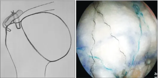
A few explanations are given in the literature for how patch augmentation may reduce the occurrence of retears.50) For ex- ample, it has been suggested to reduce the tension applied to the torn tendon by pulling the edges of torn tendons in massive cuff tears toward the lateral footprint (Fig. 2). Taking the same action (that is, pulling the torn tendons as laterally as possible to cover the rotator cuff footprint) in medial row repairs would ex- acerbate tension overloading because massive rotator cuff tears usually have much retracted and stiffened tendons. Thus, patch graft repairs in contrast would allow less lateralization of the torn tendon ends, resulting in less tension.
Postoperative complications differentially appear by graft type; for instance, xenografts are often associated with infection- induced immune responses. The more recent types—allografts and autografts—are commonly associated with bursitis, deep infection, skin rash, severe inflammatory reaction, humeral frac- ture, and cystic changes of the humeral head. Besides these, complications that generally occur with rotator cuff repairs have all been reported with grafting.
Consensus for Patch Grafts
For the treatment of massive or irreparable cuff tears, treat- ment modalities other than patch augmentation may be indi-
Fig. 2. Patch augmentations can decrease the initial high tension applied to repair sites in massive rotator cuff tears.
cated; a few examples are conservative treatment, subacromial debridement and decompression, partial repair, muscle transfer, superior capsular reconstruction, subacromial spacer, and re- verse shoulder arthroplasty. Each treatment modality has been reported with favorable clinical outcomes.51-56)
Controversy as to whether patch augmentation is the most ideal treatment for massive or irreparable cuff arthopathies re- mains, and some studies have attempted to answer this. In the American Academy of Orthopaedic Surgeons (AAOS)’s guide- lines,57) xenografts are not recommended, while partial repairs, debridement, and muscle transfer are weakly recommended.
And whilst allografts are not recommended, the guidelines states there is not sufficient evidence to back their claim. In a survey that asked ASES members whether they agreed to the AAOS guidelines,58) 86.5% of respondents agreed that partial repairs are appropriate for irreparable cuff tears and 64.6% agreed that debridement is appropriate. Only 11.2% and 45.4% of respon- dents said that allografts should be used for primary treatment and for revision treatment, respectively. Taking these findings into account, it seems that there is yet insufficient evidence, both empirically and consensually, that justifies the use of patch grafts.
Conclusions
No gold standard of treatment exists for massive rotator cuff tears, especially for irreparable cuff tears. These tears have been shown to be challenging to treat in young and active patients.
The antecedent xenografts for cuff tear arthropathies were as- sociated with unfavorable clinical outcomes and postoperative complications. For these reasons xenografts are no longer used in the clinic. The more current patch augmentation such as through human dermal allografts has been reported to have favorable outcomes, but well-designed, long-term randomized control studies are required to evaluate the cost-effectiveness of this treatment. Because there is a wide range of options for irreparable rotator cuff tears besides patch augmentation, such as debridement, partial repair, superior capsular reconstruction, and reverse arthroplasty, the choice of treatment should be care- fully selected on the basis of the relative benefits and limitations of each method.
References
1. Zumstein MA, Jost B, Hempel J, Hodler J, Gerber C. The clinical and structural long-term results of open repair of massive tears of the rotator cuff. J Bone Joint Surg Am. 2008;90(11):2423-31.
2. Melillo AS, Savoie FH 3rd, Field LD. Massive rotator cuff tears: debridement versus repair. Orthop Clin North Am.
1997;28(1):117-24.
3. Moser M, Jablonski MV, Horodyski M, Wright TW. Functional outcome of surgically treated massive rotator cuff tears: a com-
parison of complete repair, partial repair, and debridement.
Orthopedics. 2007;30(6):479-82.
4. Burkhart SS, Barth JR, Richards DP, Zlatkin MB, Larsen M. Ar- throscopic repair of massive rotator cuff tears with stage 3 and 4 fatty degeneration. Arthroscopy. 2007;23(4):347-54.
5. Galatz LM, Ball CM, Teefey SA, Middleton WD, Yamaguchi K.
The outcome and repair integrity of completely arthroscopi- cally repaired large and massive rotator cuff tears. J Bone Joint Surg Am. 2004;86(2):219-24.
6. Boileau P, Brassart N, Watkinson DJ, Carles M, Hatzidakis AM, Krishnan SG. Arthroscopic repair of full-thickness tears of the supraspinatus: does the tendon really heal? J Bone Joint Surg Am. 2005;87(6):1229-40.
7. Cole BJ, McCarty LP 3rd, Kang RW, Alford W, Lewis PB, Hayden JK. Arthroscopic rotator cuff repair: prospective func- tional outcome and repair integrity at minimum 2-year follow- up. J Shoulder Elbow Surg. 2007;16:579-85.
8. Coleman SH, Fealy S, Ehteshami JR, et al. Chronic rotator cuff injury and repair model in sheep. J Bone Joint Surg Am.
2003;85(12):2391-402.
9. Fuchs B, Weishaupt D, Zanetti M, Hodler J, Gerber C. Fatty degeneration of the muscles of the rotator cuff: assessment by computed tomography versus magnetic resonance imaging. J Shoulder Elbow Surg. 1999;8(6):599-605.
10. Goutallier D, Postel JM, Lavau L, Bernageau J. Impact of fatty degeneration of the suparspinatus and infraspinatus msucles on the prognosis of surgical repair of the rotator cuff. Rev Chir Orthop Reparatrice Appar Mot. 1999;85(7):668-76.
11. Bond JL, Dopirak RM, Higgins J, Burns J, Snyder SJ. Ar- throscopic replacement of massive, irreparable rotator cuff tears using a graftjacket allograft: technique and preliminary results. Arthroscopy. 2008;24(4):403-9.e1.
12. Derwin KA, Baker AR, Spragg RK, Leigh DR, Iannotti JP. Com- mercial extracellular matrix scaffolds for rotator cuff tendon repair. Biomechanical, biochemical, and cellular properties. J Bone Joint Surg Am. 2006;88(12):2665-72.
13. Soler JA, Gidwani S, Curtis MJ. Early complications from the use of porcine dermal collagen implants (Permacol) as bridging constructs in the repair of massive rotator cuff tears. A report of 4 cases. Acta Orthop Belg. 2007;73(4):432-6.
14. Ferguson DP, Lewington MR, Smith TD, Wong IH. Graft utilization in the augmentation of large-to-massive rota- tor cuff repairs: a systematic review. Am J Sports Med.
2016;44(11):2984-92.
15. Thangarajah T, Pendegrass CJ, Shahbazi S, Lambert S, Alexander S, Blunn GW. Augmentation of rotator cuff repair with soft tissue scaffolds. Orthop J Sports Med.
2015;3(6):2325967115587495.
16. Steinhaus ME, Makhni EC, Cole BJ, Romeo AA, Verma NN.
Outcomes after patch use in rotator cuff repair. Arthroscopy.
2016;32(8):1676-90.
17. Wong I, Burns J, Snyder S. Arthroscopic graftjacket repair of rotator cuff tears. J Shoulder Elbow Surg. 2010;19(2 Sup- pl):104-9.
18. Nasca RJ. The use of freeze-dried allografts in the manage- ment of global rotator cuff tears. Clin Orthop Relat Res.
1988;(228):218-26.
19. Rotini R, Marinelli A, Guerra E, et al. Human dermal matrix scaffold augmentation for large and massive rotator cuff re- pairs: preliminary clinical and MRI results at 1-year follow-up.
Musculoskelet Surg. 2011;95 Suppl 1:S13-23.
20. Gilbert TW, Freund JM, Badylak SF. Quantification of DNA in biologic scaffold materials. J Surg Res. 2009;152(1):135-9.
21. Cheung EV, Silverio L, Sperling JW. Strategies in biologic aug- mentation of rotator cuff repair: a review. Clin Orthop Relat Res. 2010;468(6):1476-84.
22. Bedi A, Maak T, Walsh C, et al. Cytokines in rotator cuff de- generation and repair. J Shoulder Elbow Surg. 2012;21(2):218- 27.
23. Sano H, Kumagai J, Sawai T. Experimental fascial autografting for the supraspinatus tendon defect: remodeling process of the grafted fascia and the insertion into bone. J Shoulder Elbow Surg. 2002;11(2):166-73.
24. Cho NS, Yi JW, Rhee YG. Arthroscopic biceps augmentation for avoiding undue tension in repair of massive rotator cuff tears. Arthroscopy. 2009;25(2):183-91.
25. Mori D, Funakoshi N, Yamashita F. Arthroscopic surgery of irreparable large or massive rotator cuff tears with low- grade fatty degeneration of the infraspinatus: patch autograft procedure versus partial repair procedure. Arthroscopy.
2013;29(12):1911-21.
26. Tachiiri H, Morihara T, Iwata Y, et al. Characteristics of donor and host cells in the early remodeling process after transplant of Achilles tendon with and without live cells for the treat- ment of rotator cuff defect: what is the ideal graft for the treat- ment of massive rotator cuff defects? J Shoulder Elbow Surg.
2010;19(6):891-8.
27. Barber FA, Burns JP, Deutsch A, Labbé MR, Litchfield RB. A prospective, randomized evaluation of acellular human der- mal matrix augmentation for arthroscopic rotator cuff repair.
Arthroscopy. 2012;28(1):8-15.
28. Ciampi P, Scotti C, Nonis A, et al. The benefit of synthetic ver- sus biological patch augmentation in the repair of posterosu- perior massive rotator cuff tears: a 3-year follow-up study. Am J Sports Med. 2014;42(5):1169-75.
29. Encalada-Diaz I, Cole BJ, Macgillivray JD, et al. Rotator cuff re- pair augmentation using a novel polycarbonate polyurethane patch: preliminary results at 12 months’ follow-up. J Shoulder Elbow Surg. 2011;20(5):788-94.
30. Hirooka A, Yoneda M, Wakaitani S, et al. Augmentation with a Gore-Tex patch for repair of large rotator cuff tears that cannot be sutured. J Orthop Sci. 2002;7(4):451-6.
31. Visuri T, Kiviluoto O, Eskelin M. Carbon fiber for repair of the rotator cuff. A 4-year follow-up of 14 cases. Acta Orthop Scand. 1991;62(4):356-9.
32. Walton JR, Bowman NK, Khatib Y, Linklater J, Murrell GA.
Restore orthobiologic implant: not recommended for aug- mentation of rotator cuff repairs. J Bone Joint Surg Am.
2007;89(4):786-91.
33. Badhe SP, Lawrence TM, Smith FD, Lunn PG. An assessment of porcine dermal xenograft as an augmentation graft in the treatment of extensive rotator cuff tears. J Shoulder Elbow Surg. 2008;17(1 Suppl):35S-9S.
34. Venouziou AI, Kokkalis ZT, Sotereanos DG. Human dermal allograft interposition for the reconstruction of massive ir- reparable rotator cuff tears. Am J Orthop (Belle Mead NJ).
2013;42(2):63-70.
35. Audenaert E, Van Nuffel J, Schepens A, Verhelst M, Verdonk R.
Reconstruction of massive rotator cuff lesions with a synthetic interposition graft: a prospective study of 41 patients. Knee Surg Sports Traumatol Arthrosc. 2006;14(4):360-4.
36. Ozaki J, Fujimoto S, Masuhara K, Tamai S, Yoshimoto S. Re- construction of chronic massive rotator cuff tears with synthetic materials. Clin Orthop Relat Res. 1986;(202):173-83.
37. Metcalf MH, Savoie FH, Kellum B. Surgical technique for xe- nograft (SIS) augmentation of rotator-cuff repairs. Oper Tech Orthop. 2002;12(3):204-8.
38. Burkhead WZ Jr., Schiffern SC, Krishnan SG. Use of graft jacket as an augmentation for massive rotator cuff tears. Semin Arthroplast. 2007;18(1):11-8.
39. Phipatanakul WP, Petersen SA. Porcine small intestine submu- cosa xenograft augmentation in repair of massive rotator cuff tears. Am J Orthop (Belle Mead NJ). 2009;38(11):572-5.
40. Nada AN, Debnath UK, Robinson DA, Jordan C. Treatment of massive rotator-cuff tears with a polyester ligament (Da- cron) augmentation: clinical outcome. J Bone Joint Surg Br.
2010;92(10):1397-402.
41. Gupta AK, Hug K, Berkoff DJ, et al. Dermal tissue allograft for the repair of massive irreparable rotator cuff tears. Am J Sports Med. 2012;40(1):141-7.
42. Petrie MJ, Ismaiel AH. Treatment of massive rotator-cuff tears with a polyester ligament (LARS) patch. Acta Orthop Belg.
2013;79(6):620-5.
43. Proctor CS. Long-term successful arthroscopic repair of large and massive rotator cuff tears with a functional and degradable reinforcement device. J Shoulder Elbow Surg.
2014;23(10):1508-13.
44. Lenart BA, Martens KA, Kearns KA, Gillespie RJ, Zoga AC, Wil- liams GR. Treatment of massive and recurrent rotator cuff tears augmented with a poly-l-lactide graft, a preliminary study. J Shoulder Elbow Surg. 2015;24(6):915-21.
45. Giannotti S, Ghilardi M, Dell’osso G, et al. Study of the por- cine dermal collagen repair patch in morpho-functional recov-
ery of the rotator cuff after minimum follow-up of 2.5 years.
Surg Technol Int. 2014;24:348-52.
46. Cho CH, Lee SM, Lee YK, Shin HK. Mini-open suture bridge repair with porcine dermal patch augmentation for massive ro- tator cuff tear: surgical technique and preliminary results. Clin Orthop Surg. 2014;6(3):329-35.
47. Petri M, Warth RJ, Horan MP, Greenspoon JA, Millett PJ.
Outcomes after open revision repair of massive rotator cuff tears with biologic patch augmentation. Arthroscopy.
2016;32(9):1752-60.
48. Modi A, Singh HP, Pandey R, Armstrong A. Management of irreparable rotator cuff tears with the graftjacket allograft as an interpositional graft. Shoulder Elbow. 2013;5(3):188-94.
49. Iannotti JP, Codsi MJ, Kwon YW, Derwin K, Ciccone J, Brems JJ. Porcine small intestine submucosa augmentation of surgical repair of chronic two-tendon rotator cuff tears. A randomized, controlled trial. J Bone Joint Surg Am. 2006;88(6):1238-44.
50. Yoon JP, Chung SW, Kim JY, et al. Outcomes of combined bone marrow stimulation and patch augmentation for massive rotator cuff tears. Am J Sports Med. 2016;44(4):963-71.
51. Burkhart SS. Partial repair of massive rotator cuff tears: the evo- lution of a concept. Orthop Clin North Am. 1997;28(1):125-32.
52. Drake GN, O’Connor DP, Edwards TB. Indications for reverse total shoulder arthroplasty in rotator cuff disease. Clin Orthop Relat Res. 2010;468(6):1526-33.
53. Lo IK, Lind CC, Burkhart SS. Glenohumeral arthroscopy por- tals established using an outside-in technique: neurovascular anatomy at risk. Arthroscopy. 2004;20(6):596-602.
54. Namdari S, Voleti P, Baldwin K, Glaser D, Huffman GR. Latis- simus dorsi tendon transfer for irreparable rotator cuff tears: a systematic review. J Bone Joint Surg Am. 2012;94(10):891-8.
55. Senekovic V, Poberaj B, Kovacic L, Mikek M, Adar E, Dekel A. Prospective clinical study of a novel biodegradable sub- acromial spacer in treatment of massive irreparable rotator cuff tears. Eur J Orthop Surg Traumatol. 2013;23(3):311-6.
56. Moore DR, Cain EL, Schwartz ML, Clancy WG Jr. Allograft reconstruction for massive, irreparable rotator cuff tears. Am J Sports Med. 2006;34(3):392-6.
57. Tashjian RZ. AAOS clinical practice guideline: optimizing the management of rotator cuff problems. J Am Acad Orthop Surg. 2011;19(6):380-3.
58. Paxton ES, Matzon JL, Narzikul AC, Beredjiklian PK, Abboud JA. Agreement among ASES members on the AAOS clinical practice guidelines. Orthopedics. 2015;38(3):e169-77.