대한소화기학회지 2008;52:203-205 □ IMAGE OF THE MONTH □
연락처: 천재희, 120-752, 서울시 서대문구 신촌동 134 연세대학교 의과대학 내과학교실
Tel: (02) 2228-1990, Fax: (02) 393-6884 E-mail: [email protected]
Correspondence to: Jae Hee Cheon, M.D.
Department of Internal Medicine, Yonsei University College of Medicine, 134, Shinchon-dong, Seodaemun-gu, Seoul 120- 752, Korea
Tel: +82-2-2228-1990, Fax: +82-2-393-6884 E-mail: [email protected]
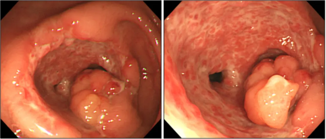
Fig. 1. Initial colonoscopic find- ings. A protruding polypoid mass with surface nodularity is noted at the ileocecal area. On adjacent mucosa, discrete and deep ulcer- ations are accompanied.
대장암으로 오인된 장결핵
연세대학교 의과대학 내과학교실, 소화기병연구소
박재준ㆍ천재희
Intestinal Tuberculosis Mimicking Colon Cancer
Jae Jun Park, M.D. and Jae Hee Cheon, M.D.Department of Internal Medicine and Institute of Gastroenterology, Yonsei University College of Medicine, Seoul, Korea
증례: 43세 남자 환자가 약 4개월간의 복부 통증을 주소로 내원하였다. 특이 과거력 없던 환자는 내원 한 달 전 하복부 통증 있어 대장내시경 검사를 받았으며 상행결장에 궤양을 동반한 종괴가 관찰되어 대장암 의심하에 추가 검사와 치료 를 위해 전원되었다. 내원 당시 혈압 100/60 mmHg, 맥박 78 회/분, 호흡수 20회/분, 체온 37.1oC였다. 환자의 전신상태는 양호하였으며 의식은 명료하였다. 복부 진찰에서 우하복부에 경미한 압통이 있었다. 말초혈액검사에서 혈색소 14.5 g/dL, 백혈구 수 7,730/mm3, 호중구 65.2%, 혈소판 289,000/mm3, 혈 액 침강속도 16 mm/hr였고, 혈액응고검사와 소변검사는 정 상이었다. 생화학검사에서 혈액요소질소 7.8 mg/dL, 크레아 티닌 0.6 mg/dL, 혈당 81 mg/dL, 칼슘 8.7 mg/dL, 총 빌리루
빈 0.4 mg/dL, ALT 31 IU/L, AST 21 IU/L, 총 단백 7.5 g/dL, 알부민 3.9 d/dL, 요산 4.3 mg/dL이었고, 혈청 검사에서 CRP 는 0.650 mg/dL이었다. 흉부 X선 검사와 단순복부 촬영에서 이상 소견은 없었다.
대장내시경검사에서 회맹부위에 2×4 cm 크기의 표면에 궤양 및 소결절 변화를 동반한 융기 병변이 관찰되었다(Fig.
1). 내시경으로는 베체트장염, 장결핵, 대장암이 감별 진단 이었다. 복부전산화단층촬영에서 상행결장 근위부에 돌출 종괴와 결장주위 지방층의 침윤, 국소 림프절 종대와 복막 비후, 골반강 내의 소량의 복수가 관찰되었다(Fig. 2). 영상 진단은 주위 림프절 전이를 동반한 상행결장암이었다. 처음 시행한 종괴의 조직병리검사에서 비특이적인 궤양 소견만
204 대한소화기학회지: 제52권 제4호, 2008
Fig. 2. Abdominal CT scan shows a polypoid mass at the proximal ascending colon. Pericolic fat infiltration, lymph node enlargement, peritoneal thickiening, and small amounts of ascites are noted.
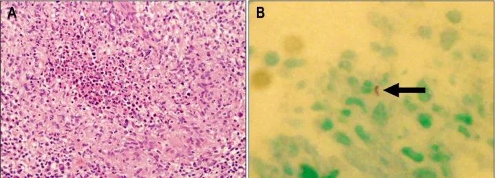
Fig. 3. Histologic findings. (A) It shows ill-defined chronic granulomatous inflammation with caseation necrosis (H&E stain, ×200). (B) An acid-fast bacilli is seen on Ziehl-Neelsen stain (arrow).
관찰되어 조직검사를 재시행하였으며 두 번째 조직검사에 서 건락 괴사를 동반한 만성 육아종 염증소견과 Ziehl- Neelsen 염색에서 항산균이 관찰되었다(Fig. 3). 진단은?
진단: 궤양비후형 병변으로 발현하여 대장암과 감별이 필 요한 대장 결핵
환자는 항결핵제 투여를 시작하였고 6개월째 시행한 추적 내시경검사에서 이전에 보였던 상행결장의 종괴는 크기가 감소하였고 주변의 궤양도 호전되어 반흔과 변형이 관찰되 었다(Fig. 4).
장결핵은 폐외결핵 감염 중 아주 높은 빈도를 보이는 질 환으로1 원발 또는 속발로 발생한다. 감염 경로는 혈행 파 종, 활동 폐결핵 환자에서 오염된 객담의 삼킴, 오염된 우유 나 음식 섭취, 인접한 주위 장기로부터의 전파 등으로 설명 된다.2 과거에는 장결핵이 활동 폐결핵과 관련이 있었으나 근래에는 장결핵 환자의 50% 이하에서만 폐결핵이 발견된
다.2-4 장결핵은 위장관의 어느 곳이나 침범할 수 있으나 호 발하는 부위는 회맹부이며 회맹부 이외의 장결핵 호발 부위 는 주로 상행결장과 횡행결장으로 분절 장염 양상으로 나타
난다.3,5-7 임상 증상으로는 복통이 가장 흔한 증상이며 그 외
증상으로 발열, 식욕 감퇴, 설사, 체중 감소, 변비, 출혈 등이 있다.8,9 내시경에서는 점막 궤양과 결절, 아프타 궤양, 부종 점막 주름, 협착, 가폴립, 관내강 협착 등 다양한 소견을 보 일 수 있다.10 병변의 형태에 따라 크게 궤양형, 궤양비후형, 비후형의 세 가지 범주로 나눌 수 있으며 궤양형이 가장 흔 한 형태이다.2,3 궤양비후형이나 비후형의 경우는 암과의 감 별이 어려울 수 있다. 복부전산화단층촬영에서는 광범위한 림프절 종대, 비장 및 간 종대, 복수 등의 소견을 보일 수 있으나 비특이적이다.11
진단은 기본적으로 임상증상, 영상 검사, 내시경 검사 등 을 종합하여 조직 검체에서 건락 육아종을 확인하거나, 항 산균이 보이거나, 조직 배양에서 결핵균이 배양되면 진단
박재준 외 1인. 대장암으로 오인된 장결핵 205
Fig. 4. Colonoscopic findings after 6 months of anti-turberculous treat- ment. The size of the polypoid mass is reduced and active ulcer is healed into scar.
할 수 있으나 이러한 검사에서 양성을 보이는 경우는 50%
미만이며,12,13 배양 결과와 조직 검사 결과가 음성이라도 임 상적으로 장결핵이 의심되고 항결핵 치료에 반응이 있는 경 우 역시 장결핵으로 진단하여 치료가 가능하다.3,7 조직검사 시 육아종은 주로 점막하에서 발견되므로 궤양 변연과 궤양 의 심층부에서 다발 조직생검을 해야한다.14
이번 증례는 내시경에서 상행결장의 궤양 비후 종괴와 복 부전산화단층촬영에서 결장주위 지방층의 침윤, 국소림프 절종대와 복막 비후, 골반강 내의 소량의 복수 등 악성 종양 과 유사한 소견을 보였으나 반복하여 시행한 조직 검체에서 건락 육아종 소견과 항산균 염색양성 소견으로 장결핵이 확 진되었고 항결핵제 투여 후 호전되었다.
참고문헌
1. al Karawi MA, Mohamed AE, Yasawy MI, et al. Protean manifestation of gastrointestinal tuberculosis: report on 130 patients. J Clin Gastroenterol 1995;20:225-232.
2. Horvath KD, Whelan RL. Intestinal tuberculosis: return of an old disease. Am J Gastroenterol 1998;93:692-696.
3. Marshall JB. Tuberculosis of the gastrointestinal tract and peritoneum. Am J Gastroenterol 1993;88:989-999.
4. Kim KM, Lee A, Choi K, Lee KY, Kwak JJ. Intestinal tuber- culosis: clinicopathologic analysis and diagnosis by endo- scopic biopsy. Am J Gastroenterol 1998;93:606-609.
5. Singh V, Kumar P, Kamal J, Prakash V, Vaiphei K, Singh K.
Clinicocolonoscopic profile of colonic tuberculosis. Am J Gastroenterol 1996;91:565-568.
6. Chen WS, Leu SY, Hsu H, Lin JK, Lin TC. Trend of large bowel tuberculosis and the relation with pulmonary tuberculosis. Dis Colon Rectum 1992;35:189-192.
7. Shah S, Thomas V, Mathan M, et al. Colonoscopic study of 50 patients with colonic tuberculosis. Gut 1992;33:347-351.
8. Choi SM, Yang SK, Jung HY, et al. Clinical features of in- testinal tuberculosis with special reference to risk factors for complications. Korean J Gastroenterol 1997;30:462-471.
9. Yang US, Cho M, Song GA, et al. Endoscopic findings of colonic tuberculosis. Korean J Gastrointest Endosc 1996;16:
724-732.
10. Ferentzi CV, Sieck JO, Ali MA. Colonoscopic diagnosis and medical treatment of ten patients with colonic tuberculosis.
Endoscopy 1988;20:62-65.
11. Hulnick DH, Megibow AJ, Naidich DP, Hilton S, Cho KC, Balthazar EJ. Abdominal tuberculosis: CT evaluation. Radio- logy 1985;157:199-204.
12. Gan HT, Chen YQ, Ouyang Q, Bu H, Yang XY. Differ- entiation between intestinal tuberculosis and Crohn's disease in endoscopic biopsy specimens by polymerase chain reac- tion. Am J Gastroenterol 2002;97:1446-1451.
13. Amarapurkar DN, Patel ND, Amarapurkar AD, Agal S, Baigal R, Gupte P. Tissue polymerase chain reaction in diag- nosis of intestinal tuberculosis and Crohn's disease. J Assoc Physicians India 2004;52:863-867.
14. Youn JE, Park IB, Kwon SY, et al. Follow-up colonoscopy at 3 months of therapy in patients with tentative diagnosis of in- testinal tuberculosis. Korean J Intern Med 1996;50:227-234.