culosis according to the Amount of Pleural Effusion Specimens
Department of Internal Medicine, Ewha Womans University College of Medicine, Seoul, Korea Jin Wook Moon, M.D.
흉막 결핵의 진단에 있어서 흉수 검체양에 따른 중합효소연쇄반응 검사의 민감도
문 진 욱
이화여자대학교 의과대학 내과학교실
연구 배경: 결핵성 흉막염의 진단에 있어서 흉수에 대한 중합효소연쇄반응 검사는 매우 낮은 민감도를 보였으며, 이는 흉수 내의 결핵균의 수가 적은 데에 기인하는 것으로 추정된다. 이번 연구에서는 흉수의 검체양을 증가시켰을 때 결핵성 흉수에 대한 중합효소연쇄반응 검사의 민감도가 향상되는지를 알아보고자 하였다.
방 법: 결핵성 흉막염에 대한 감별 진단이 필요하였던 53명의 환자에 대하여 전향적인 연구를 시행하였다. 각각의 환자로 부터 얻은 흉수를 10 ml, 25 ml, 50 ml로 양을 달리 하여 결핵균에 대한 중합효소연쇄반응 검사를 시행하였으며, Cobas Amplicor MTB (Roche Diagnostic Systems)를 이용하였다. 결핵성 흉막염은 흉수에서 결핵균이 배양된 경우, 흉막 생검에 서 결핵이 진단된 경우, 가래에서 결핵균이 배양된 경우 및 기타 원인이 배제된 상태에서 임상적으로 결핵성 흉수가 의심 되며 항결핵제 투약으로 흉수가 호전된 경우를 포함하였다.
결 과: 53명의 환자 중 26명이 결핵성 흉막염으로 진단되었다. 흉수에 대한 항산균 바름질, 결핵균 배양, 아데노신 디아미 나아제 측정 및 흉막 생검의 민감도는 각각 3.8%, 15.4%, 88.5%, 84.6%이었다. 10 ml, 25 ml, 50 ml의 흉수를 이용한 결핵 균 중합효소연쇄반응 검사는 각각 3명(11.5%), 4명(15.4%), 3명(11.5%)에서 양성이었으며, 통계적으로 유의한 차이를 보이 지 않았다(p>0.05, symmetry exact 검사).
결 론: 결핵균의 수가 극히 적을 것으로 예상되는 흉수 등의 검사물에 대해서는 결핵균 중합효소연쇄반응 검사의 임상적 유용성이 극히 제한적이며, 검사물의 양을 증가시키더라도 민감도는 향상되지 않는다.
(Tuberc Respir Dis 2007; 62: 184-191)
Key words: Amplicor, Pleural effusion, Polymerase chain reaction (PCR), Sensitivity, Tuberculosis.
Address for correspondence: Jin Wook Moon, M.D.
Department of Internal Medicine, Ewha Womans University College of Medicine, 911-1 Mok-6-dong, Yangcheon-gu, Seoul, Korea.
Phone: 82-2-2650-2507, Fax: 82-2-2655-2076 E-mail: [email protected]
Received: Feb. 8. 2007 Accepted: Feb. 28. 2007
INTRODUCTION
The recent increase in tuberculosis cases and the emergence of multi-drug resistant strains have called for more rapid and sensitive methods in the laboratory diagnosis than the conventional diagnostic techniques
1-3. Although the culture of Mycobac- terium tuberculosis (MTB) continues to be the gold standard for the diagnosis of tuberculosis, the results are neither satisfactorily sensitive nor rapid.
The mycobacterial culture takes at least two weeks
or longer depending on culture media used
4. The nucleic acid amplification tests (NATs) including polymerase chain reaction (PCR) improved the accuracy as well as the time for the diagnosis of tuberculosis in respiratory specimens. For non- respiratory specimens like pleural effusion, however, previous studies have shown highly variable results about the usefulness of NAT because they used different in-house NAT methods, rather small study populations, and diverse criteria of pleural tuberculosis.
The Cobas Amplicor MTB test (Roche Diag-
nostic Systems, Inc., Branchburg, NJ, USA) is a
well-established and commercially available PCR
technique commonly used for the direct detection of
M. tuberculosis in clinical samples. The test uses
biotinylated genus-specific primers (KY18 and
KY75) to amplify the 584-base-pair sequence
within the 1500-base-pair region encoding 16s rRNA of M. tuberculosis. It combines five instruments into one (thermal cycler, automatic pipettor, incubator, washer and reader). Because it is fully automated for the amplification and detection steps on a single instrument, it is thought to be able to minimize inter-individual variabilities.
Furthermore, since it binds M. tuberculosis-specific oligonucleotide probes to the amplified sequences, it can increase the overall specificity.
Tuberculous pleural effusion occurs in up to 30%
of patients with tuberculosis
5and occupies the major portion of the extrapulmonary tuberculosis morbidity
6. However, the number of organisms in the pleural effusion from most cases of tuberculous pleuritis is relatively low, with positive cultures found in less than 25% of cases. Even the pleural biopsy shows granulomatous inflammation only in approximately 60% of patients
7. PCR has been used to detect M. tuberculosis in pleural fluid samples, with highly variable sensitivities (11 to 81%) in the previous studies using different in-house PCR methods
8-13. Our previous study has, however, demonstrated very low sensitivity of PCR technique for the diagnosis of pleural tuberculosis (sensitivity 17.5%, specificity 98.1%) using the kit developed for the diagnosis of pulmonary tuberculosis
14. The purpose of this investigation is to determine whether the sensitivity of PCR technique for the diagnosis of pleural tuberculosis can be improved when increasing the amount of pleural effusion specimens.
MATERIALS AND METHODS
Study population and samples
Fifty three patients older than 18 years of age, for whom the exclusion of the possibility of tuberculous pleural effusion was necessary, were prospectively
analyzed for one year. The suspicion of pleural tuberculosis was based on the unilateral pleural effusion with clinical manifestations suggestive of tuberculosis such as productive cough, chronic low-grade fever, weight loss, anorexia, night sweating, etc. All of the 53 pleural effusion specimens were sent for routine analysis, acid-fast bacilli (AFB) smear, mycobacterial culture, adeno- sine deaminase (ADA) level, Gram stain, bacterial culture, and cytologic examination. For the comparison of the sensitivities of pleural effusion PCR test according to the amount of pleural effusion specimens, M. tuberculosis PCR was performed with 10, 25, and 50 ㎖ of each pleural effusion sample, respectively. The sputum was collected in the morning from each patient and was sent for AFB smear and mycobacterial culture.
Pleural biopsy and pathologic examination were not performed in 36 out of the 53 patients due to a small amount of pleural effusion and/or patient's refusal.
Diagnostic criteria of pleural tuberculosis Cases of pleural tuberculosis were defined as those with one of the following: positive M.
tuberculosis culture of pleural fluid, and/or histopathologic finding consistent with tuberculosis on pleural biopsy, and/or positive M. tuberculosis culture of sputum, and/or positive M. tuberculosis culture of other biologic specimens, and/or positive response to anti-tuberculous medication without other possible causes of pleural effusion.
AFB smear and mycobacterial culture
For a microscopic examination, Ziehl-Neelsen
staining was performed. After being deconta-
minated by an equal volume of 4% sodium
hydroxide (NaOH) solution, each of the collected
sputum and pleural effusion samples was inoculated
onto two slopes of Ogawa media containing 3%
potassium dihydrogen phosphate (KH
2PO
4) (3%
Ogawa media)
15. The inoculated medium was incubated at 35-37℃ until the growth was observed or was discarded as negative after eight weeks.
Determination of adenosine deaminase (ADA) activity in pleural effusion
ADA activity was determined using 2 ml of pleural fluid by the colorimetric method described by Giusti
16. ADA level below 45 IU/L was considered as negative.
Tuberculin skin test
Tuberculin skin test was not included in this study because there were difficulties in interpreting the results in Korea where Mycobacterium bovis BCG vaccination program covers more than 90% of the population.
Nucleic acid amplification and detection techniques
(i) Specimen preparation: pleural fluid specimens were decontaminated using the equal amount of 4%
NaOH solution and were centrifuged at 3,000 x g for 20 minutes to collect the sediments. 100 ㎕ of sediment from each pleural effusion sample was transferred to a microcentrifuge tube containing 500
㎕ of washing solution, and centrifuged at 12,500 x g for 10 minutes. The supernatant was discarded and 100 ㎕ of specimen lysis reagent was added to extract DNA template. The mixture was vortexed and incubated at 60℃ in a dry heat block for 45 minutes. 100 ㎕ of specimen neutralization reagent was added. Then a 50-㎕ aliquot of the DNA extract was transferred to a PCR tube containing 50
㎕ of amplification mixture.
(ii) Amplification, hybridization, detection, and interpretation were performed according to the manufacturer's instructions.
Statistical analysis
The relationship between the amount of pleural fluid specimen and the MTB PCR-positive rate was assessed using symmetry exact test. SPSS version 11.0 (SPSS, Inc., Chicago, IL, USA) was used.
RESULTS
Subjects' characteristics
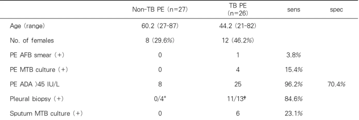
Of the 53 patients, 26 (49.1%) were diagnosed with pleural tuberculosis. The cases of pleural tuberculosis (n=26) consisted of those with positive M. tuberculosis culture of pleural fluid (n=4), those with histopathologic finding consistent with tuberculosis on pleural biopsy (n=11), those with positive M. tuberculosis culture of sputum (n=6), and/or those with a positive response to anti-tuberculous medication (n=12) without other possible causes of pleural effusion. The mean age was 44.2 years for the pleural tuberculosis patients and 60.2 years for the non-tuberculous pleural effusion patients. The number of female patients was 12 (46.2%) and 8 (29.6%) in each group (Table 1). The etiologies of the non-tuberculous pleural effusion included malignancy (n=10, 37.0%), bacterial pneumonia (n=11, 40.8%), sepsis (n=2, 7.4%), intra-abdominal infection (n=1, 3.7%), and undetermined origin (n=3, 11.1%).
Sensitivity and specificity of pleural effusion AFB smear, MTB culture, ADA activity, pleural biopsy pathology, and sputum MTB culture
Of the 26 tuberculous pleural effusion specimens,
AFB smear-positive was one, MTB culture-
positive were four, and ADA-positive (above 45
IU/L) were 25. Considering the combination of
MTB culture, pleural pathology and clinical
diagnosis as the reference method of diagnosing
Non-TB PE (n=27) TB PE
(n=26) sens spec
Age (range) 60.2 (27-87) 44.2 (21-82)
No. of females 8 (29.6%) 12 (46.2%)
PE AFB smear (+) 0 1 3.8%
PE MTB culture (+) 0 4 15.4%
PE ADA >45 IU/L 8 25 96.2% 70.4%
Pleural biopsy (+) 0/4* 11/13† 84.6%
Sputum MTB culture (+) 0 6 23.1%
ADA: adenosine deaminase; AFB: acid-fast bacilli; MTB: Mycobacterium tuberculosis;
Non-TB PE: non-tuberculous pleural effusion; sens: sensitivity; spec: specificity.
*Pleural biopsy was performed to four patients.
†11 out of the 13 biopsy-performed patients showed the histology consistent with pleural tuberculosis.
Table 1. Baseline characteristics of study population and sensitivity/specificity of pleural effusion acid-fast bacilli smear, Mycobacterium tuberculosis culture, adenosine deaminase, pleural biopsy pathology, sputum Mycobacterium tuberculosis culture in tuberculous pleural effusion.
Amount of PE sample
No. of patients with positive PE MTB PCR
10 ml 3/26 (11.5%)
25 ml 4/26 (15.4%)
50 ml 3/26 (11.5%)
(p >0.05, symmetry exact test)
PE: pleural effusion; MTB: Mycobacterium tuber- culosis; PCR: polymerase chain reaction.
Table 2. Sensitivities of pleural effusion Mycobac- terium tuberculosis polymerase chain reaction test according to the amount of pleural effusion specimens.
pleural tuberculosis, the sensitivities were 3.8%, 15.4%, and 96.2%, respectively. The pleural biopsy was performed in 13 of the 26 pleural tuberculosis patients, and 11 of the 13 pleural biopsy-performed patients showed the histologic findings consistent with pleural tuberculosis. Of the 26 pleural tuberculosis patients, six showed the sputum MTB culture-positive findings. The sensitivities of pleural biopsy and sputum MTB culture were 84.6%
and 23.1%, respectively. Of the 27 non-tuberculous
pleural effusion specimens, ADA-positive were eight. The specificity of pleural effusion ADA activity, therefore, was 70.4% (Table 1).
Sensitivities of pleural effusion MTB PCR test according to the amount of pleural effusion specimens
Of the 26 tuberculous pleural effusion specimens, MTB PCR-positive were three, four, and three when using 10 ml, 25 ml, and 50 ml of pleural fluid samples, respectively. The sensitivities of pleural effusion MTB PCR test were 11.5%, 15.4%, and 11.5%, respectively, which did not show statistically significant differences (p>0.05, symmetry exact test) (Table 2).
Of the four pleural effusion MTB PCR-positive
patients, pleural effusion AFB smear-positive was
one, pleural effusion MTB culture-positive were
three, pleural biopsy pathology-positive were three
(pleural biopsy was not performed to the remaining
one), and sputum MTB culture-positive were two.
DISCUSSION
As for tuberculous pleurisy, early in the course of tuberculous infection, a few organisms may gain access to the pleural space and cause a hyper- sensitivity response in the presence of cell- mediated immunity
17,18. Commonly, this form of tuberculous pleuritis goes unnoticed, and it resolves spontaneously. In some patients, however, the tuber- culous involvement of pleura is manifested as an acute illness with fever and pleuritic pain. The effusion is generally small and unilateral. In approximately 30% of patients, there is no radiographic evidence of involvement of the lung parenchyma in spite of the presence of lung parenchymal lesions in most cases as evidenced by findings of lung dissections
19. In the absence of concurrent pulmonary tuberculosis, the diagnosis of pleural tuberculosis requires thoracentesis and, in most cases, even pleural biopsy
7.
For tuberculous pleural effusion, the number of organisms in the pleural fluid is very small, so the conventional methods for the detection of M.
tuberculosis are often of no use. Even though the combination of microscopic examination and culture of pleural biopsy specimens was reported to increase the diagnostic rate up to 90%, it is time-consuming
20. Thus many physicians request nucleic acid amplification tests (NATs) including PCR for pleural effusion specimens to obtain a rapid and accurate diagnosis of pleural tuberculosis.
Several commercial and in-house NATs to detect MTB in clinical specimens, have been developed.
These tests amplify various targets in DNA or RNA sequences that are genus- or species-specific, which is followed by the detection step using gel electrophoresis or hybridization method. Currently there are four commercial NATs for the detection of MTB: 1) the Amplicor MTB test and its automated
version, the Cobas Amplicor MTB test (Roche Diagnostic Systems, Inc., Branchburg, NJ), 2) the Enhanced Amplified Mycobacterium Tuberculosis Direct Test (E-AMTDT) (Gen-Probe, Inc., San Diego, CA), 3) the BDProbe Tec ET test (Becton Dickinson, Sparks, MD), and 4) the INNO-LiPA- Rif. TB test (Innogenetics N. V., Zwijndrecht, Belgium). Of these, the E-AMTDT was approved by the FDA for the direct detection of MTB in both smear-positive and smear-negative respiratory specimens from the patients suspected of having tuberculosis, and the Amplicor MTB test was approved only for smear-positive respiratory specimens
21-23. As for in-house NATs, diverse methods using different primers have been used to detect MTB in pleural fluid samples, with highly variable sensitivities (11 to 81%) in the previous studies
8-13.
As for commercial NATs, the sensitivities have also been variable (20 to 100%) because the number of the patients with pleural tuberculosis was rather small and, in some studies, only the MTB culture-positive cases were included in the pleural tuberculosis group
24-30. Because the cases with MTB culture-positive pleural effusion occupy a relatively small portion of the whole pleural tuberculosis cases and the sensitivity of the pleural effusion MTB PCR test largely depends on the bacillary load, the sensitivity from the study group of patients with MTB culture-positive pleural effusion does not reflect the exact one from the whole group of patients with pleural tuberculosis.
From these reasons, there has been no consensus made about the usefulness of the MTB PCR test in the diagnosis of tuberculous pleural effusions.
In our previous study, we have demonstrated the
low sensitivity of MTB PCR test for the diagnosis
of pleural tuberculosis (sensitivity 17.5%, specificity
98.1%), using the commercially available Cobas
Amplicor MTB test which can minimize the inter-tester variabilities and maximize the speci- ficities due to the automated specimen processing and detection step and employing the combination of MTB culture, pleural pathology and clinical diagnosis as the reference method of diagnosing pleural tuberculosis
14.
In this study, we have examined whether the sensitivity of pleural effusion MTB PCR test for the diagnosis of pleural tuberculosis can be improved when increasing the amount of pleural effusion specimens. The results again showed that MTB PCR test of pleural effusion has a lower sensitivity compared with mycobacterial culture of pleural effusion sample and pleural biopsy pathology. The sensitivities of pleural effusion MTB PCR test using a different amount of pleural fluid samples, did not show statistically significant differences.
These results indicate that the sensitivity of MTB PCR test cannot be improved even with an increased amount of pleural effusion specimens. The sensitivity of NAT depends not only on the number of mycobacteria but also on their homogenous distribution in the specimen, the presence of the amplification inhibitor in the sample, and the type of the primers
31. When applying MTB PCR test to paucibacillary specimens, therefore, these all aspects should also be considered.
Although MTB PCR assay provides a rapid result and has a potential role in confirming tuberculous pleuritis, in conclusion, it has limitations in itself.
Our results suggest that the pleural effusion MTB PCR using the Cobas Amplicor MTB test has a low sensitivity and hence does not seem to be useful in excluding the disease. Therefore, it cannot replace the conventional diagnostic methods including culture techniques and histopathologic examina- tions. Furthermore, the results of the pleural effusion MTB PCR test need to be interpreted in
conjunction with those of the conventional methods and the clinical findings to reach the final diagnosis of pleural tuberculosis.
SUMMARY
Background: For the diagnosis of pleural tuberculosis, polymerase chain reaction (PCR) of pleural effusion specimens has shown very low sensitivity, which might be due to the small number of bacilli in the samples. The purpose of this investigation is to determine whether the sensitivity of PCR testing can be improved when increasing the amount of pleural effusion specimens.
Methods: We prospectively analyzed pleural effusion specimens obtained from 53 patients for whom the exclusion of the possibility of tuberculous pleural effusion was necessary. We performed Mycobacterium tuberculosis PCR testing using the Cobas Amplicor MTB test (Roche Diagnostic Systems) with three different amounts (10ml, 25ml, and 50ml) of pleural effusion specimen in each patient. Pleural tuberculosis was defined as having one of the following: culture-positive pleural fluid sample, histopathologic finding consistent with tuberculosis on pleural biopsy, culture-positive sputum specimen, and/or positive response to anti-tuberculous medication without other possible causes of pleural effusion.
Results: Of the 53 patients, 26 received the diagnosis of pleural tuberculosis. The sensitivities of AFB smearing, Mycobacterium tuberculosis culture of pleural effusion specimen, pleural biopsy, and measurement of ADA were 3.8%, 15.4%, 84.6%, and 88.5%, respectively. The results of PCR testing were positive for 3 (11.5%), 4 (15.4%), and 3 (11.5%) of the 26 patients when using 10ml, 25ml, and 50ml of pleural effusion specimens, respectively.
These results did not show a statistically significant
difference in the sensitivity of PCR testing when increasing the amount of pleural effusion samples (p>0.05, symmetry exact test).
Conclusion: For specimens such as pleural effusion, in which the bacillary load is very low, the clinical utility of PCR testing seems highly limited with the kits designed for the diagnosis of pulmonary tuberculosis. An increased amount of pleural effusion sample does not improve the sensitivity of PCR testing.
ACKNOWLEDGMENTS
This work was supported by the Ewha Womans University Research Grant of 2005.
I, the author, would like to thank Dr. Sung Kyu Kim, Dr. Joon Chang, Dr. Sang Nae Cho, Dr. Se Kyu Kim, Dr. Young Sam Kim, Dr. Jung Hyun Chang, Dr. Jin Hwa Lee, Dr. Yun Su Sim, Dr. Yoon Hee Jun, and Dr. So Yeon Lim for their sincere assistance and instruction. Each individual provided insights that guided and challenged my thinking, substantially improving my finished product.
REFERENCES
1. Menzies D. Issues in the management of contacts of patients with active pulmonary tuberculosis. Can J Public Health 1997;88:197-201.
2. Behr MA, Warren SA, Salamon H, Hopewell PC, Ponce de Leon A, Daley CL, et al. Transmission of Mycobacterium tuberculosis from patients smear- negative for acid-fast bacilli. Lancet 1999;353:444-9.
3. Toman K. Tuberculosis case-finding and chemo- therapy: questions and answers. Geneva, Switzerland:
World health organization; 1979.
4. Al Zahrani K, Al Jahdali H, Poirier L, Rene P, Gennaro ML, Menzies D. Accuracy and utility of commercially available amplification and serologic tests for the diagnosis of minimal pulmonary tuberculosis. Am J Respir Crit Care Med 2000;
162:1323-9.
5. Ogawa K, Koga H, Hirakata Y, Tomono K, Tashiro T,
Kohno S. Differential diagnosis of tuberculous pleurisy by measurement of cytokine concentrations in pleural effusion. Tuber Lung Dis 1997;78:29-34.
6. Iseman MD. A clinician's guide to tuberculosis.
Philadelphia, PA: Lippincott Williams & Wilkins;
2000.
7. American thoracic society. Diagnostic standards and classification of tuberculosis in adults and children.
Am J Respir Crit Care Med 2000;161:1376-95.
8. Kolk AH, Schuitema AR, Kuijper S, van Leeuwen J, Hermans PW, van Embden JD, et al. Detection of Mycobacterium tuberculosis in clinical samples by using polymerase chain reaction and a nonradioactive detection system. J Clin Microbiol 1992;30:2567-75.
9. de Wit D, Maartens G, Steyn L. A comparative study of the polymerase chain reaction and conventional procedures for the diagnosis of tuberculous pleural effusion. Tuber Lung Dis 1992;73:262-7.
10. de Lassence A, Lecossier D, Pierre C, Cadranel J, Stern M, Hance AJ. Detection of mycobacterial DNA in pleural fluid from patients with tuberculous pleurisy by means of the polymerase chain reaction:
comparison of two protocols. Thorax 1992;47:265-9.
11. Querol JM, Minguez J, Garcia-Sanchez E, Farga MA, Gimeno C, Garcia-de-Lomas J. Rapid diagnosis of pleural tuberculosis by polymerase chain reaction. Am J Respir Crit Care Med 1995;152:1977-81.
12. Villegas MV, Labrada LA, Saravia NG. Evaluation of polymerase chain reaction, adenosine deaminase, and interferon-gamma in pleural fluid for the differential diagnosis of pleural tuberculosis. Chest 2000;118:
1355-64.
13. Nagesh BS, Sehgal S, Jindal SK, Arora SK.
Evaluation of polymerase chain reaction for detection of Mycobacterium tuberculosis in pleural fluid. Chest 2001;119:1737-41.
14. Moon JW, Chang YS, Kim SK, Kim YS, Lee HM, Kim SK, et al. The clinical utility of polymerase chain reaction for the diagnosis of pleural tuberculosis. Clin Infect Dis 2005;41:660-6.
15. World Health Organization. Laboratory Services in Tuberculosis Control. Part III: Culture WHO/TB/
98.258. Geneva, Switzerland: World Health Organi- zation; 1998.
16. Giusti G, Galanti B. Methods of enzyme analysis.
New York, NY: Academic Press; 1983.
17. Berger HW, Mejia E. Tuberculous pleurisy. Chest 1973;63:88-92.
18. Ellner JJ. Pleural fluid and peripheral blood lymphocyte function in tuberculosis. Ann Intern Med 1978;89:932-3.
19. Stead WW, Eichenholz A, Stauss HK. Operative and
pathologic findings in twenty-four patients with syndrome of idiopathic pleurisy with effusion, presumably tuberculous. Am Rev Tuberc 1955;71:
473-502.
20. Light RW. Pleural diseases. Philadelphia, PA: Lea &
Febiger; 1983.
21. Nightingale SL. From the Food and Drug Admi- nistration. JAMA 1996;275:585.
22. Centers for Disease Control and Prevention. From the Centers for Disease Control and Prevention. Update:
Nucleic acid amplification tests for tuberculosis.
JAMA 2000;284:826.
23. Shamputa IC, Rigouts And L, Portaels F. Molecular genetic methods for diagnosis and antibiotic resis- tance detection of mycobacteria from clinical speci- mens. APMIS 2004;112:728-52.
24. Ehlers S, Ignatius R, Regnath T, Hahn H. Diagnosis of extrapulmonary tuberculosis by Gen-Probe ampli- fied Mycobacterium tuberculosis direct test. J Clin Microbiol 1996;34:2275-9.
25. Pfyffer GE, Kissling P, Jahn EM, Welscher HM, Salfinger M, Weber R. Diagnostic performance of amplified Mycobacterium tuberculosis direct test with cerebrospinal fluid, other nonrespiratory, and res- piratory specimens. J Clin Microbiol 1996;34:834-41.
26. Vlaspolder F, Singer P, Roggeveen C. Diagnostic value of an amplification method (Gen-Probe) compared
with that of culture for diagnosis of tuberculosis. J Clin Microbiol 1995;33:2699-703.
27. Carpentier E, Drouillard B, Dailloux M. Diagnosis of tuberculosis by Amplicor Mycobacterium tuberculosis test: a multicenter study. J Clin Microbiol 1995;
33:3106-10.
28. Shah S, Miller A, Mastellone A. Rapid diagnosis of tuberculosis in various biopsy and body fluid specimens by the AMPLICOR Mycobacterium tuber- culosis polymerase chain reaction test. Chest 1998;113:1190-4.
29. Levidiotou S, Vrioni G, Galanakis E, Gesouli E, Pappa C, Stefanou D. Four-year experience of use of the Cobas Amplicor system for rapid detection of Mycobacterium tuberculosis complex in respiratory and nonrespiratory specimens in Greece. Eur J Clin Microbiol Infect Dis 2003;22:349-56.
30. Reischl U, Lehn N, Wolf H, Naumann L. Clinical evaluation of the automated COBAS AMPLICOR MTB assay for testing respiratory and nonrespiratory specimens. J Clin Microbiol 1998;36:2853-60.
31. Ruiz-Manzano J, Manterola JM, Gamboa F. Detection of Mycobacterium tuberculosis in paraffin-embedded pleural biopsy specimens by commercial ribosomal RNA and DNA amplification kits. Chest 2000;118:
648-55.