GW Kim, et al
70 Ann Dermatol
Received July 27, 2010, Revised October 7, 2010, Accepted for publication October 7, 2010
*This study was supported by a grant of the Korean Heath Technology R&D Project, Ministry of Health and Welfare, Republic of Korea (A070001).
Corresponding author: Moon-Bum Kim, M.D., Ph.D., Department of Dermatology, School of Medicine, Pusan National University, 1-10 Ami-dong, Seo-gu, Busan 602-739, Korea. Tel: 82-51-240-7338, Fax:
82-51-245-9467, E-mail: [email protected]
This is an Open Access article distributed under the terms of the Creative Commons Attribution Non-Commercial License (http://
creativecommons.org/licenses/by-nc/3.0) which permits unrestricted non-commercial use, distribution, and reproduction in any medium, provided the original work is properly cited.
Ann Dermatol Vol. 24, No 1, 2012 http://dx.doi.org/10.5021/ad.2012.24.1.70
CASE REPORT
Delayed Diagnosis of Scrofuloderma Misdiagnosed as a Bacterial Abscess
Gun-Wook Kim, M.S.
1, Hyun-Je Park, M.S.
1, Hoon-Soo Kim, M.S.
1, Su-Han Kim, M.S.
1,
Hyun-Chang Ko, M.S.
1,2, Byung-Soo Kim, Ph.D.
1, Moon-Bum Kim, M.D., Ph.D.
1,2, Eun-Kyung Sim
31Department of Dermatology, School of Medicine, 2Medical Research Institute, Pusan National University,
3Department of Beauty Care, College of Health, Social Welfare and Education, Tongmyong University, Busan, Korea
An 82-year-old woman presented with a four-month history of an ulcerative plaque overlying her left neck. This lesion had developed as a subcutaneous nodule, gradually increased in size, and evolved into ulcers. Before visiting our Dermatology clinic, the patient had been diagnosed as having a bacterial abscess, but treatments with antibiotics were unsuccessful. The presence of a purulent discharge and prominent ulceration caused further confusion as bacterial abscess, and radiologic evaluation on computed tomography also led to the possibilities of secondary lesions from an abscess or malignancy. However, the characteristic appearance of her lesion allowed us to discern cutaneous tuberculosis, especially scrofuloderma. Based on clinical examinations, staining for acid-fast bacilli, and positive findings of polymerase chain reaction, a quick diagnosis of scrofulo- derma was made. After that, she was treated successfully with anti-tuberculosis therapy and the ulcer healed. Our case highlights the problem of delayed diagnosis of scrofulo- derma presenting as a bacterial abscess. In conclusion, having a high index of suspicion is needed to diagnose cutaneous tuberculosis correctly. (Ann Dermatol 24(1) 70∼
73, 2012)
-Keywords-
Bacterial abscess, Scrofuloderma
INTRODUCTION
Cutaneous tuberculosis is a rare form of extrapulmonary tuberculosis, and it is often misdiagnosed because of a low index of suspicion1. Scrofuloderma results from the involvement and break-down of the skin overlying a tuberculous focus2. It begins as a painless subcutaneous nodule, called a cold abscess. With the continuous pro- pagation of an underlying infection, it is characterized by fistula formation and pus discharge from a lymph node.
The cervical lymphatic chains are most frequently invol- ved2-4. Herein we report a case of scrofuloderma misdiag- nosed as a bacterial abscess and note the importance of maintaining a high level of awareness.
CASE REPORT
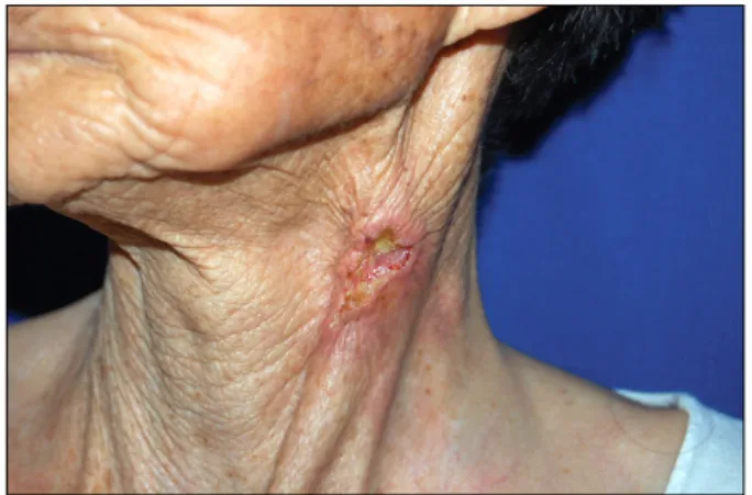
An 82-year-old woman presented with a four-month his- tory of an ulcerative plaque overlying her left neck. This lesion had developed as a subcutaneous nodule, which gradually increased in size and evolved into ulcers. The physical examination revealed a 3×5 cm, erythematous suppurative plaque on the surface of her neck. It was well- demarcated with elevated borders and deep undermined edges. The lesion also had central ulceration covered by a scant, cheesy, and purulent discharge (Fig. 1). There was no history of trauma. Systemic examination did not reveal any abnormalities, and there was no recent weight loss or night sweats.
Before visiting our Dermatology clinic, the patient had been diagnosed as having a bacterial abscess and was
Delayed Diagnosis of Scrofuloderma Misdiagnosed as a Bacterial Abscess
Vol. 24, No. 1, 2012 71 Fig. 1. Erythematous suppurative plaques with central ulceration
on the surface of her neck.
Fig. 2. Epithelioid cell granulomas with inflammatory cell infiltrations in the dermis (H&E, ×100), Inset: Acid-fast bacilli on Ziehl-Neelsen staining (H&E, ×1,000).
Fig. 3. Positive results on polymerase chain reaction for the detection of Mycobacterium tuberculosis. Lane M: molecular weight marker, Lane PC: positive control, Lane NC: negative control, Lane 3: patient’s sample.
treated with antibiotics at two local clinics and by an ENT doctor at our hospital. Earlier therapeutic attempts with antibiotics (Banan; Meiact; cefditoren pivoxil) were unsuccessful and the lesion was sometimes reduced but never had disappeared entirely. Further investigations such as a computed tomography (CT) study of the neck, and fine needle aspiration cytology (FNAC) were done by the ENT doctor of our hospital to rule out malignancy.
Also, she was referred to our clinic for the evaluation of chronic dermatologic disease. At that time, the characteristic appearance of her lesion allowed us to discern cutaneous tuberculosis, especially scrofuloderma and further workups including skin biopsy, polymerase chain reaction (PCR) for Mycobacterium tuberculosis, cul- tures (bacterial, fungal, and mycobacterial), and chest X-ray were carried out.
Histological examinations revealed epithelioid cell granu- lomas with inflammatory cell infiltrates in the dermis.
Ziehl-Neelsen stain demonstrated acid-fast bacilli (Fig. 2).
Cultures from the skin biopsy showed growth of Myco- bacterium tuberculosis after 8 weeks. Fungal and standard bacterial cultures from the skin biopsy were negative. A PCR study for the detection of Mycobacterium tuberculosis was conducted on a biopsy specimen and turned out positive (Fig. 3). However, FNAC from the skin lesions on the neck showed only neutrophils and fibrin exudates.
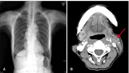
A radiograph of the chest revealed 1 cm, small, calcified nodules on the right upper pulmonary lobe, and pulmo- nary tuberculosis was suspected (Fig. 4A). CT of the neck, which demonstrated peripheral enhancing low density lesions with extension to the cutaneous area, was found and ruled out an abscess or malignancy (Fig. 4B).
Based on clinical examinations, staining for acid-fast bacilli, and positive PCR findings, a rapid diagnosis of scrofulo- derma was made. The patient was quickly started on an anti-tubercular treatment regimen that included isoniazid, rifampicin, ethambutol and pyrazinamide. The cutaneous lesion regressed in a slow but complete fashion. Her treatment with anti-tuberculosis therapy was successful and the ulcer healed after 18 months.
DISCUSSION
Cutaneous tuberculosis remains a rare infection, with an incidence of 3.5% reported among patients with organ tuberculosis5. Among them, scrofuloderma is a common form of cutaneous tuberculosis; it is seen in many deve- loping countries and in some European countries5,6. It is
GW Kim, et al
72 Ann Dermatol
Fig. 4. 1 cm small calcified nodules on the right upper pulmonary lobe (A), and peripheral enhancing low density lesions (arrow) on the left neck (B).
caused by a lesion in the lymph node, bone, muscle or tendon that continuously spreads to the skin2-4.
The skin lesion first presents as painless, subcutaneous nodules, which is called a cold abscess. Then, the reddish- blue induration suppurates and breaks down, forming an ulcer with granulating tissue at the base. Sometimes sinu- ses may develop and communicate directly with areas of deep infection. Progression and scarring produce irregular adherent masses, densely fibrous in places, and fluctuant or discharging in others2-4. The disease is usually slow- growing, and has an insidious course and an unusual pre- sentation; a chronically discharging sinus or a non-healing ulcer are possible4.
Scrofuloderma remains a diagnostic and therapeutic chal- lenge because the clinical appearance of the lesions is not always characteristic, and a positive culture result is not always obtained1,7. This case highlights delayed diagnosis of scrofuloderma presenting as a bacterial abscess. Al- though our case presents typical features of scrofuloderma, the presence of purulent discharge, prominent ulceration, or features suggestive of a carbuncle can cause further confusion and misdiagnosis as a bacterial abscess7. More- over, radiologic evaluation on CT also led to the possi- bility of secondary lesions from an abscess or malignancy.
The delay in the diagnosis emphasizes the importance of maintaining a high level of suspicion.
In our case, scrofuloderma was confirmed by the presence of acid fast bacilli on AFB staining and a positive culture for Mycobacterium tuberculosis. Supportive evidence in- cluded the presence of epithelioid granulomas on histo- pathologic examination and a positive result on PCR for mycobacteria.
Scrofuloderma may be a manifestation of systemic tuber- culous disease and can result from direct extension or
hematogeneous spread of the infection6. The incidence of systemic involvement in adults with scrofuloderma has been reported to be as high as 35%6. Of note, using PCR to identify the infectious agents has increased the sensi- tivity and improved the overall detection rate among those with a diagnosis of cutaneous tuberculosis7,8. A further advantage of PCR is the possibility of earlier initiation of treatment7,8.
Histology may show marked caseation necrosis, with numerous bacteria usually in the deeper structures2,9. The center of the lesion is dominated by necrosis and abscess material with inflammatory infiltrates. In the epidermis, scarring and atrophic changes often predominate. Tuber- cle bacilli are usually easy to identify in the purulent discharge or biopsy tissue3.
Chemotherapy still remains the treatment of choice2,10. It aims to cure the disease as rapidly as possible in order to prevent relapses and the emergence of resistant strains.
The standard 6-month regimen for adults includes isonia- zid, rifampin, pyrazinamide, and ethambutol for the initial 2 months followed by isoniazid and rifampin for a further 4 months in the continuation phase2,10. When a therapeu- tic trial is undertaken in a case of cutaneous tuberculosis, 6 weeks of therapy with four drugs appears adequate to confirm the diagnosis11. The response to anti-tuberculous treatment alone as a primary therapy is usually good, but some authors have recommended wide surgical removal of all diseased tissue, combined with pharmacologic anti- tuberculous treatment2,3.
Our case serves as a reminder that chronic neck ulcers may be due to cutaneous tuberculosis and that pulmonary tuberculosis may be a focus for tuberculous infection. A chronic, non-healing ulcer is a common presentation of scrofuloderma. Nevertheless, scrofuloderma with concomi-
Delayed Diagnosis of Scrofuloderma Misdiagnosed as a Bacterial Abscess
Vol. 24, No. 1, 2012 73 tant bacterial infection frequently occur, preventing early
diagnosis of tuberculosis if not suspected1,7. Therefore, a high index of suspicion is needed for the prompt diagnosis and management of this uncommon skin disorder.
When a therapeutic trial is undertaken in a case of cuta- neous tuberculosis, 6 weeks of therapy with four drugs appears adequate to prove (or disprove) the diagnosis.
REFERENCES
1. Lantos G, Fisher BK, Contreras M. Tuberculous ulcer of the skin. J Am Acad Dermatol 1988;19:1067-1072.
2. Tappeiner G. Tuberculosis and infections with atypical my- cobacteria. In: Wolff K, Goldsmith LA, Katz SI, Gilchrest BA, Paller AS, Leffell DJ, editors. Fitzpatrick's dermatology in general medicine. 7th ed. New York: McGraw-Hill Medical, 2008:1768-1778.
3. Barbagallo J, Tager P, Ingleton R, Hirsch RJ, Weinberg JM.
Cutaneous tuberculosis: diagnosis and treatment. Am J Clin Dermatol 2002;3:319-328.
4. Sehgal VN, Wagh SA. Cutaneous tuberculosis. Current con-
cepts. Int J Dermatol 1990;29:237-252.
5. Kivanç-Altunay I, Baysal Z, Ekmekçi TR, Köslü A. Incidence of cutaneous tuberculosis in patients with organ tuberculo- sis. Int J Dermatol 2003;42:197-200.
6. Yates VM, Ormerod LP. Cutaneous tuberculosis in Black- burn district (U.K.): a 15-year prospective series, 1981-95. Br J Dermatol 1997;136:483-489.
7. Tan WP, Tang MB, Tan HH. Scrofuloderma from the acro- mioclavicular joint presenting as a chronic ulcer in an immu- nocompetent host. Singapore Med J 2007;48:e243-245.
8. Hsiao PF, Tzen CY, Chen HC, Su HY. Polymerase chain reaction based detection of Mycobacterium tuberculosis in tissues showing granulomatous inflammation without demon- strable acid-fast bacilli. Int J Dermatol 2003;42:281-286.
9. Vashisht P, Sahoo B, Khurana N, Reddy BS. Cutaneous tuberculosis in children and adolescents: a clinicohistolo- gical study. J Eur Acad Dermatol Venereol 2007;21:40-47.
10. Handog EB, Gabriel TG, Pineda RT. Management of cuta- neous tuberculosis. Dermatol Ther 2008;21:154-161.
11. Ramam M, Mittal R, Ramesh V. How soon does cutaneous tuberculosis respond to treatment? Implications for a thera- peutic test of diagnosis. Int J Dermatol 2005;44:121-124.