임상병리검사과학회지
:
제 28권 제 1 호1996.
골수천 자액 에 서 의 결 핵 균 검 출과 Competitive PCR에 의한 정량방법의 소개
서울중앙병원 임상병리과
조은주·문애란
Detection of Mycobacterium tuberculosis in Bone Marrow Blood and Introduction of Semi -quantitation
Method by Competitive PCR
Cho , Eun Ju. , Moon , Ae Ran
Dept. of Clinical
Path여ogy,Asan Medical Center , Seoul , K orea
The conventional methods for the detection of
.1\ψ'Cobacteriumtuberculosis are the AFB staining and cultivation method. But cultivation method spends 2 to 8 weeks to identify and in the case of sputum specimens, only 28 to 70% numbers of overall bacteria is able to be cultivated due to the loss of viability resulting in pretreatment of sputum specimen. And the bacteria is rarely cultivated in BM aspirate , peripheral blood , or many kinds of body fluids as a specimens.
In order to overcome these problems , we introduced PCR method to detect M. tubercu- losis. In this study , we reported an example of the detection of this bacteria in BM as- pirate of a patient and described semi -quantitative method using competitive PCR. We used the cloned PCR product as a positive control element and the newly developed DNA fragment as an internal control that is derived from v- erbB gene and has the same primer binding regions as positive control at the 5 ’ and 3 ’ end and about 120bp size difference.
ln the competitive PCR , we put the various known amounts of internal control and
the constant amounts of the genomic DNA extracted from BM aspirate in one tube and
amplified the 156110 fragment of M. tuberculosis with thermal cycle r. And then the PCR
products were electrophoresed and stained with EtBr. After photographed, we scanned
the photographs using the Image Master program of Phamacia Biotech to compare the
amounts of intensity of amplified target DNA and the amplified internal contro l. We
found the region that represented the equal amount of intensity of target DNA and in-
ternal contro l. At that region, we could calculate the amount of target DNA from the amount of internal contro l. As a result , it was estimated that 2ml of BM aspirate of a patient contained about 37 cells of M. tuberculosis.
The detection of M. tuberculosis by PCR is very useful in BM aspirates , bloods , or several kinds of body fluids as specimens. This method could be used f or semi - quantitation and it could quantitate more accurate if we applied intimately controlled a- mounts of internal contro l. We might also apply to EL1SA system to estimate the a- mounts of amplified products using each specific probes of target DNA and internal contro l.
Key words : Mvcobacterium tubercu/osis. bone marrow aspirate, competitive PCR,
seπ1i - quantitation
1 .
서 론Mcobacterium
tuberculosis를 검 출하기 위 한 방 법으로는 일반적으로 항산성 염색법과 결핵균 배 양방법을 주로 이용하고 있으나 근래에는poly- merase chain
reaction(PCR) 을 이용한 결핵균 검출방법을 많이 이용하고 있는 추세다 1)PCR방법은 민감도가 높아 배양방법을 통해 잘 검출 되지않는 혈액이나 체강액, 소변 등에 서 결핵균을 검출할 수 있어 객담을 받기 어려 운 환자나 소아에서 결핵균 검출에 유용하며,
검출까지 소요되는 시간이 빠르다는 장점을 가
지고 있다 2-10)
이에 저자들은 결핵균 감염이 의심되는 한 환자의 골수세포로부터 PCR을 이용한 결핵균 검출 방법 및 정량방법을 소개하고자 한다.
11.
실험재료 및 방법1)
환자 및 검체1996년 3월에 서울중앙병원 소아과에 입원 한 13세 여자 환자로서 골수검사 소견으로 경 미한 반응성형질세포 및 조직구증식증을 보였 으며 결핵균감염을 의심하여 항산성염색을 시 행한 결과 음성을 나타냈다. 검체는 EDTA에 채취한 골수천자액
2
ml를 사용하였다.2) DNA
추출골수천 자액 을
Ficoll- Paque @( Pharmacia Bio- tech ,
Sweden) 을 이용하여1 , 500
rpm에서 30분 간 원심분리한 후 단핵구를 수거하여phosphate buff ered saline (PBS
)으로 1 회 세척하였다. 수거 된 단핵구에lysis buffer 1(0.14 M NaCl , 1. 5 mM MgCl 2 , 10 mM Tris- HCl , pH 8.6 , 0.5% NP- 40 , 1 mM DTT) 200
μl와Tris- EDTA(TE) buffer 100
띠를 넣 은 후10 mg/ml lysozyme (Boehringer Mannheim , Germany) 10
μl를 넣 어 370C 에서 1 시간 반응 시켰다.Lysis buffer
II (0.2 M Tris - HCl , pH 8.0 , 25 mM EDT A , pH 8.0, 0.3 M NaCl, 2 % SDS) 200
야를 첨 가 하여 완전 히lysis
시 킨 후20 mg/ml protein- ase K (Boehringer Mannheim , Germany)
를10
μ1 넣어 550C 에서 5 분간 반응시켰다. 여기에 동량의
phenol : chlorof orm (1 :
1) 을 넣고 완전 히 혼합한 후4
0C 15 , 000
rpm에서 8분간 원침 하고 상충액만을 취하여 2.5배의100% cold
ethanol을 첨가하였다. -20oC 에서 1 시간 이상 방치시킨 후
4
0C , 15 , 000
rpm에서 15 분간 원침 하고70% cold
ethanol로 1 회 세척하였으며,여분의 ethanol을 증발 건조시키고, 적당량의 증류수에 DNA를 녹여
PCR
반응에 사용하였 다.3) Oligonucleotide
primer의 구성PCR에 사용된 pnmer는
Mycobacterium tu berculosis
complex에 존재 하는insertion se
quence 인 lS6110 의 일부분에 해당하며
Myccr bacterium tuberculosis
한 마리 당 평 균8 copy
정 도 존재 한다고 한다 11 , 12, 13). Oligonucleotide pri
mer는 한국 생공에 주문하여 합성하였으며 그 염기서열은 아래와 같다 14)
sense(PT8);5' GTGCGGATGGTCGCAGAGAT 3' antisense(PT9); 5' CTCGATGCCCTCACGGTTCA 3'
4)
중합효소연해반응 (polymerasechain re- action , PCR)
10X
반응 완충액 (250mM KCl , 200 mM Tris - HCl , pH 8.3 , 0.5 % Tween 20 , 1 mg/ml gel atin) 5
띠,25 mM MgC1 2 6.25
띠,deoxynu- cleoside
triphosphate( 각각2.5 mM, Pharmacia, USA) 5
띠,10
pmole/띠primer
PT8과 PT9 를 각각 5 띠씩 넣고 검체로부터 추출한 DNA를 5 띠 넣어 혼합한 뒤 총량이 50 띠가 되도록 증 류수를 첨가하였다. 자동 온도 조절기 (GeneAmp 9600 , Perkin-Elmer ,
USA) 에 서94
0C 3
분간 반응시켜 DNA를 변성시킨 후 800C 에서
Taq DNA polymerase 1. 25 unit( 5
unit/띠,Boe- hringer Mannheim , Germany)
을 첨 가 하 였 다.PCR
반응 조건은94
0C
1 분,68
0C
3 초,72
0C 1
분으로서
40
주기로 증폭시켰다.PCR
산물은ethidium
bromide(EtBr) 를 포함한2.5% Meta-
DNA를
EcoR 1
제한 효소로 처리한 후PT8
과
PT9
primer를 이용해 증폭시키고JET sorb Gel extraction kit(Genomed,
USA) 를 이 용하여agarose
gel로부터 PCR산물 만을 정 제하였다.음성 대조 물질로는 증류수를 사용하였다.
6)
제 한효소 (restrictionenzyme)
처 리 증폭된PCR
결과물이 비특이적 반응 결과 물이 아닌 결핵균의lS6110
중의 한 단편임을 확인하기 위해 제한효소Sma 1 (Boehringer Ma nnheim ,
Germany) 으로 처리하였다. 제한효소 반응은Sma 1 (1 0
U/μ1)1
띠,10X reaction bu ffer(33 mmol/
~Tris-acetate , 66 mmol/
~K -acetate, 10 mmol/
~Mg-acetate, 0.5 mmol /
~DTT , pH 7.9 , BM 1ncubation buffer A) 1
띠,PCR
결과물8
μl를 취하여 300C 에서 1 시 간 방치시켰다.7) Mycobaclerium tuberculosis
정 량Competitive
PCR을 이용하였으며, 내부지시물질은
PCR mimic
kit를 사용하여 만들었으며, 이 물질의 내부는
u-erbB
gene의 일부분 으로 구성되어있으며,5'
말단과3'
말단에 각 각PT8 , PT9
primer의 결합부위를 가지고 있 어서 결핵균의lS6110
단편과 함께 증폭될 수 있게 제작하였다(그림1.)
phor agarose gel(FMC Bio products, USA) 에 A 많회 lS6110 I PT 9 I 541 bp
서 전기영동 (5
V /cm)
하여541bp
크기의band
를 확인하였다 B.
I PT 8 I u - erb B I PT 9 426 bp
5)
양성, 음성 대조 물질양성 대조 물질은 배양된 인형 결핵균의
ge nomic
DNA를 PT8과 PT9 를 이 용하여 증폭시 키고 이를 cloning하여 사용하였다. 증폭산물 을TA cloning ™Kit (l nvitrogen ,
USA) 를 이 용해cloning
하 고cloning
된E.
coli로 부터plasmid
DNA를 추출하였다 15) 추출한plasmid
Figure 1. Representation of PCR targets , IS6110 fragment (A) and internal control element (B).
PCR
반응조건은 결핵균 정성검사와 같은 조건으로 하였으나 증폭주기 는plateau effect
에 도달하기 전인
30
주기로 하였다. 일정량의 내부지시물질과 검체로부터 추출한 DNA 를 동일한 tube에서 증폭시키고,
PCR
결과물을 전 기영동한 후, 정량하고자하는 결핵균의PCR
산물과 내부지시물질의PCR
산물이 EtBr에 염색된 intensity를 비교하였다.1ntensity
측정1 2 3
은
1mage Master program (Pharmacia , USA)
51검빠t'yp~• 541 bp
• 399 bp
을 이 용하여refractive
density를 측정하였으며, 결핵균의
PCR
산물이 541bp이며 내부지시 물질의PCR
산물이 426bp인 점을 감안하여 크기에 따른 염색의 정도를 고려한 보정값으로 보정하였다. 이렇게해서 측정한 intensity가 같 은 점에서 두PCR
산물의 양이 동일한 것으 로 보고,PCR
실시 전의 양도 동일한 것으로 추정했다.ill.
결 과1) M.
tuberculosis의 검 출방법에서 기술한 대로 골수 천자액으로부터 단핵 세포를 분리하고
genomic
DNA 를 추출하 였다. 이genomic
DNA의 일부를 취하여PCR
을 실시하였고, 이를 전기 영동하여 양성 대조1 2 3 4
517 , 506bp • - - • 541 bp
Figure 2. Agarose gel electrophoresis of the am plified products
Lane 1: DNA size marker( lkb ladder , Gibco
•BRL)
Lane 2 : Amplified products of positive control Lane 3 : Amplified products of genomic DNA ex
tracted from M aspirate of the patient Lane 4 : Amplified products of negative control
154 bp • • 142 bp
Figure 3. Sma 1 treatment of PCR products Lane 1: DNA size markerOkb ladder , Gibco-
BRL)
Lane 2 : PCR products , untreated
Lane 3 : Sma 1 treatment of PCR products
물질의 541bp와 같은 크기의
DNA
band를 확 인할 수 있었다. 이것이 결핵균의 IS6110의 일 부에 해당하는 지를 알아보기 위하여 제한 효 소Sma
1으로 처리하여 확인하였다(그림2 ,
그 림3).
그림에 의하면, 환자 검체에서 추출한 DNA와 양성 대조 물질의PCR
산물이 제한 효소에 의해 동일한 크기의 band로 절단되는 것을 관찰하였으며, 이로서 환자 검체에도 결 핵균이 존재한다고 할 수 있다. 이 방법은 PCR에 의해 결핵균 DNA의 일부를 증폭시켜 서, 결핵균이 존재하는 지를 검사하는 것이며 PCR이 끝난 후 제한 효소 처리를 통하여 그 특이 도를 높이 고자 하였다.2) Competitive
PCR에 의 한 정 량검 사Competitive
PCR에 의한 정량 검사는pnm-
er의 결합 부위가 같은 두 물질, 즉, 알고자하 는 결핵균의 DNA혹은 검체로부터 추출한 DNA와 내부 지시물질을 동시에 반응시켜 pnmer가 두 물질에 대하여 서로 경쟁적으로 결합하게 하고 이 때의PCR
산물의 양을 비 교하여 애초에 존재하는 양을 추정하는 것이 다. 즉, PCR 에 의 해 서 증폭되 는 DNA는PCR
의 반응이plateau
effect에 도달하기 전에서 일정한 증가의 양상을 보이며 이런 현상을 이 용하여PCR
산물을 비 교함으로서PCR
이 전에 검체내에 존재하는 DNA의 양을 측정할 수 있는 것이다. 이때, PCR에 의해 증폭되는 두 물질의 양이 동일한 시점에서 검체내 결핵균 DNA양과 내부 지시물질의 양이 동일하므로 그 양을 측정할 수 있게 된다. 이런 원리를 기 본으로 하여
plateau
effect에 도달하기 전의PCR
증폭 횟수를 실험을 통하여 30회로 정하 였고, 이미 알고 있는 양의 내부 지시물질 일 정량을 환자 검체에서 추출한 DNA와 혼합하 여 PCR을 실시하였다. PCR이 끝난 후,PCR
산물을2.5 % agarose
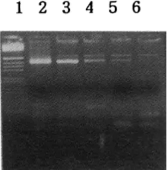
gel에서 전기영동하였다 (그림4.).
내부 지시물질의 양이 훨씬 많이 존재하는 경우에는 결핵균의 DNA는 전혀 증폭이 되지 못하다가 내부 지시물질의 양이 조금씩 감소함 에 따라 결핵균의 DNA가 조금씩 증폭되며 두 물질의 증폭된 양이 비슷해 보이는 영역이
1 2 3 4 5 6
Figure 4. Agarose gel electrophoresis of competi- tive PCR products.
Competitive PCR was carried out using 5야 of genomic DNA solution extracted from BM blood of a patient and various known amounts of inter- nal control DNA as templates.
Lane 1: DNA size marker (l kb ladder , Gibco- BRL)
Lane 2 : 1500 molecules of internal control DNA Lane 3 : 150 molecules of internal control DNA Lane 4 : 15 molecules of internal control DNA Lane 5": 1. 5 molecules of internal control DNA Lane 6 : 0 molecules of internal control DNA
존재하는 것을 볼 수 있다. 이것을
Pharmacia
Biotech의Image Master
Program을 이 용하여 분석하고 두 물질의 크기에 따른 보정값으로 보정한 결과 골수 천자액 2ml에 포함된 결핵 균의 양은 약 37마리에 해당하는 것으로 산출 되었다. 같은 검체를 slide에 도말하여AFB
staining을 한 경우에는 결핵균은 검출되지 않 았으며, 정량검사로 산출된 결핵균의 양과sli
de에smearmg
되었을 검체의 양을30"'60
띠 라고 고려해 보면AFB
staining에서는 결핵균 이 검출될 가능성이 희박해보인다.이런 결과로 미루어보면, PCR에 의한 결핵 균의 검출방법은 검체의 처리방법이나 처리할 수 있는 양의 한계에서 올 수 있는 여러 가지 단점들을 줄일 수 있는 방법이라 여겨진다. 이 실험에서 저자들은 내부 지시물질의 양을
10
배씩 증가시키는 방법을 썼으나, 그 범위를 줄 인다면 좀 더 정확한 결과를 보여줄 수 있을 것 으로 보이 며, digoxigenin
혹은 biotin이 표지 된 probe를 이용하여PCR
산물의 양을 측정하 는 방법 을 도입 하면 microplate에 서 반응을 시키고
ELISA
reader로 그 양을 측정할 수 있을것이다.
N.
고 찰최근들어 후진국에서 뿐만 아니라 유럽, 미 국 등 선진국에서도
AIDS
등 면역결핍증 환 자, 알콜중독환자 등의 증가로 인한 결핵균 감염이 다시 증가되고 있는 추세이다 16, 17, 18)
현재 결핵균의 검출은 대부분 항산성 염색법 과 결핵균 배양법에 의존하고 있으나 항산성 염색법은 낮은 민감도와 특이도를 나타내어 주 로 예 비 진 단
( preliminary diagnosis)
에 사용되 고 있으며, 배양법은 최종진단까지 2"'8주 정 도의 긴 시간이 소요될 뿐만 아니라 객담이외 의 혈액이나 체강액, 소변 등에서의 검출률이 다소 낮은 단점을 가지고 있다. 이를 보완할 수 있는 방법으로 PCR을 이용한 결핵균 검출 방법이 많이 이용되고 있는데 이 방법은 빠르고 민감도가 높아, 객담뿐만 아니라 혈액이나 체강액, 소변 듀 결핵균의 수가 적어 잘 배양 되지 않는 검체로부터 결핵균을 검출할 수 있 으므로, 객담을 받기 어려운 환자나 소아의 결 핵균 검출에 많이 이용되고 있다.
이에 저자들은 결핵균 감염이 의심되는 환자 의 골수 천자액으로부터 PCR을 시행하여 결 핵균을 검출하였으며, 검체내의 결핵균의 양을 정량하므로써 활동성 결핵균의 진단과 치료경 과 관찰에 도움이 되고자 하였다.
v. 결 료르
1..-
PCR에 의한 결핵균 검출은 항산성 염색법 이나 배양방법으로는 그 검출률이 낮은 혈액,
체강액, 소변 등에서 결핵균을 검출하기에 매 우 유용한 방법으로 여겨지며,
competitive
PCR을 이용한 정량검사는 지속적인 경과 관찰 에도 매우 유용할 것으로 보인다. 또한 객담의 경우에 전처리 과정에서 사멸될 수 있는 결핵균
이 28'"'-'70% 인 점을 감안하면 19, 20, 21 ) 배양이 잘
되지 않는 객담에서도 PCR에 의한 검출은 매 우 유용할 것으로 보인다. 이 경우, 검출되는 결핵균이 활동성인지를 알아보기 위해서 시차 적인 정량검사를 실시하여 확인할 수 있을 것이다 22)
또, 앞서 실시한 정량방법에선
PCR
산물의 intensity를 비교하여 정량하였는데, 보다 객관 적인 결과를 위해서는 probe를 이용한ELISA
방법을 도입할 수도 있으며 3 , 현재 추진 중에 있다. 또한 정량의 정확도를 강화하기 위해서 는 내부지시물질의 첨가량을 좀더 세분화 하여 거의 동일한 intensity를 보이는 지점을 찾아내 는 것이 효과적일 것이다.
REFERENCES
1. Wilson SM , McNerney R , Nye PM , Godfrey- Faussett PD, Stoker NG, and Voller A. Progress toward a simplified
polymerase chain reaction and its applica- tion to diagnosis of tubeculosis. J. Clin.
Microbio l., 31 : 776-782 , 1993.
2. Brisson - Noël , Gicquel AB , Chureau C , Nguyen S , Pierre C , Bartoli M , Bonete R , Pialoux G , Gicquel B , and Garrigue G. , Diagnosis of tuberculosis by DNA amplifi- cation in clinical practice evaluation. Lan- cet 338 : 364-366, 199 1.
3. Clarridge JE , m , Shawar RM , Shinnick TM , and Plikaytis BB. , Large - scale use of polymerase chain reaction f or detection of Mycobacterium tuberculosis in a routine mycobacteriology laboratory. J. Clin.
Microbio l. 31 : 2049- 2056 , 1993.
4. Cousins DV , Wilton SD , Francis BR , and Gow BL. Use of polymerase chain reac- tion for rapid diagnosis of tuberculosis. J.
Clin. Microbio l. 30 : 255 - 258 , 1992.
5. Forbes BA , and Hicks KES. Direct detec- tion of Mycobacterium tuberculosis in res- piratory specimens in a clinical laboratory by polymerase chain reaction. J. Clin.
Microbio l. 31 : 1688-1694, 1993.
6. Manjunath N , Shankar P , Rajan L , Bhargava A , Saluja S , and Shriniwas.
Evaluation of a polymerase chain reaction for the diagnosis of tuberculosis. Tubercle , 72:21-27 , 199 1.
7. Miyazaki Y , Koga H , Kohno S , and Kaku M. , Nested polymerase chain reaction for detection Mycobacterium tuberculosis in clinical sample., J. Clin. Microbio l. 31:
2228- 2232 , 1993.
8. Pierre C , Lecossier D , Boussougant Y , Bacart D , Jdiy V , Yeni P , and Hance AJ. Use of a reamplification
protoc이lm- proves sensitivity of detection of
Mycσbacterium tuberculosis in clinical samples
by amplification of DNA. J. Clin. Micro-
bio l. 29 : 712-717, 199 1.
9.
Sail이RK , Gelfand DH , Stoffel S , Scharf SJ , Higuchi R , Horn GT , Mullis KB , and Erlich HA. Primer directed enzymatic am- plification of DNA with a thermostable DNA polymerase. Science 239 : 487 -491 , 1988.
10. Sritharan U , Mecklenburg M , Andersen AB , and Miorner H. Polymerase chain re action for the detection of Mycobacterium tuberculosis. J. Clin. Microbio l. 28 : 2200 2204 , 1990.
11. Otal 1 , Martin C , Vincent - Lé vy - Fré- bault V , Thierry D , Gicquel B. Restriction fragment length polymorphism analysis using IS6110 as epidemiological marker in tuberculosis. J. Clin. Microbio l. 29: 1252 -1254 , 199 1.
12. Thierry D , Brisson-Noël A , Vincent-Lé-
vy-Fr깅bault