A Characteristic EEG Pattern of Angelman Syndrome
Joong-Soo Yoon1, Woon-Heung Song2, and Hwa-Sik Choi2
Brain-Neuro Laboratory, Seoul National University Children's Hospital, Seoul 110-744, Korea1 Department of Clinical Laboratory Science, ShinHeung College University, Uijeongbu 480-701, Korea2
The two new female cases of Angelman syndrome (AS) were described, which diagnosed on the basis of clinical features (dysmorphic facial features, severe mental retardation with absent speech, peculiar jerky movements, ataxic gait and paroxysms of inappropriate laughter) and neurophysiological findings. Failure to detect the deletion of the long arm of chromosome 15 or the absence of epileptic seizure were not considered sufficient to exclude a diagnosis of AS. Feeding problems, developmental delay and early signs of ataxia, especially tremor on handling objects and unstable posture when seated, proved effective as the clinical markers for early diagnosis of AS. Most of the authors agreed about the existence of three main EEG patterns in AS which may appear in isolation or in various combinations in the same patient. The most frequently observed pattern in children has prolonged runs of high amplitude rhythmic 2-3 Hz activity predominantly over the frontal region with superimposed interictal epileptiform discharges. High amplitude rhythmic 4–6 Hz activity, prominent in the occipital regions, with spikes, which can be facilitated by eye closure, is often seen in children under the age of 12 years. The EEG findings are characteristic of AS when seen in the appropriate clinical context and can be helpful to identify AS patients at an early age when genetic counselling may be particularly important.
Received 27, APR 2010 / Returned 14, OCT 2010 / Accepted 10, DEC 2010 Key Words : Angelman syndrome, Epilepsy, Chromosome 15q-llq13 deletion, EEG
I. Introduction
In 1965, Harry Angelman, a Lancashire paediatrician, described three unrelated, mentally retarded children with a typical facial appearance and unusual jerky movements of the limbs, which he likened to those of a string puppet : microcephaly with occipital flattening, frequent tongue protrusions, apparent prognathism, particular ocular
Corresponding author : Joong-Soo Yoon. Brain-Neuro Laboratory, Seoul National University Children's Hospital, 101 Daehak-Ro, Chongno-Gu, Seoul 110-744, Korea.
TEL : 02-2072-3464,
E-mail : [email protected]
features such as blue eyes, chorioidal pigment hypoplasia, strabismus, and easily provoked, prolonged paroxysms of laughter, epileptic seizures, and EEG abnormalities (Angelman, 1965).
It is known that different genetic mechanisms involving the chromosome 15q11-13 region cause AS: chromosome microdeletion, paternal uniparental disomy (UPD), imprinting defects (ID), and point mutations or small deletions within the ubiquitin-protein ligase (UBE3A) gene located in the 15q11.2 region (Mayo et al, 1973;
Minassian et al, 1998). These distinct genetic mechanisms can inactivate or disrupt the maternally derived UBE3A.
The unique epileptic patterns including EEG abnormalities
Patient A (1 yr, F) Patient B (12 yr, F)
Developmental delay Developmental delay
1st baby of twin, dizygote diagnostic work up
46XX at routine chromosome sedation
early feeding - good spontaneity Table 1. Patients chief Complaint
and distinct behavioral features in AS may be related to the multiple actions of the UBE3A gene. Bower and Jeavons (1967) described two further cases and suggested the term "Happy Puppet syndrome" thus emphasizing the easily provoked and prolonged periods of laughter.
To date, can be genetically confirmed in 85-90% of patients clinically diagnosed as having this disorder. The incidence of Angelman syndrome is estimated to be between 1 in 10,000 and 1 in 40,000 (Clayton-Smith and Laan, 2003). Neurophysiological studies reported variety of EEG abnormalities in Angelman syndrome patients (Pampiglione and Martinez, 1983; Boyd et al, 1988;
Matsumoto et al, 1992; Casara et al, 1995; Buoni et al, 1999). It is fascinating to note that EEGs of Angelman syndrome patients (in contrast to many other epilepsies with infantile onset such as West syndrome, Lennox-Gastaut syndrome, and others) do not have a standard paradigm of abnormalities with regard to both the background activity and the epileptiform discharges in the same patient (Boyd et al, 1988; Laan et al, 1997).
Moreover, changes in EEG abnormalities with age mean that one unified neurophysiological pattern for AS patients cannot be found (Laan et al, 1997). Nevertheless, epileptic seizures in combination with suggestive EEG abnormalities are indispensable for reaching an early diagnosis of AS and providing appropriate genetic counselling.
We report on 2 patients with confirmed Angelman's syndrome who demonstrated the characteristic EEG features.
II. Material and Method
Two children (2 girls) had the clinical diagnosis of Angelman syndrome, agreed by neurologist. Patient files were collected from Seoul National University Children's Hospital (Table 1).
All patients had undergone EEG examination, by using the routine 19-channel digital EEGs of international 10-20 system, with a Grass-Telefactor, Twin EEG system (Grass technologies, West Warwick, Rhode Island). EEG was recorded continuously for more than 20 minutes. EEG findings were classified into six patterns according to the previous report by Matsumoto et al (1992): N (no spike, including focal slow waves), HVS (diffuse high-voltage slow burst with or without spikes), F (focal spikes or multifocal spikes), S (diffuse spike and waves), C (continuous diffuse spike and waves), Hy (hypsarrythmia or hypsarrhythmia like waves). When focal slow waves were seen in the frontal or other areas, they were classified into N (no spike, including focal slow waves).
If high voltage slow waves were seen diffusely, they were classified into HVS (diffuse high- voltage slow burst with or without spikes). When diffuse spikes and waves continued for more than 20 s, they were defined as C (continuous diffuse spike and waves).
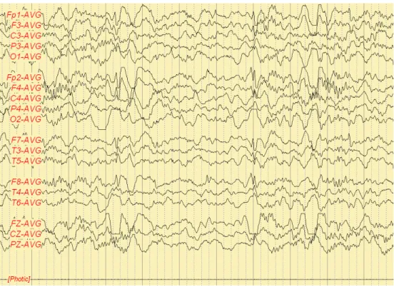
Fig. 1. Intermittent, sometimes rhythmic high amplitude delta activity (Patient A).
Fig. 2. Occasional spike or sharp wave discharges from both frontal and temporal area (Patient A).
Fig. 3. Frequent synchronous or independent spike discharges from Fp1 and Fp2 (Patient B).
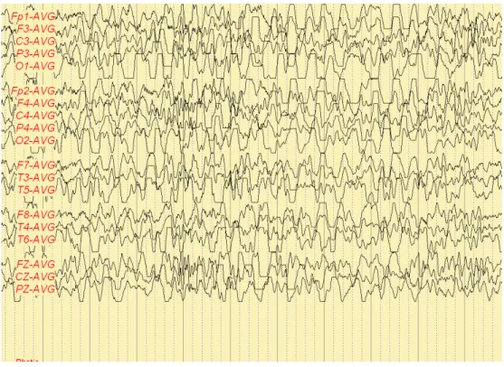
Fig. 4. Anterior dominant diffuse high amplitude delta activity (Patient B).
III. Results
1. Patient A
Very large amplitude slow activity at 2-3/s, often rhythmic, usually occurring in prolonged runs reaching over 200 μV (sometimes over 500 μV) and often more prominent anterior (Fig. 1). Spikes or sharp waves were sometimes associated with these slow waves forming ill-defined spike-wave complexes (Fig. 2).
This recording is suggestive of diffuse cerebral dysfunction and consistent with partial seizure.
2. Patient B
The pattern of patient B shows frequent synchronous or independent spike discharges (Fig. 3) and anterior dominant diffuse high amplitude delta activity (Fig. 4).
This record is indicative of diffuse cerebral dysfunction and consistent with partial seizure.
Ⅳ. Discussion
We found characteristic features of epilepsy in 2 patients with AS with a chromosome 15q11-13 deletion.
The following three characteristic EEG abnormalities in childhood AS patients have been reported by many authors (Mayo et al, 1973; Sugimoto et al, 1992; Laan et al, 1996; Laan et al, 1997; Minassian et al, 1998): (1) rhythmic 4-6 Hz activity, more than 200 mV, often generalized, not associated with drowsiness. (2) Rhythmic triphasic 2-3 Hz, high voltage (200-500 mV) activity, mixed with spikes or sharp waves with a maximum over the frontal regions (Laan et al, 1997). (3) Spikes mixed with 3–4 Hz components, usually of more than 200 mV mainly posterior and facilitated by eye closure (Boyd et al, 1988). Our results were similar to (1) and (2).
In conclusion, our study of the 2 patients described here confirms the particular evolution of AS. EEG is
particularly useful, and is one of the most specific parameters for early diagnosis of the syndrome. The combination of certain (non-specific) clinical findings of early onset and the EEG features described above, can give rise to a relatively reliable diagnosis of suspected AS. This finding should lead to high resolution cytogenetic investigations, if these should prove negative, then molecular biology must be used to check for any submicroscopic deletions.
Reference
1. Angelman H. “Puppet” Children. A report of three cases. Dev Med Child Neurol 7:681-687, 1965.
2. Buoni S, Grosso S, Pucci L, Fois A. Diagnosis of Angelman syndrome: clinical and EEG criteria. Brain Dev 21:296–302, 1999.
3. Bower BD, Jeavons PM. The “Happy Puppet”
syndrome. Arch Dis Child 42:298-302, 1967.
4. Boyd SG, Harden A, Patton MA. The EEG in early diagnosis of the Angelman (happy puppet) syndrome.
Eur J Pediatr 147:508-513, 1988.
5. Casara GL, Vecchi M, Boniver C, Drigo P, Baccichetti C, Artifoni L, et al. Electroclinical diagnosis of Angelman syndrome: a study of seven cases. Brain Dev 17:64-68, 1995.
6. Clayton-Smith J, Laan LAEM. Angelman syndrome : a review of the clinical and genetic aspects. J Med Genet 40:87–95, 2003.
7. Laan LA, den Boer AT, Hennekam RC, Renier WO, Brouwer OF. Angelman syndrome in adulthood. Am J Med Genet 66:356-360, 1996.
8. Laan LAEM, Renier WO, Arts WFM, Buntinx IM, vd Burgt IJAM, Stroink H, et al. Evolution of epilepsy and EEG findings in Angelman syndrome. Epilepsia 38:195–199, 1997.
9. Matsumoto A, Kumagai T, Miura K, Miyazaki S,
Hayakawa C, Yamanaka T. Epilepsy in Angelman syndrome associated with chromosome 15q deletion.
Epilepsia 33:1083–1090, 1992.
10. Mayo O, Nelson MM, Townsend HR. Three more
“happy puppets”. Dev Med Child Neurol 15:63-74, 1973.
11. Minassian BA, DeLorey TM, Olsen RW, Philippart M, Bronstein Y, Zhang Q, et al. Angelman syndrome:
correlation between epilepsy phenotypes and genotypes.
Ann Neurol 43:485-493, 1998.
12. Pampiglione G, Martinez A. Evolution of Angelman syndrome: follow up of 3 new cases. Electroencephalogr Clin Neurophysiol 56:72S, 1983.
13. Sugimoto T, Yasuhara A, Ohta T, Nishida N, Saitoh S, Hamabe J, et al. Angelman syndrome in three siblings: characteristic epileptic seizures and EEG abnormalities. Epilepsia 33:1078-1082, 1992.