Hemangiopericytoma is a rare mesenchymal tumor originating from the pericytes of Zimmermann that are normally present in the basal membrane of the capillar- ies and postcapillary venules. The function of the peri- cytes is not well understood, although they appear to be involved in the regulation of vessel caliber. As all capil- laries are enveloped by pericytes, hemangiopericytomas can be found virtually anywhere in the body (1, 2).
However, this neoplasm more commonly affects the soft tissues of the extremities, pelvis, and retroperi- toneum. Pancreatic involvement of a hemangiopericy- toma is extremely rare. We report a case of a metastatic hemangiopericytoma involving the pancreas and its ra- diological findings on CT, MR imaging including MR an- giography, endoscopic retrograde cholangiopancreatog-
raphy (ERCP), and conventional angiography.
Case Report
A 48-year-old female was referred to our institution for further examination and treatment of a palpable ab- dominal mass. The patient complained of vague abdom- inal discomfort and dyspepsia for a duration of four months but without weight loss. Twenty years ago, the patient had undergone surgery for varicose veins in the left leg; 10 years later, the patient underwent excision of a new mass at the previous surgery site in the left leg.
The pathological diagnosis of the new mass was a he- mangiopericytoma. A physical examination showed a palpable, non-tender mass in the right upper quadrant of the abdomen. The results of routine laboratory tests, including measurement of the levels of serum amylase, lipase and bilirubin, and tumor markers, including car- cinoembryonic antigen (CEA) and carbohydrate antigen 19-9 (CA 19-9), were within normal ranges. An ab- dominal CT demonstrated three masses arising from the pancreas head and body. The longest diameters of the
J Korean Radiol Soc 2007;57:261-264
─ 261 ─
Pancreatic Metastases from a Hemangiopericytoma of the Leg: A Case Report1
Hye Jeon Hwang, M.D., Jae Ho Byun, M.D., Seong Ho Park, M.D., Moon-Gyu Lee, M.D.
1Department of Radiology and Research Institute of Radiology, University of Ulsan College of Medicine, Asan Medical Center, Seoul 138-736, Korea Received June 19, 2007 ; Accepted July 31, 2007
Address reprint requests to : Jae Ho Byun, M.D., Ph.D., Department of Radiology and Research Institute of Radiology, University of Ulsan College of Medicine, Asan Medical Center, 388-1 Pungnap2-dong, Songpa-gu, Seoul 138-736, Korea.
Tel. 82-2-3010-4400 Fax. 82-2-476-4719 E-mail: jhbyun@amc.seoul.kr
Hemangiopericytoma of the pancreas has rarely been described, and its radiological findings have never been described in the radiological literature. We report a case of a metastatic hemangiopericytoma involving the pancreas in a 48-year-old woman. CT, MR, and angiography showed three, well-demarcated, heterogeneously enhancing masses with necrosis and hemorrhage in the pancreas.
Index words :Pancreas
Pancreatic neoplasms
Tomography, X-Ray Computed Magnetic resonance (MR) Hemangiopericytoma
Hye Jeon Hwang, et al: Pancreatic Metastases from a Hemangiopericytoma of the Leg
─ 262 ─
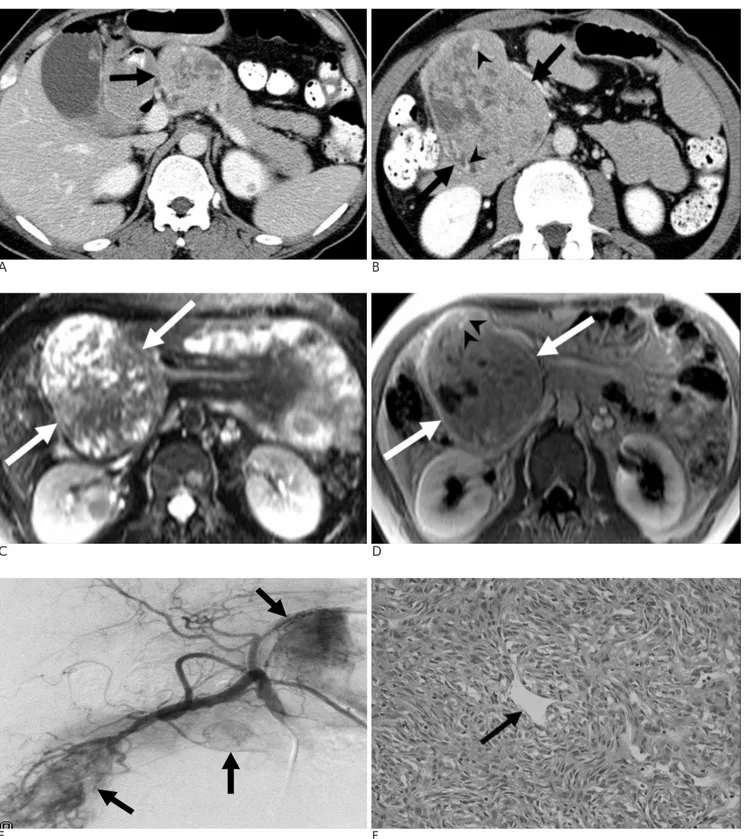
A B
C D
E F
Fig. 1. Metastatic pancreatic hemangiopericytomas in a 48-year-old woman.
A, B. Contrast-enhanced axial CT scans show two, well-marginated, heterogeneously enhancing masses (arrows) with low-attenua- tion portions, indicating necrosis or cystic degeneration as well as intratumoral vessels (arrowheads in B) in the pancreatic head and body.
C. On a T2-weighted MR image, the mass (arrows) of the pancreas head shows heterogeneous, intermediate- and high-signal inten- sity and some bright signal intensity, indicating cystic degeneration or necrosis.
D. On an axial gadolinium-enhanced T1-weighted MR image, the mass (arrows) in the pancreas head shows heterogeneous en- hancement with nonenhancing portions, indicating necrosis or cystic degeneration, as well as intratumoral vessels (arrowheads).
E. Conventional angiography shows three hypervascular masses (arrows) with intratumoral vessels in the pancreas.
F. A photomicrograph of the histological specimen shows thin-walled, endothelial-lined, ramifying vascular channels (arrow).
Reticulin and collagen fibers enmesh the tumor cells outside the vascular structure (Hematoxylin and eosin staining, × 400).
masses were 9 cm, 7 cm and 2 cm, respectively. On un- enhanced CT, the three masses showed a similar attenu- ation to the normal surrounding pancreas parenchyma with multifocal, low-attenuation portions without calci- fications. On contrast-enhanced CT, the masses were well-demarcated and showed heterogeneous enhance- ment with multifocal, low-attenuation portions and some intratumoral vessels (Figs. 1A, B). The common bile duct was mildly dilated without pancreatic duct di- latation (Fig. 1A). There were no enlarged lymph nodes in the abdomen. ERCP revealed the upward displace- ment of the distal common bile duct and mild dilatation of the intra- and extrahepatic bile ducts. Compared with the normal pancreas parenchyma, the masses showed heterogeneous, iso- and low-signal intensity on T1- weighted MR images and demonstrated heterogeneous, intermediate- and high-signal intensity and some bright signal intensity portions, representing necrosis or cystic degeneration, on T2-weighted MR images (Fig. 1C). On gadolinium-enhanced MR images, the masses showed heterogeneous enhancement with intratumoral vessels and some nonenhancing portions, indicating necrosis, cystic degeneration or old hemorrhage (Fig. 1D). MR an- giography and conventional angiography showed the hypervascularity of the masses (Fig. 1E). The patient un- derwent a pylorus-preserving pancreaticoduodenecto- my and total pancreatectomy with a splenectomy. On the gross specimens, the masses were well-demarcated and firm. The cut surfaces of the masses were yellowish with partial hemorrhage and necrosis. Histology of the specimen showed thin-walled, endothelial-lined, rami- fying vascular channels. Reticulin and collagen fibers enmeshed the tumor cells outside of the vascular struc- ture (Fig. 1F). By immunohistochemical staining, CD 34 and CD 99 were positive. The final histopathological di- agnosis of the masses was as hemangiopericytomas.
Two years following the surgery, several new masses were detected in the left psoas muscle and in both kid- neys. The patient underwent excision of these masses that were also diagnosed as hemangiopericytomas. Two years after the second surgery, a follow-up CT showed multiple, recurrent masses in the left kidney, pelvic mesentery, liver, and lung.
Discussion
An hemangiopericytoma is a rare mesenchymal tu- mor that accounts for less than 2% of all mesenchymal tumors. It was first described and named by Stout and
Murray in 1942 (2). It may occur at all ages, but is most common in the fifth and sixth decades. It is equally com- mon in men and women (1, 2). Although there is no de- finitive pathological grading system to determine the malignancy potential, the prominent mitotic activity, necrosis, hemorrhage, and increased cellularity are all suggestive of a malignancy. The prognosis of this tumor is also related to its size. The relative 10-year survival rate of patients with tumors larger than a median size of 6.5 cm in the longest diameter is 63 %, whereas the 10- year survival rate of patients with tumors smaller than 6.5 cm in the longest diameter is 92% (1).
The most common site of a hemangiopericytoma is the lower extremity, especially the thigh. In a series of 106 patients reported by Enzinger and Smith (1), 37 tu- mors (35%) affected the lower extremity and 26 tumors (25%) were found in the pelvis or retroperitoneum. Of the other 43 patients in that study, 17 tumors were found in the head and neck region, 15 tumors involved the trunk, and 11 tumors were located in the upper ex- tremity (1). Most abdominal hemangiopericytomas arise in the retroperitoneum, omentum or intestinal wall (3).
An hemangiopericytoma originating from the pancreas is very rare. On reviewing the English language litera- ture, we found four cases of a primary pancreatic he- mangiopericytoma (4) and six cases of a metastatic pan- creatic hemangiopericytoma that had arisen from an in- tracranial hemangiopericytoma (5). Unfortunately, there was only a description of the radiological findings with- out images of the pancreatic hemangiopericytoma in these ten case reports.
In this case, metastatic hemangiopericytomas of the pancreas appeared as well-demarcated, hypervascular masses with heterogeneous enhancement and multifo- cal cystic portions indicating necrosis, cystic degenera- tion or old hemorrhage. Goldman et al. (6) and Alpern et al. (7) reported that retroperitoneal hemangiopericy- tomas appeared as large, lobulate, enhancing, soft-tissue masses with cystic low-attenuation zones consistent with necrosis, hemorrhage or cystic degeneration.
Alpern et al. (7) reported the presence of speckled calci- fications in a retroperitoneal hemangiopericytoma. The incidence of calcification in hemangiopericytoma varied from one (1%) of 106 cases on plain radiography to five (71%) of seven patients on CT (1, 7). On MR imaging, a pelvic hemangiopericytoma showed intermediate to low signal intensity on T1-weighted images and intermedi- ate to high signal intensity on T2-weighted images. On gadolinium-enhanced MR images, the solid portion of
J Korean Radiol Soc 2007;57:261-264
─ 263 ─
the tumor showed strong enhancement (8). Shin et al. (9) reported that a hepatic hemangiopericytoma was sharply delineated and the extent of liver involvement could be more accurately evaluated on MR images. On angiography, hemangiopericytomas appeared as very hypervascular tumors as there was a clear increase in the number and caliber of arteries and veins of the tu- mor. Therefore, embolization might be considered pre- operatively because of the extreme vascularity of he- mangiopericytomas (6). The radiological features of he- mangiopericytomas seem to be similar in the present study and in previous reports.
The differential diagnosis of a well-demarcated, hy- pervascular pancreatic mass with cystic portions should include a non-functioning islet cell tumor as these tu- mors show strong enhancement with cystic degenera- tion and necrosis on contrast-enhanced CT. Non-func- tioning islet cell tumors share similar radiological find- ings with the findings of hemangiopericytomas. A more common hypervascular metastasis such as a renal cell carcinoma should be included in the differential diagno- sis of multiple hypervascular masses in the pancreas, because metastasis tends to repeat the imaging pattern of the primary tumor (10).
In conclusion, a metastatic hemangiopericytoma in the pancreas appears as a well-demarcated, hypervascu- lar mass with cystic degeneration or necrosis. A correct radiological differential diagnosis distinguishing it from a nonfunctioning islet cell tumor or other hypervascular
metastasis remains difficult, if the diagnosis or history of the primary extrapancreatic hemangiopericytoma is un- clear.
References
1. Enzinger FM, Smith BH. Hemangiopericytoma. An analysis of 106 cases. Hum Pathol 1976;7:61-82
2. McMaster MJ, Soule EH, Ivins JC. Hemangiopericytoma. A clini- copathologic study and long-term follow-up of 60 patients. Cancer 1975;36:2232-2244
3. Binder SC, Wolfe HJ, Deterling RA Jr. Intra-abdominal heman- giopericytoma. Report of four cases and review of the literature.
Arch Surg 1973;107:536-543
4. Bardaxogou E, Manganas D, Landen S, Ramee MP, Chareton B, Maddern GJ, et al. Hemangiopericytoma of the pancreas: report of a case and review of the literature. Hepatogastroenterology 1995;42:172-174
5. Koyama H, Harada A, Nakao A, Nonami T, Kurokawa T, Kaneko T, et al. Intracranial hemangiopericytoma with metastasis to the pancreas: case report and literature review. J Clin Gastroenterol 1997;25:706-708
6. Goldman SM, Davidson AJ, Neal J. Retroperitoneal and pelvic he- mangiopericytomas: clinical, radiologic, and pathologic correla- tion. Radiology 1988;168:13-17
7. Alpern MB, Thorsen MK, Kellman GM, Pojunas K, Lawson TL.
CT appearance of hemangiopericytoma. J Comput Assist Tomogr 1986;10:264-267
8. Kehagias D, Gouliamos A, Vlahos L. MR appearance of pelvic he- mangiopericytoma. Eur Radiol 1999;9:163-165
9. Shin MS, Koehler RE, Stanley RJ, Barton JC, Ho KJ. Malignant he- mangiopericytoma: computed tomography and magnetic reso- nance imaging. J Comput Assist Tomogr 1987;11:297-300
10. Ferrozzi F, Bova D, Campodonico F, Chiara FD, Passari A, Bassi P.
Pancreatic metastases: CT assessment. Eur Radiol 1997;7:241-245 Hye Jeon Hwang, et al: Pancreatic Metastases from a Hemangiopericytoma of the Leg
─ 264 ─
대한영상의학회지 2007;57:261-264
췌장에 생긴 전이성 혈관주위세포종의 영상 소견1
1울산의대 서울아산병원 영상의학과 황혜전・변재호・박성호・이문규
췌장의 혈관주위세포종은 매우 드물게 보고되었으며, 그 영상소견은 영상의학지에 보고된 경우가 없다. 저자들은 48세 여자 환자에서 췌장에 생긴 전이성 혈관주위세포종의 영상소견을 보고하고자 한다. 복부전산단층촬영, 자기 공명영상, 그리고 혈관조영술에서 췌장에 세 개의 경계가 좋고 불균일한 조영증강을 보이는 종괴로 괴사와 출혈을 동반하였다.