D I A B E T E S & M E T A B O L I S M J O U R N A L
This is an Open Access article distributed under the terms of the Creative Commons At- tribution Non-Commercial License (http://creativecommons.org/licenses/by-nc/3.0/) which permits unrestricted non-commercial use, distribution, and reproduction in any medium, provided the original work is properly cited.
Comparison of EGF with VEGF Non-Viral Gene Therapy for Cutaneous Wound Healing of
Streptozotocin Diabetic Mice
Junghae Ko1,2, Haejung Jun1,2, Hyesook Chung1, Changshin Yoon1, Taekyoon Kim1,2, Minjeong Kwon1,2, Soonhee Lee1,2, Soojin Jung3, Mikyung Kim4, Jeong Hyun Park1,2
1Molecular Therapy Laboratory, Paik Memorial Institute for Clinical Research, Departments of 2Internal Medicine, 3Pathology, Inje University College of Medicine,
4Department of Internal Medicine, Maryknoll Medical Center, Busan, Korea
Background: To accelerate the healing of diabetic wounds, various kinds of growth factors have been employed. It is the short half-life of administered growth factors in hostile wound beds that have limited wide-spread clinical usage. To overcome this limitation, growth factor gene therapy could be an attractive alternative rather than direct application of factors onto the wound beds. We administered two growth factor DNAs, epidermal growth factor (EGF) and vascular endothelial growth factor (VEGF) into a cutaneous wound on diabetic mice. We compared the different characteristics of the healing wounds.
Methods: Streptozotocin was injected intraperitoneally to induce diabetes into C57BL/6J mice. The ultrasound micro-bubble destruction method with SonoVue as a bubbling agent was used for non-viral gene delivery of EGF828 and VEGF165 DNAs. Each gene was modified for increasing efficacy as FRM-EGF828 or minicircle VEGF165. The degree of neoangiogenesis was assessed us- ing qualitative laser Doppler flowmetry. We compared wound size and histological findings of the skin wounds in each group.
Results: In both groups, accelerated wound closure was observed in the mice receiving gene therapy compared with non treated diabetic control mice. Blood flow detected by laser doppler flowmetry was better in the VEGF group than in the EGF group.
Wound healing rates and histological findings were more accelerated in the EGF gene therapy group than the VEGF group, but were not statistically significant.
Conclusion: Both non-viral EGF and VEGF gene therapy administrations could improve the speed and quality of skin wound healing. However, the detailed histological characteristics of the healing wounds were different.
Keywords: Epidermal growth factor; Gene therapy; Non-viral; Skin wound; Vascular endothelial growth factor
Corresponding author: Jeong Hyun Park
Department of Internal Medicine, Pusan Paik Hospital, Inje University College of Medicine, 633-165 Gaegeum 2-dong, Busanjin-gu, Busan 614-735, Korea
INTRODUCTION
Wound healing, which proceeds through a series of consecu- tive, but overlapping stages, is characterized by the sequential movement of different cell populations into the wound site [1].
At the molecular level, the acute wound healing response is characterized by changes in the composition and organization of the extracellular matrix (ECM) as well as the local profile of
growth factors [2,3].
Blood components are released into the wound site activat- ing the clotting cascade. The resulting clot induces hemostasis and provides a matrix for the influx of inflammatory cells. Plate- lets degranulate releasing alpha granules, which secrete growth factors such as: epidermal growth factor (EGF), platelet-de- rived growth factor (PDGF), and transforming growth factor- beta (TGF-β) [4].
pISSN 2233-6079 · eISSN 2233-6087
With the assistance of platelet released vascular endothelial growth factor (VEGF) and fibroblast growth factor (FGF), en- dothelial cells proliferate and angiogenesis ensues. This pro- cess is essential for the synthesis, deposition, and organization of a new ECM [5].
EGF, originally reported by Dr. Stanley Cohen [6,7], is se- creted by platelets, macrophages, and fibroblasts. This growth factor acts in a paracrine fashion on keratinocytes [8,9]. In vi- tro studies have shown that EGF is up-regulated after acute in- jury significantly accelerating reepithelialization [10] and in- creasing tensile strength of the wound [11].
VEGF is produced by endothelial cells, keratinocytes, fibro- blast, smooth muscle cells, platelets, neutrophils, and macro- phages [12-17]. It is important in wound healing because it promotes the early events in angiogenesis, particularly endo- thelial cell migration [18-20] and proliferation [21-25] as seen in several in vitro studies. Chronic wounds have areas of local skin ischemia making VEGF a possible therapeutic modality.
However, high blood glucose levels impair both granulocyte and neutrophil function, and chemotaxis, resulting in increased risk of infection. Inflammation is prolonged, angiogenesis is impaired and there is decreased synthesis of collagen [1].
There are changes in the cellular infiltrate and extracellular matrix, with prolonged expression of fibronectin. Finally, there are fewer T cells and more macrophages persisting beyond the initial stages of healing [3].
We have recently shown that the minicircle-VEGF165 is ef- fective for the healing of the skin wound of the diabetic mice [26].
After the above study, we administered EGF using another gene therapy method. Therefore, this study was thus designed to report the different characteristics of two genes on the skin wound of diabetic mice.
METHODS
Plasmid construction
Human EGF cDNA was a kind gift from Dr. Siu-Yuen Chan, University of Hong Kong. The pβ-EGF159 was constructed by using human mature EGF plasmid and human full length EGF.
We did not construct the minicircle DNA (cDNA) by using of pβ-EGF159. In the case of VEGF165, p2øC31-β-VEGF165 was constructed for the production of minicircle DNA. The DNA fragment only contains the chicken β-actin promoter, VEGF165 cDNA. The SV40 poly adenylation signal sequence was excised
with BglII and ClaI from the pβ-VEGF165, and then bluntly li- gated between the attB and attP sites of the p2øC31 plasmid, which was a kind gift from Dr. Mark A. Kay (Department of Genetics, Stanford University, CA, USA).
Preparation of DNAs
E. coli DH5α (Invitrogen, Carlsbad, CA, USA) was transformed by p2øC31-β-VEGF165. A single colony of the transformants was grown at 37°C overnight (OD600=4.5–5.0). The 1 L of bacterial culture in the steady state was spun down in a clinical centrifuge (rotor JA-14, J2-MC centrifuge; Beckman, Fuller- ton, CA, USA) at 20°C, 1,300×g for 15 minutes. The pellet was re-dissolved with 1 L of fresh LB broth (pH 7.0) containing 1%
L-(+)-arabinose. The resuspended bacteria were incubated at 30°C with constant shaking at 225 rpm for 2 hours. Subsequent- ly, 1 L of fresh LB broth (pH 9.0) containing 1% L-(+)-arabi- nose was added to the culture and the bacteria were cultivated for additional 2 hours at 37°C for the activation of the restric- tion enzyme I-SceI. Super-coiled minicircle DNA was prepared from the culture, using plasmid purification kits from the Qia- gen (Valencia, CA, USA). The contaminated endotoxin in the DNA preparation was removed by the AffinityPak Detoxi-Gel (Pierce, Rockford, IL, USA).
Cell culture and in vitro transfection
The human embryonic kidney (HEK293) cells, were purchased from ATCC (Manassas, VA, USA). HEK293 cells were main- tained in Dulbecco’s Modified Eagle Medium (DMEM) sup- plemented with 10% fetal bovine serum (FBS) in a 5% CO2 in- cubator.
SonoVue
SonoVue microbubbling echo contrast agent that utilizes sul- fur hexafluoride was purchased from Bracco Diagnostics, Inc.
(Bracco, UK).
For the assessment of transfection by sonoporation, the cells were detached by trypsin-EDTA, washed twice in phosphate- buffered saline, and resuspended with serum free media at 2.0×10⁵ cells/well in 48-well plate (Nunc, Rochester, NY, USA).
The diameter of each well was 12.0 mm. After addition of mi- crobubble, each plasmid encoding human EGF and VEGF was mixed with the cell supernatant (2 μg/well). Immediately after the addition of the mixture of plasmids and microbub- bles, the cells were exposed to ultrasound with a 20% duty cy- cle (Sonitron 1000; Rich Mar Inc., Inola, OK, USA) for 30 sec-
onds, using a 6.0 mm in diameter ultrasound probe. The ultra- sound probe was immersed directly into the cell suspension without any contact to the surface of well. After exposure, the cell suspensions were harvested, separated by centrifugation, and plated in 6-well dishes. The cells were incubated for 48 hours before ELISA.
Comparison of minicircle VEGF with a typical plasmid with bacterial backbone
To assay the efficiency of transfection by minicircle VEGF, branched polyethylenimine (BPEI, 25kDa; Sigma–Aldrich, St.
Louis, MO, USA) was used as a gene carrier. The plasmid/BPEI complexes were prepared at a 10/1 N/P ratio and incubated for 30 minutes at room temperature. The cells were washed twice with serum-free medium, and then 2 mL of fresh serum-free medium was added. The plasmid/BPEI complex was added to each well. The cells were then incubated for 4 hours at 37°C in a 5% CO2 incubator. After 4 hours, the transfection mixtures were removed and 2 mL of fresh medium containing FBS was added. The cells were incubated in a CO2 incubator for 48 hours.
The cells and media were harvested for ELISA.
ELISA
EGF protein was measured using Quantikine human EGF ELI- SA kit (R&D Systems, Minneapolis, MN, USA). EGF protein was measured using Quantikine human EGF ELISA kit (R&D Systems).
The secreted VEGF protein in the media was measured us- ing Quantikine human VEGF ELISA kit (R&D Systems).
Animals
All animal procedures were approved by the Experimental An- imal Committee at the Inje University. Two-week-old male C57BL/6J mice (20 to 30 g) were purchased from the Samtako (Animal Breeding Center, Osan, Korea) and housed in the pathogen-free condition and had ad libitum access to water and the standard mouse chow. Diabetes mellitus was induced by the single intraperitoneal injection (200 mg/kg) of strepto- zotocin (STZ) [27]. Random blood glucose levels were mea- sured 1 week after the first STZ injection using a glucometer (Accu-Check; Roche Diagnostics, Indianapolis, IN, USA). Mice that showed blood glucose levels over 200 mg/dL were consid- ered as diabetic [28]. The mice were divided into four groups:
control group (n=5), diabetic control group (n=5), EGF treat- ed group (n=5), and VEGF treated group (n=5), and the pro-
cess was performed in triplicate.
Skin wounding
Each animal was subjected to wounding (6 mm in diameter) by punch biopsy (Stiefel, Bad Oldesloe, Germany) on the skin of the back of the mouse. Briefly, prior to wounding, general anesthesia was induced by the administration of ketamine hy- drochloride (100 mg/kg). The hair was shaved with the stan- dard blade (no. 10). Then, the skin was disinfected with the povidone–iodine solution and wiped with sterile water.
Gene delivery into diabetic mouse wound
Diabetic C57BL/6J mouse with the back skin wound received a subcutaneous injection of PBS containing the mixture of minicircle-VEGF165 (20 μg) and pβ-EGF828 (20 μg) with mi- crobubble solution (100 μg). Immediately after the injection, ultrasound (frequency, 1.0 MHz; duty, 20%; intensity, 2.0 W/
cm2; time, 30 seconds) was applied to the injection site of the wound edge using a Sonitron 2000 ultrasonicator (Rich-Mar, Inola, OK, USA) with a probe of diameter 6 mm.
Measurement of wound area and laser doppler imaging (LDI)
The areas of the wounds were evaluated by densitometry (Multi Gauge V3.0 software; Fujifilm Life Science, Tokyo, Japan). The percentage wound closure was calculated as a ratio of final wound area to initial wound area. For the estimation of the skin blood flow, the wounds were scanned with a Laser Dop- pler Imager (Periscan PIM II Laser Doppler Perfusion Imager;
Perimed AB, Järfälla, Sweden) under the brief general anes- thesia with inhalation of 1% to 2% isoflurane on the days 2, 6, and 12. The mice were placed on a light absorbing dark green background. The distance between the scanner head and the object was 20 cm. The Min and Max values were set at 0 and 8 V, respectively. The perfusion scan image color scale displayed the lowest value in dark blue and the highest value in red, which represent the relative amount of skin perfusion. All mice were placed on a warming pad to maintain core body temperature between 36.8°C and 37.8°C during the scanning.
Immunohistochemistry
The tissue samples adjacent to the wounds were embedded in OCT compound (Tissue Tek; Sakura Finetek, Torrance, CA, USA). Transverse sections of 5 μm thickness were placed on polyl-lysine-coated slides.
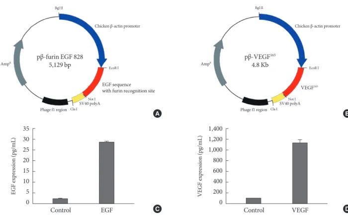
polyadenylation sequence. For the secretion of translated EGF, we added a furin protease recognition site to the N-terminal of EGF sequence (Fig. 1A). Likewise, we cloned the cDNA se- quence of human VEGF165 to generate pβ-VEGF (Fig. 1B). All plasmids DNA were transformed into DH5α, cultured to liter scale, and prepared using DNA maxi-prep kit (Qiagen, Valen- cia, CA, USA).
Gene delivery to HEK293 cells by sonoporation
The human embryonic kidney (HEK) 293 cells in the presence of 2 μg of each plasmid were exposed to 1 MHz ultrasound with microbubble (10 mg/mL). With proper exposure intensi- ty (2.0 W/cm2) and duty (20%) the cell membrane was caused to be in a transiently porous state under ultrasound irradiation called “sonoporation.” The gene delivery by ultrasound has a major advantage of safety to the cell or tissue that exert the therapeutic effect of transgene. As shown in Fig. 1C and D, EGF and VEGF were measured from cell culture media (25 pg/mL Histology
On day 12, wound tissues were fixed in 4% phosphate buffered formalin and then embedded in paraffin. From the paraffin- embedded tissue blocks, 5 μm sections were cut and stained with hematoxylin and eosin (H&E) for the histological analy- sis.
Statistical analysis
All the presented data were expressed as mean±standard de- viation. The statistical significance was analyzed by the Stu- dent’s t-test and ANOVA using the SPSS PC program (SPSS Inc., Chicago, IL, USA).
RESULTS
Plasmid construction
The cDNA sequence of human mature EGF was inserted into pβ-plasmid, which had a chicken β-actin promoter and a SV40
Control EGF
35 30 25 20 15 10 5 0
EGF expression (pg/mL)
Control VEGF
1,400 1,200 1,000 800 600 400 200 VEGF expression (pg/mL) 0 C
A
D B
Fig. 1. Plasmid constructions and their expressions by ultrasound mediated gene delivery in human embryonic kidney (HEK) 293 cells. (A) cDNA encoding human epidermal growth factor (EGF) with a N-terminal furin recognition sequence was inserted between EcoR I and Not I restriction sites under chicken β-actin promoter. (B) cDNA of human vascular endothelial growth fac- tor 165 (VEGF165) was inserted to produce pβ-VEGF165. (C, D) Ultrasound mediated gene delivery in HEK 293 cells. The cells were exposed to 1 MHz US in the presence of 2 μg of each plasmid with a concentration of microbubble (10 mg/mL). Ultrasound intensity was 2.0 W/cm2; ultrasound exposure time was 30 seconds; duty cycle was 20%.
Bgl II
pβ-furin EGF 828 5,129 bp
EGF sequence with furin recognition site
EcoR I
Not I Cla I
Chicken β-actin promoter
AmpR
Phage f1 region SV40 polyA
pβ-VEGF165 4.8 Kb
VEGF165
EcoR I
Not I Cla I
Chicken β-actin promoter Bgl II
AmpR
Phage f1 region SV40 polyA
and 1.2 ng/mL, respectively) 48 hours after gene delivery in HEK293 cells.
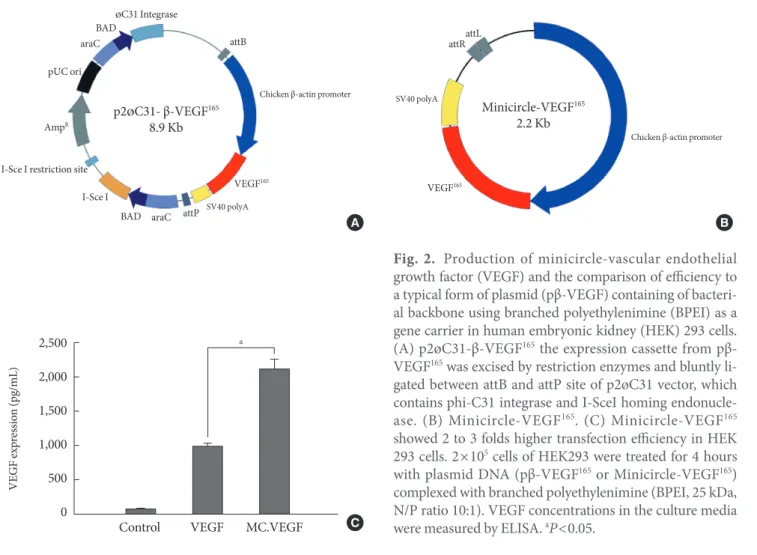
Production of minicircle-VEGF
For the healing of chronic diabetic wound model, the initial rapid generation of blood circulation is known to be critical for the attraction of neutrophiles and monocytes to advance the repair process [29]. In the case of gene delivery of VEGF, we hypothesized that the earlier blood vessel formation and the initial increase of blood perfusion in the wound tissue would be necessary to accelerate the whole wound healing process. Therefore, we constructed a parent plasmid (p2øC31- VEGF) to induce a minicircle form of DNA encoding VEGF that has minimal size of expression machinery to transgene expression of VEGF (Fig. 2A). By adding L-(+)-arabinose and adjusting pH and temperature to pH 9.0 and 37°C, respective- ly, minicircle-VEGF DNA was recombinated out of the parent plasmid in DH5α. The remaining circular bacterial backbone
DNA including an antibiotics resistance gene and a replication origin is linearized by homing endonuclease I-Sce I and de- graded by bacterial exonucleases. The outcome of these pro- cesses is the production of supercoiled minicircle-VEGF DNA (2.2 kb) that has largely reduced the size in comparison with a typical form of plasmid pβ-VEGF (4.8 kb) (Fig. 2B). The trans- gene expression using minicircle-VEGF was 2 to 3 fold higher than a typical form of plasmid pβ-VEGF (Fig. 2C). This is con- sidered to provide more rapid movement of minicircle DNA across the cytosol to the nuclear membrane than previously possible. Furthermore, minicircle DNA has no CpG island that stimulates unnecessary immune responses resulting in transgene quiescence.
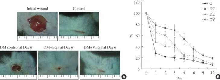
Measurement of wound area
The results for the changes of the percentages of wound closure in both gene therapy groups are shown in Fig. 3. By day 6 post operation, all EGF and VEGF treated groups demonstrated
Fig. 2. Production of minicircle-vascular endothelial growth factor (VEGF) and the comparison of efficiency to a typical form of plasmid (pβ-VEGF) containing of bacteri- al backbone using branched polyethylenimine (BPEI) as a gene carrier in human embryonic kidney (HEK) 293 cells.
(A) p2øC31-β-VEGF165 the expression cassette from pβ- VEGF165 was excised by restriction enzymes and bluntly li- gated between attB and attP site of p2øC31 vector, which contains phi-C31 integrase and I-SceI homing endonucle- ase. (B) Minicircle-VEGF165. (C) Minicircle-VEGF165 showed 2 to 3 folds higher transfection efficiency in HEK 293 cells. 2×10⁵ cells of HEK293 were treated for 4 hours with plasmid DNA (pβ-VEGF165 or Minicircle-VEGF165) complexed with branched polyethylenimine (BPEI, 25 kDa, N/P ratio 10:1). VEGF concentrations in the culture media were measured by ELISA. aP<0.05.
Control VEGF MC.VEGF C 2,500
2,000 1,500 1,000 500 0
VEGF expression (pg/mL)
a
p2ØC31- β-VEGF165 8.9 Kb
Chicken β-actin promoter
SV40 polyA VEGF165 attB
attP ØC31 Integrase BAD
araC pUC ori
AmpR
I-Sce I I-Sce I restriction site
BAD araC
Chicken β-actin promoter
Minicircle-VEGF165 2.2 Kb
attRattL
SV40 polyA
VEGF165
A B
complete closure of the wounds in contrast to diabetic control with a mean percent closure below 70%. The differences among the groups were not statistically significant.
LDI
The differences of wound perfusion were monitored by non invasive LDI at day 2 and 6 (Fig. 4). In comparison with the EGF group, the minicircle-VEGF DNA treated diabetic mice group showed increased blood flow in the wound area. Skin perfusions were gradually decreased after day 12 (data not shown).
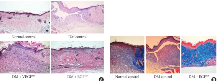
Histological findings
Both EGF and VEGF gene therapy could improve the speed and quality of skin wound healing. The detailed histological characteristics of healing wounds were different. The histology of biopsied wound tissue at day 12 in each group is shown in Fig. 5A. The wound of diabetic control shows severe edema and a disorganized pattern with heavy infiltration of inflam- matory cells. In minicircle-VEGF treated group, wound resto- ration is considered to be caused mainly by new blood vessel formation. Contrary to minicircle-VEGF, the EGF treated group, demonstrated epidermal reorganization that has com- pletely restored the normal wound microarchitectures (Fig.
5B) without observation of excessive increase of blood perfu- sion (Fig. 4).
DISCUSSION
Diabetic foot ulcers (DFUs), one of the most common compli- cations of diabetes, are a leading cause of hospitalization for diabetic patients. For a patient living with diabetes, the lifetime risk of developing a DFU is estimated to be 25% [30,31]. The prevalence of DFUs causes significant health care resources to be spent on the management of wounds. The interventions in- clude emergency room visits, antibacterial medications, am- putations, and a multitude of other therapies directed at non healing wounds. Therefore, many growth factors affecting wound healing are studied.
In 2004, one growth factor, topically applied recombinant PDGF, is currently approved by the US Food and Drug ad- ministration for the treatment of diabetic foot ulcers. [30]
However, its clinical success is limited in part by the need for daily application. There is a hostile proteolytic wound environ- ment in which the protein is applied. A study about the sus- tained effect and therapeutic potency is being performed.
There were many previous studies using VEGF and EGF.
Angiogenesis is necessary for granulation tissue formation as well as for providing oxygen and nutrition to wounds [32]. In- adequate angiogenesis in diabetic patients inhibits wound healing. VEGF is one of the most potent growth factors in stimulating angiogenesis. It triggers endothelial cell division, chemotaxis, and vascular permeability [33]. VEGF acts in a
120 100 80 60 40 20
0 0 1 2 3 4 6 9 12
Day
C DC DE DV Initial wound Control
DM control at Day 6 DM+EGF at Day 6 DM+VEGF at Day 6
A B
Fig. 3. Wound size comparison by ultrasound mediated gene delivery of epidermal growth factor (EGF) and vascular endothe- lial growth factor (VEGF). (A) Growth factor gene delivery by sonoporation enhanced the progress of wound closure in diabetic mice to non-diabetic normal control group. (B) Average areas (in pixels) of wound that received ultrasound mediated gene deliv- ery of EGF and VEGF were significantly reduced to non-diabetic control mice by day 12 post operation. C, control; DC, Diabetic (DM) control; DE, DM+EGF; DV, DM+VEGF.
%
paracrine manner on dermal microvessels and endothelial cells. It also promotes the production of nitric oxide, which enhances collagen deposition. Previous studies with VEGF have demonstrated its efficacy in ischemic heart disease [34].
However, the increase in reepithelialization by VEGF165-treated wounds has been reported [35]. Data from this study, con- firmed by histology, showed the improvement in reepithelial- ization. In previous studies, the more rapid maturation of granulation tissue in the VEGF165-treated wound was shown to have more organized collagen fiber bundles deposited in gran- ulation tissue. However, in this study, the greater cell density in the granulation tissue and healed tissue was observed in the EGF-treated group.
In addition, there were many studies using recombinant hu- man EGF to enhance the healing effect on the chronic diabetic foot in human [36]. Various routes of administration have been used for delivering genes to the chronic wound. The ad- vent of new topical agents has broadened the treatment ap-
proaches to wound healing. However, in the case of a full thick- ness wound, the wound healing process might be delayed by using only topical application. When an intralesional infiltra- tion of EGF was done, the wound size reduction time was ac- celerated [36]. Abundant functional capillaries started to emerge and vascular endothelial nuclei were clearly less hy- pertrophic.
In the present study, the improvement of wound healing time and wound reepithelization were similar in both groups.
However, the detailed characteristics were different. In the VEGF treated diabetic group, LDI showed improved blood perfusion at the wound site, but ECM reorganization was more immature than the EGF treated group.
On the other hand, blood perfusion was not different from the control group in the EGF treated group, but ECM reorga- nization was markedly improved. The EGF treated group demonstrated an increased amount of mature collagen com- pared to the VEGF group.
Control DM control DM + EGF828 DM + VEGF165
2 days
6 days
8 days
Fig. 4. Blood perfusion in the wound tissue was significantly increased by ultrasound mediated gene delivery of growth factors to diabetic mice. The laser doppler imager (PeriScan) used in this study was employed for the visualization of blood perfusion.
Red color represents high blood perfusion in the wound site. At the 6th day post operation, a significant increase of blood perfu- sion was observed in diabetic (DM) mice group that received vascular endothelial growth factor (VEGF) gene delivery.
Jazwa et al. [37] demonstrated that combined VEGF and EGF gene transfer into the wound improves wound healing in diabetic mice. In this study, authors concluded that VEGF might increase the migration of diabetic fibroblasts cultured in high glucose concentration through FGF4-mediated up- regulation of one of the VEGF receptors, Flt-1 (VEGF-Recep- tor 1). We did not try the combined gene transfer into the wound, but it needs further evaluation. The combined gene therapy could be explained as a synergistic effect or a side ef- fect.
We used a nonviral gene delivery system. When choosing appropriate biomaterials during the design of a delivery vehi- cle, several factors must be considered [38,39]. These factors include predictability, accessibility, and safety issues. We fo- cused on the safety issues. The efficacy of nonviral gene deliv- ery is lower than the viral system, but the safety is most impor- tant factor.
One of the limitations of our study is that we used different delivery system in each group. There is the structural difference between VEGF and EGF DNAs. Second, we administrated the same dose of DNAs but we could not identify that the dose acts with the same efficacy. Therefore, if we injected the same dose, the effect could be different. Third, we regret that we did not perform the collagen stain in the VEGF group, since we
were unable to compare the efficacy of reepithelization between two groups.
In conclusion, VEGF enhances the neoangiogenesis, but was not effective on maturation of organized reepithelization.
The neoangiogenesis induced by EGF was not dominant, but it enhanced the maturation of organized reepithelization.
CONFLICTS OF INTEREST
No potential conflict of interest relevant to this article was re- ported.
ACKNOWLEDGMENTS
This study was supported by a grant from the Korea Health- care Technology R&D Project, Ministry for Health, Welfare &
Family Affairs, Republic of Korea (A090258).
REFERENCES
1. Bennett SP, Griffiths GD, Schor AM, Leese GP, Schor SL. Growth factors in the treatment of diabetic foot ulcers. Br J Surg 2003;
90:133-46.
2. Petri JB, Konig S, Haupt B, Haustein UF, Herrmann K. Molec-
Normal control DM control
DM + VEGF165 DM + EGF828 Normal control DM control DM + EGF828
A B
Fig. 5. Histology of wound tissues. (A) Histology of wound tissues at day 12 post operation (H&E stain, ×100). After the gener- ation of skin wound, non-treated diabetic (DM) control showed severe edema, disorganized micro-architectures and the heavy infiltration of inflammatory cells. The wound tissues that received ultrasound mediated gene delivery of epidermal growth factor (EGF) and vascular endothelial growth factor (VEGF) showed a highly restored well organized state of tissue compared to the non-diabetic normal control. (B) Tissue reorganization of wound tissues at day 12 post operation (M&T, ×100). In contrast to non-treated diabetic control, the wound tissues that received gene delivery of EGF showed more collagen accumulation and ap- peared as a organized wound matrix.
ular analysis of different phases in human wound healing. Exp Dermatol 1997;6:133-9.
3. Loots MA, Lamme EN, Zeegelaar J, Mekkes JR, Bos JD, Mid- delkoop E. Differences in cellular infiltrate and extracellular matrix of chronic diabetic and venous ulcers versus acute wounds. J Invest Dermatol 1998;111:850-7.
4. Barrientos S, Stojadinovic O, Golinko MS, Brem H, Tomic-Ca- nic M. Growth factors and cytokines in wound healing. Wound Repair Regen 2008;16:585-601.
5. Hantash BM, Zhao L, Knowles JA, Lorenz HP. Adult and fetal wound healing. Front Biosci 2008;13:51-61.
6. Cohen S, Elliott GA. The stimulation of epidermal keratiniza- tion by a protein isolated from the submaxillary gland of the mouse. J Invest Dermatol 1963;40:1-5.
7. Carpenter G, Cohen S. Epidermal growth factor. J Biol Chem 1990;265:7709-12.
8. Shiraha H, Glading A, Gupta K, Wells A. IP-10 inhibits epider- mal growth factor-induced motility by decreasing epidermal growth factor receptor-mediated calpain activity. J Cell Biol 1999;146:243-54.
9. Schultz G, Rotatori DS, Clark W. EGF and TGF-alpha in wound healing and repair. J Cell Biochem 1991;45:346-52.
10. Brown GL, Curtsinger L 3rd, Brightwell JR, Ackerman DM, Tobin GR, Polk HC Jr, George-Nascimento C, Valenzuela P, Schultz GS. Enhancement of epidermal regeneration by biosyn- thetic epidermal growth factor. J Exp Med 1986;163:1319-24.
11. Brown GL, Curtsinger LJ, White M, Mitchell RO, Pietsch J, Nordquist R, von Fraunhofer A, Schultz GS. Acceleration of tensile strength of incisions treated with EGF and TGF-beta.
Ann Surg 1988;208:788-94.
12. Namiki A, Brogi E, Kearney M, Kim EA, Wu T, Couffinhal T, Varticovski L, Isner JM. Hypoxia induces vascular endothelial growth factor in cultured human endothelial cells. J Biol Chem 1995;270:31189-95.
13. Nissen NN, Polverini PJ, Koch AE, Volin MV, Gamelli RL, DiPietro LA. Vascular endothelial growth factor mediates an- giogenic activity during the proliferative phase of wound heal- ing. Am J Pathol 1998;152:1445-52.
14. Banks RE, Forbes MA, Kinsey SE, Stanley A, Ingham E, Wal- ters C, Selby PJ. Release of the angiogenic cytokine vascular endothelial growth factor (VEGF) from platelets: significance for VEGF measurements and cancer biology. Br J Cancer 1998;
77:956-64.
15. Gaudry M, Bregerie O, Andrieu V, El Benna J, Pocidalo MA, Hakim J. Intracellular pool of vascular endothelial growth fac-
tor in human neutrophils. Blood 1997;90:4153-61.
16. Berse B, Brown LF, Van de Water L, Dvorak HF, Senger DR.
Vascular permeability factor (vascular endothelial growth fac- tor) gene is expressed differentially in normal tissues, macro- phages, and tumors. Mol Biol Cell 1992;3:211-20.
17. Jazwa A, Loboda A, Golda S, Cisowski J, Szelag M, Zagorska A, Sroczynska P, Drukala J, Jozkowicz A, Dulak J. Effect of heme and heme oxygenase-1 on vascular endothelial growth factor synthesis and angiogenic potency of human keratinocytes. Free Radic Biol Med 2006;40:1250-63.
18. Yebra M, Parry GC, Stromblad S, Mackman N, Rosenberg S, Mueller BM, Cheresh DA. Requirement of receptor-bound urokinase-type plasminogen activator for integrin alphavbe- ta5-directed cell migration. J Biol Chem 1996;271:29393-9.
19. Suzuma K, Takagi H, Otani A, Honda Y. Hypoxia and vascular endothelial growth factor stimulate angiogenic integrin ex- pression in bovine retinal microvascular endothelial cells. In- vest Ophthalmol Vis Sci 1998;39:1028-35.
20. Senger DR, Ledbetter SR, Claffey KP, Papadopoulos-Sergiou A, Peruzzi CA, Detmar M. Stimulation of endothelial cell migra- tion by vascular permeability factor/vascular endothelial growth factor through cooperative mechanisms involving the alphav- beta3 integrin, osteopontin, and thrombin. Am J Pathol 1996;
149:293-305.
21. Morbidelli L, Chang CH, Douglas JG, Granger HJ, Ledda F, Ziche M. Nitric oxide mediates mitogenic effect of VEGF on coronary venular endothelium. Am J Physiol 1996;270(1 Pt 2):
H411-5.
22. Pepper MS, Ferrara N, Orci L, Montesano R. Potent synergism between vascular endothelial growth factor and basic fibroblast growth factor in the induction of angiogenesis in vitro. Biochem Biophys Res Commun 1992;189:824-31.
23. Goto F, Goto K, Weindel K, Folkman J. Synergistic effects of vascular endothelial growth factor and basic fibroblast growth factor on the proliferation and cord formation of bovine capil- lary endothelial cells within collagen gels. Lab Invest 1993;69:
508-17.
24. Watanabe Y, Lee SW, Detmar M, Ajioka I, Dvorak HF. Vascular permeability factor/vascular endothelial growth factor (VPF/
VEGF) delays and induces escape from senescence in human dermal microvascular endothelial cells. Oncogene 1997;14:
2025-32.
25. Gerber HP, McMurtrey A, Kowalski J, Yan M, Keyt BA, Dixit V, Ferrara N. Vascular endothelial growth factor regulates endo- thelial cell survival through the phosphatidylinositol 3’-kinase/
Akt signal transduction pathway. Requirement for Flk-1/KDR activation. J Biol Chem 1998;273:30336-43.
26. Yoon CS, Jung HS, Kwon MJ, Lee SH, Kim CW, Kim MK, Lee M, Park JH. Sonoporation of the minicircle-VEGF(165) for wound healing of diabetic mice. Pharm Res 2009;26:794-801.
27. Ramabadran K, Bansinath M, Turndorf H, Puig MM. The hy- peralgesic effect of naloxone is attenuated in streptozotocin- diabetic mice. Psychopharmacology (Berl) 1989;97:169-74.
28. Anjaneyulu M, Ramarao P. Studies on gastrointestinal tract functional changes in diabetic animals. Methods Find Exp Clin Pharmacol 2002;24:71-5.
29. Martin P. Wound healing: aiming for perfect skin regeneration.
Science 1997;276:75-81.
30. Boulton AJ, Kirsner RS, Vileikyte L. Clinical practice. Neuro- pathic diabetic foot ulcers. N Engl J Med 2004;351:48-55.
31. Setacci C, de Donato G, Setacci F, Chisci E. Diabetic patients:
epidemiology and global impact. J Cardiovasc Surg (Torino) 2009;50:263-73.
32. Li J, Zhang YP, Kirsner RS. Angiogenesis in wound repair: an- giogenic growth factors and the extracellular matrix. Microsc Res Tech 2003;60:107-14.
33. Ferrara N. Molecular and biological properties of vascular en- dothelial growth factor. J Mol Med 1999;77:527-43.
34. Rosengart TK, Lee LY, Patel SR, Kligfield PD, Okin PM, Hack- ett NR, Isom OW, Crystal RG. Six-month assessment of a phase I trial of angiogenic gene therapy for the treatment of coronary
artery disease using direct intramyocardial administration of an adenovirus vector expressing the VEGF121 cDNA. Ann Surg 1999;230:466-70.
35. Brem H, Kodra A, Golinko MS, Entero H, Stojadinovic O, Wang VM, Sheahan CM, Weinberg AD, Woo SL, Ehrlich HP, Tomic-Canic M. Mechanism of sustained release of vascular endothelial growth factor in accelerating experimental diabet- ic healing. J Invest Dermatol 2009;129:2275-87.
36. Acosta JB, Savigne W, Valdez C, Franco N, Alba JS, del Rio A, Lopez-Saura P, Guillen G, Lopez E, Herrera L, Fernandez- Montequin J. Epidermal growth factor intralesional infiltra- tions can prevent amputation in patients with advanced dia- betic foot wounds. Int Wound J 2006;3:232-9.
37. Jazwa A, Kucharzewska P, Leja J, Zagorska A, Sierpniowska A, Stepniewski J, Kozakowska M, Taha H, Ochiya T, Derlacz R, Vahakangas E, Yla-Herttuala S, Jozkowicz A, Dulak J. Com- bined vascular endothelial growth factor-A and fibroblast growth factor 4 gene transfer improves wound healing in dia- betic mice. Genet Vaccines Ther 2010;8:6.
38. Andreadis ST, Geer DJ. Biomimetic approaches to protein and gene delivery for tissue regeneration. Trends Biotechnol 2006;
24:331-7.
39. Vasita R, Katti DS. Growth factor-delivery systems for tissue engineering: a materials perspective. Expert Rev Med Devices 2006;3:29-47.