pISSN 2234-3180 / eISSN 2234-2591 http://dx.doi.org/10.12771/emj.2013.36.1.43
Received: July 31, 2012, Accepted: October 15, 2012
Corresponding author: Myung-Hyun Kim, Department of Neuro- surgery, Ewha Womans University School of Medicine, 1071 Anyangcheon-ro, Yangcheon-gu, Seoul 158-710, Korea Tel: 82-2-2650-5088, Fax: 82-2-2650-5088
E-mail: [email protected]
경추 미세 신경공 확장술의 크기가 수술 성적에 미치는 영향
김 명 현
이화여자대학교 의학전문대학원 신경외과학교실
Does the Size of Anterior Microforaminotomy Affect the Surgical Outcome?
Myung-Hyun Kim
Department of Neurosurgery, Ewha Womans University School of Medicine, Seoul, Korea
Objectives: Anterior microforaminotomy (AMF) is an alternative procedure to treat unilateral cervical pathology.
Although the results have been reported favorable in the previous studies, postoperative degeneration may occur.
We analyze long-term outcome to determine the critical size of AMF.
Methods: A retrospective study was performed. Clinical data with chart review, radiologic data with picture ar- chiving and communication system (PACS) images were obtained. Long-term clinical outcomes were accessed by a questionnaire, including visual analogue scale (VAS) and neck disability index (NDI). Various clinical, radio- logical data were statistically analyzed.
Results: Eight-two patients were enrolled in this study. Main pathology was spondylotic spur (53.7%), soft disc herniation (36%). Mean age was 49 years old. There was no surgery-related complication. Mean follow-up was 6.1 years. 90.3% showed favorable clinical outcome. Mean VAS score was decreased from 8.2 to 2.9, and NDI score was decreased from 24.5 to 6.7 (P<0.05). 88.7% showed decrease of disc height (DH), and mean change was 1 mm. DH change was correlated positively with the disc invasion and AMF diameter (P<0.05). Mean diameter of AMF was 5.2 mm. According to statistical analysis, the critical diameter of AMF was 4.7 mm, directly affecting DH decrease. Any radiological parameters did not affect the clinical outcome.
Conclusion: AMF was an effective procedure to treat unilateral cervical pathology. Critical DH decreases and/or disc invasion may be the trigger of sequential degeneration. To preserve DH, AMF diameter should be small and disc invasion should be avoided. (Ewha Med J 2013;36(1):43-50)
Key Words: Cervical degeneration; Cervical radiculopathy; Disc height; Microforaminotomy
서 론
전방경유골유합술은 경추부 퇴행성 질환에 대해 가 장 많이 시행되는 수술법이지만[1-6], 수술 후 운동관 절의 소실로 인해 인접부에 퇴행성변화를 가중시킨다 고 알려져 있다[7-13]. 이러한 점을 개선하기 위해 1968
년 Verviest [14]는 경추측면접근법(lateral approach)을
제안하였고, 1976년 Hakuba [15]는 구상돌기 부위의 디
스크를 통한 접근법(transuncodiscal approach)을 제안
하였다. 하지만 모두 수술적 노출이 심하고, 적어도 일
부에서는 골유합이 필요하거나 수술 후 골유합이 진행
되었다. 1989년 Snyder와 Bernhardt [16]는 전방경유경
추디스크를 통한 감압술(anterior cervical fractional in-
terspacedecompression)을 제안했으나 이는 결국 부분
적인 전방경유디스크절제술이었다. 1996년 Jho [17]는
운동 관절을 유지하면서 신경공주변의 병변만 제거하
기 위해 전방경유미세신경공확장술(anterior micro-
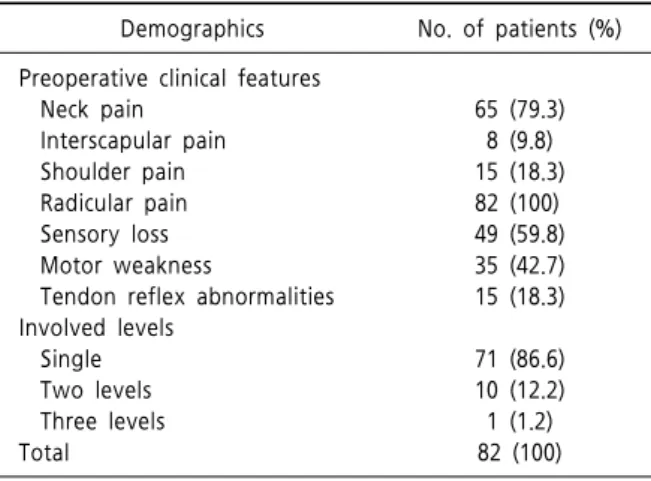
Table 1. Preoperative clinical features and levels of lesion Demographics No. of patients (%) Preoperative clinical features
Neck pain Interscapular pain Shoulder pain Radicular pain Sensory loss Motor weakness
Tendon reflex abnormalities Involved levels
Single Two levels Three levels Total
65 (79.3) 8 (9.8) 15 (18.3) 82 (100) 49 (59.8) 35 (42.7) 15 (18.3)
71 (86.6) 10 (12.2) 1 (1.2) 82 (100)
Table 2. Type of lesion and level of pathology Demographic No. of levels (%) Type of lesion
Soft disc herniation Spondylotic osteophyte Mixed
Level of pathology C3∼4
C4∼5 C5∼6 C6∼7 Total
35 (36) 52 (53.7) 10 (10.3)
8 (8.2) 19 (19.6) 50 (51.6) 20 (20.6) 97 (100)
foraminotomy, AMF)을 개발했다. 이후로 많은 저자들 이 이 수술을 시행하고 좋은 임상 결과들을 발표한 바 있다[18-27]. 일부 저자들은 AMF 후 방사선학적 관찰 을 시행하여 수술 후 가중된 퇴행성 변화의 지표인, 해 당 분절의 디스크 높이가 감소함을 발견하였다[21]. 그 러나 추적 관찰이 짧고, 광범위한 분석이 이루어지지 않아 정확하게 어떠한 요소가 디스크 높이를 감소시키 는지, 어떻게 하면 이를 피할 수 있는지 알려지지 않았 다. 이에 저자는 이 수술을 시행받은 충분한 수의 환자 를 대상으로 장기적인 관찰을 하여 해당 분절의 디스 크 퇴행성 변화를 가중시키는 인자들을 연구하였다.
방 법
1. 환자군
본원에서는 2000년 9월부터 2011년 8월까지 11년간 232명의 퇴행성 경추부 질환 환자에 대해 272예의 AMF를 시행하였다. 수술 대상은 6주 이상의 보존적인 치료에도 호전되지 않고, 임상증상이 CT, MRI로 확인 된 병변을 가진 환자들이었다. 이 중에서 전에 경추부 수술을 받지 않았고, 척수병증을 보이지 않았으며, 경 추부의 다른 수술을 시행받지 않았고, 3년 이상 임상 적, 방사선학적으로 추적관찰이 가능했으며, 마지막 관찰 시 자료분석에 동의하고 임상적, 방사선학적 검 사를 시행 받은 82명을 대상으로 하였다. 이중 71명 (86.6%)에 대해서는 한 개 분절만 수술하였고, 10명 (12.2%)에 대해서는 두 개 분절을 수술하였으며, 1명 (1.2%)은 세 개 분절에 대해 수술을 시행하여 모두 97 예의 수술을 시행하였다. Table 1은 이 환자들의 수술 전 임상상태와 수술을 시행한 분절수를 보여주고 있
으며, Table 2는 수술한 분절의 병변 종류와 부위를 보 여주고 있다. 이들은 남자 56명, 여자 26명이었으며, 수술 당시 평균연령은 48.4세(범위, 31∼76세)였다.
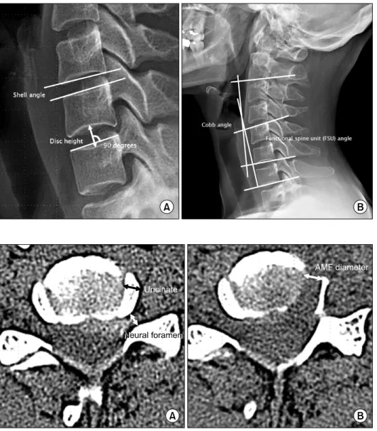
임상적인 분석은 입원 및 통원 기록지를 분석하였 고, 방사선학적 결과는 PACS 자료를 분석하였다. 입 원 당시 visual analog scale (VAS)과 neck disability in- dex (NDI) 점수를 포함한 수술 전 증상과 증세를 확인 했고, 수술 후에는 퇴원 당시와 이후 2주마다 한번씩 통원 관찰을 하여 3개월간 및 수술 후 1년에 확인하였 으며, 본 연구가 시행될 시기에 마지막으로 설문지와 전화를 통해 VAS와 NDI 점수를 얻었다. 방사선학적 검사는 수술 전 모든 환자에서 역동적 검사를 포함한 단순방사선촬영과 고해상도 CT, MRI를 시행하였고, 수술 후 AMF 범위를 확인하기 위한 고해상도 CT를 시행하였으며, 수술 후 3개월 동안 매 2주마다 역동적 검사를 포함한 단순방사선촬영을 시행하였고, 마지막 으로 연구시작 시 시행하였다. PACS workstation (Starpacs 5.0.8.1; Infinite, Seoul, Korea)을 이용하여 수 술 후 CT에서 AMF 크기를 얻었고(Fig. 1), 수술소견에 서 디스크 침범 여부를 확인하였으며, 수술 전후 단순 촬영에서 해당 관절의 Cobb angle, functional spine unit (FSU) angle, shell angle (SA), 그리고 디스크 높이 를 측정하였고(Fig. 2), 역동적 촬영에서 각각도의 운 동범위를 측정하였다. 통계적인 분석은 SPSS ver. 15.0 (SPSS Inc., Chicago, IL, USA)를 이용하였다.
2. 수술법
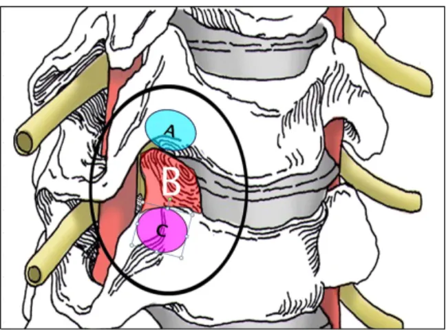
AMF는 세가지 접근법으로 세분되는데 저자는 이 중구상돌기를 통한 접근법을 시행하였다(Fig. 3) [22].
기관 삽관을 통한 전신마취 하에 누운 상태에서 수술
이 시행된다. 머리는 정중앙에 똑바로 위치시키고, 양
어깨는 수술 중 방사선 촬영이 용이하게 접착테이프
Fig. 1. (A) Measurement of disc height (DH) and shell angle (SA) on a plain radi- ograph in neutral lateral position. (B) Functional sp- ine unit (FSU) and Cobb angles.
Fig. 2. (A) Diameter of the uncinate process is mea- sured at the preoperative axial CT image showing the widest area of the disc.
Diameter of the neural for- amen was measured at the point just medial to the fossa locating the dorsal root ganglion. (B) Diameter of anterior microforamino- tomy is measured on the postoperative axial CT ima- ge. AMF, anterior microfor- aminotomy.
를 이용하여 잡아당겨 수술대에 고정한다. 수술부를 포함한 경부 전면을 소독하고 수술부만 노출한 뒤 소 독포를 덮는다. 환측에 중앙에서 3 cm 목의 피부선과 평행하게 절개하고, 피하 조직과 광경근을 동일하게 절개한다. 목빗근과 경동맥의 내측, 기도와 식도의 외 측 사이를 박리하여 경추전면을 노출한다. 정확한 수 술부를 방사선으로 확인하고, 통상적인 경추견인기를 위치하여 환측 longus colli 근육의 전면을 확인한다.
수술현미경을 위치하고, 해당 추간판의 상하경추횡돌 기의 근위부를 노출하여 구상돌기관절을 확인한다.
제6∼7 경추간 수술 시에는 척추동맥이 lonus colli 근 육 아래, 제7경추횡돌기 위에 위치하여 주의를 요한 다. 상하횡돌기 사이에 위치한 구상돌기관절을 고속
드릴을 사용하여 구멍을 만든다. 이때 경추의 측면에
서 1∼3 mm 사이에 존재하는 척추 동맥의 손상을 예
방하기 위해 구상돌기 외벽을 얇게 남기고 후종 인대
가 드러나면 후에 이 외벽을 제거해낸다. 구상돌기의
외벽을 제거하면 신경근의 압박이 풀리는 것을 확인
할 수 있고, 만일 병변이 척추증이라면 여기서 수술은
끝날 수 있다. 파열된 디스크는 후종인대를 부분적으
로 열고 제거할 수 있다. 잔존하여 신경근을 압박할
수 있는 뼈나 인대를 모두 제거하여 신경근이 완전히
감압되는 것을 확인한 후 수술을 끝낸다. 광경근과 피
부는 통상적인 방법으로 봉합하고 경부 보조구는 착
용하지 않는다.
Fig. 3. Illustration showing the various entry and resection sites of microforaminotomy. (A) Entry point of upper transcorporeal approach. (B) Resection site of transuncal approach. (C) Entry point of lower transcorporeal approach.
Fig. 4. Box-plot graph showing preoperative and postoperative changes of visual analog scale (VAS) and neck disability index (NDI) scores with mean, maximum, and minimum values.
VASpre, preoperative VAS; VASpost, postoperative VAS; NDIpre, preoperative NDI; NDIpost, postoperative NDI.
결 과
저자는 본원에서 11년간 경추부 퇴행성 질환을 가 진 환자 232명의 272분절에 대한 AMF를 시행하였으 며, 이 중 82명의 97분절에 대해 이 연구를 시행하였 다. 82명의 환자에서 추적 관찰 후, 59명(72%)은 Odom’s 기준상 뛰어난 결과를 보였고, 15명(18.3%)은 좋은 결과를 보였으며, 6명(7.3%)은 적정한 결과를 보 였고, 2명(2.4%)은 호전되지 않았다(Table 3). 수술부가 1개 분절인 경우는 71명(86.6%), 2개 분절인 경우는 10 명(12.2%), 그리고 3개 분절인 경우는 1명(1.2%)이었 다(Table 1). 주된 병변은 경추증이 52예(53.7%), 추간 판탈출증이 35예(36%)였으며, 나머지 10예(10.3%)는 두 병변이 공존하는 상태였다. 수술한 분절은 제5∼6 경추간 병변이 50예(51.6%), 제6∼7 경추간 20예 (20.6%), 제4∼5 경추간 19예(19.6%), 그리고 제3∼4 경추간 8예(8.2%)였다(Table 2). 환자의 연령을 보면 40대 33명(40.2%), 50대 26명(31.7%), 30대 14명 (17.1%), 60대 7명(8.5%), 그리고 70대가 2명(2.4%)이었 다. 수술로 인한 합병증은 없었으며, 재수술은 2명에 서 시행되었다. 둘 다 수술 후 1개월 이내에 방사통이 재발하여 재수술을 시행하였고, 수술 시 침범된 추간 판에서 작은 디스크 조각이 돌출하여 신경근을 압박 하는 소견을 보였다. 평균 추적기간은 6.1년(범위, 3.1
∼8.0년)이었다.
평균 VAS 점수는 수술 전 8.2에서 수술 후 2.9로 호 전되었으며, NDI 점수도 수술 전 24.5에서 수술 후 6.7
로 통계적으로 유의하게 호전되었다(P>0.01) (Fig. 4).
이러한 수술 전후의 VAS, NDI 점수나 그 변화는 연 령, 성별, 수술 부위, 수술 전 퇴행성 변화여부, AMF 크기, 수술 전후의 추간판 높이 변화, 다양한 각도 변 화 및 운동범위와 무관하였다. 수술 중 디스크 침범은 64예(65.9%)에서 보였다.
평균적인 AMF 크기는 5.2 mm (범위, 2.6∼9.6 mm)
였으며, AMF 크기는 디스크 침범, 수술 후 추간판 높
이 감소와 유의한 상관관계를 보였다(P<0.05). 디스
크 침범이 있는 경우에는 수술 후 추간판 높이 감소는
물론이고, 수술 후 FSU와 SA 각도 및 FSU의 운동 범
위 등의 감소, 수술 분절의 전방 골극 변화발생에 의
미 있는 차이를 보였다(P<0.05). 수술 전 추간판 높이
는 5.8 mm (범위, 1.9∼8.4 mm)였으며, 수술 후 추간
판 높이는 4.7 mm (범위, 1.0∼7.1 mm)였고, 수술 전
후의 추간판 높이 감소는 평균 1.1 mm (범위, 0∼3.1
mm)로 유의한 수술 전후 차이를 보였다(P<0.01). 16
예(16.5%)에서는 추간판 높이 감소가 없었고, 3예에서
는 50% 이상의 추간판 높이 감소를 보였다. 수술 후
추간판 높이가 감소하면 FSU, SA, 그리고 FSU의 운동
범위가 감소하였다(P<0.05). AMF 크기와 수술 후 추
간판 높이 감소의 상관관계 도표를 그려서 추세선을
확인하여 결정적인 AMF 크기는 4.7 mm임을 추정할
수 있었다(Fig. 5). 환자군을 AMF 크기 4.7 mm를 기준
으로 하여 두 군으로 나누면, 두 군 사이에 수술 중
추간판 침범 여부, 추간판 높이 감소, 그리고 SA 각도
가 유의한 차이를 보였다(P<0.05). 두 군간에 수술 전
Fig. 5. Graph showing correlation between the anterior microforaminotomy (AMF) diameter and changes in posto- perative disc height (DH). Curvilinear lines indicate the trend line with 95% confidence lines extracted by quadratic regre- ssion analysis. The critical AMF diameter is 4.7 mm, above which the DH difference will increase remarkably as the AMF diameter increases.
Fig. 6. Preoperative CT and sagittal MR images. (A∼C) Sagittal T1 and T2 MRI and CT images showing disc protrusion with spur at the C5-6 level. (D, E) Axial CT images showing combined disc and spur compression at the right neural fora- men.
후 전체 경추부 각도나 해당 분절의 각도 및 각운동 범위의 차이는 없었다. 수술 전후 Cobb, FSU, SA 각도 는 유의한 차이가 없었으며, 각각도의 운동 범위도 유 의한 차이가 없었다(P<0.05). 추적 검사상 측만증이 나 굴곡 변형을 보인 예는 없었다.
전형적인 예를 소개하면 다음과 같다.
51세 여자가 내원하기 3개월 전부터 우측 제6신경
근 지배부의 방사통을 주소로 내원하였다. 신경학적
검사상 운동 약화나 건반사 감소는 없었으며, 우측 엄
지와 검지의 감각 저하와 이상 감각을 보였다. 수술
전 CT, MRI에서 우측 제5∼6 경추간 추간판 돌출과
경추증이 공존하며 신경근을 압박하는 소견을 보였다
(Fig. 6). 저자는 우측으로 접근하여 제5∼6 경추간 4.5
mm 크기의 AMF를 시행하였다. 수술 중 AMF 내측에
서 일부 추간판 침범이 있었다. 수술 중 신경근 감압
이 확인되었고, 수술 후 방사통과 감각증상은 바로 호
전되었으며, CT에서 신경근 감압이 확인되었다. 5년
추적 검사상 추간판 높이는 수술 전의 5.8 mm에서
Fig. 7. Postoperative axial CT images and dynamic plain radiographs. (A) Im- mediate postoperative axial CT image showing forami- notomy on the right side.
(B) Axial CT image show- ing new bone formation around the margin of the foraminotomy at a follow- up of 5 years. (C, D) Plain extension and flexion X- rays showing well- preser- ved cervical motion.
4.2 mm로 감소하였다. 이차적인 경추 불안정, 후만- 측만 변형, 또는 골극 변화는 없었으며, 경추 운동 범 위는 잘 유지되었다(Fig. 7).
고 찰
저자와 동일한 방법으로 수술을 시행한 대부분의 저자들은[17,18,20-24,28] 수술 후 91%에서 99%의 환 자에서 임상 증상의 호전을 보였다고 보고하였으며, 한 저자는 단지 52%에서만 호전을 보였다고 하였다 [29]. 그러나 이 연구들은 모두 상세한 방사선학적 결 과가 결여되어 진정한 수술 결과라고 하기에는 미흡 했다. 본 연구에서는 90.3%의 환자에서 임상 증상의 호전을 보였으며, VAS와 NDI 점수는 97.6%에서 유의 한 호전을 보였다. 방사선학적 결과에서도 수술 후 추
가적인 변형없이 해당 분절이나 경추 전반에 걸쳐 정 적인 모양이나 운동 범위가 잘 유지되는 모습을 보였 다. 지금까지 발표된 합병증은 7% [22]에서 22% [29]
였으나 본 연구에서는 4.9% (2예)의 추간판 재발로 인 한 재수술 외에는 합병증이 없었다.
신경공확장술로 인해 손상되는 구상돌기-척추간
관절은 경추 불안정과 운동에 있어서 상당히 중요할
수 있다. Kotani 등[30]과 Chen 등[31]은 사체에서 얻
은 경추 분절을 이용하여 구상돌기-척추 관절을 제거
한 후 생역학적 실험을 하고, 신경공에 접한 후방부위
가 해당 분절의 안정성과 운동성에 중요하다고 하였
다. Hacker와 Miller [29]는 23명의 환자에서 신경공
확장술을 시행하여 30%의 환자에서 추가적인 수술이
필요했으며, 단지 12명에서만 좋은 결과를 얻을 수 있
었다고 하였고, 구상돌기-척추 관절의 제거가 지나친
운동성을 초래하여 불안정하고 통증을 유발하게 된다 고 하였다. 본 연구에서는 신경공 확장술 크기가 4.7 mm 이상이거나 수술 시 추간판 침범이 있는 경우에 는 해당 분절의 각도 변화만 상관관계를 보였고, 이로 인해 1 mm가 넘는 추간강 감소가 있었던 경우에만 수술 후 해당 분절과 경추 전반의 각도 변화와 상관관 계를 보였다.
신경공확장술을 시행하여 추적 관찰한 학자들은 수 술 후 환자 모두는 아니더라도 추간판의 높이가 감소 하는 경우를 발표하였다[21]. 추간판의 높이 감소는 수술 후 해당 분절의 추간판에 퇴행성 변화가 가속화 됨을 의미한다. 이러한 변화는 추간판의 기능 저하와 운동성 감소를 초래할 수 있다. 하지만 수술 시 어떠 한 인자가 추간판 높이 감소에 관여하는지에 대한 연 구는 없었다. 본 연구에서 조금이라도 추간판 높이가 감소한 경우는 수술을 시행한 총 97분절 중 81분절 (83.5%)였고, 평균적인 추간판 높이 감소는 1 mm였 다. 신경공확장술의 크기와 추간판 높이 감소의 상관 관계 그래프를 분석하여 추세선을 이용하면 신경공확 장술의 크기가 4.7 mm를 넘으면 신경공확장술 크기 에 비례하여 추간판 높이가 감소하였다. 신경공확장 술 크기 4.7 mm를 기준으로 두 군으로 나누었을 때 두 군간 추간판 높이의 감소와 디스크 침범 여부에 있 어서 의미있는 차이를 보였으나, 수술 전후 전체 경추 부 각도나 해당 분절의 각도 및 각운동범위의 차이는 없었다.
경추부의 퇴행성 변화가 진행하면, 추간판의 변성 변화, 추간강 감소, 추체골극 변화, 연골판의 경화증, 후방으로 추간판 돌출, 경막의 압박, 그리고 신경공의 협착이 발생한다[32,33]. 수술 시 추간판에 작은 손상 이 발생할 경우에도 이러한 퇴행성 변화가 발생할 수 있다. Osti 등[34]은 양에서 추간판에 실험적 손상을 준 후 시간 경과를 두고 관찰하여 추간판이 급격하게 퇴행성 변화를 보이고 1∼18개월에 걸쳐 추간판 높이 가 감소하는 것을 발표하였다. Nassr 등[11]은 수술 시 정확한 수술 부위를 확인하기 위해 추간판에 바늘을 찌르는 것 조차 추간판 퇴행성 변화를 초래할 가능성 이 3배 정도 증가한다고 하였다. 본 연구에서 수술 시 추간판 침범이 있는 경우는 97분절 중 64분절(67%)이 었으며, 추간판침범이 있는 경우, 없는 경우에 비교하 여 수술 후 추간판 높이 감소는 물론이고, 수술 후 FSU와 SA 각도 및 FSU의 운동 범위 등의 감소, 나아 가서는 수술 분절의 전방 골극 변화 발생에 의미있는 차이를 보였다. 이는 신경공확장술의 크기도 중요하
지만 신경공확장술의 크기와 관계없이 수술 시 추간 판을 침범할 경우 해당 분절의 퇴행성 변화가 뚜렷하 게 나타난다는 것을 의미한다.
신경공확장술은 경추부 퇴행성 질환에 대한 안전하 고, 효과적인 치료법이다. 하지만 수술 후 장기적인 관찰시 추간판 높이가 감소하여, 이로 인해 일련의 퇴 행성 변화가 유발될 수 있다. 따라서 장기적으로 좋은 수술 결과를 얻기 위해서는 가능한 한 그 크기를 작게 하고, 추간판 침범을 피해야 한다.
참고문헌
1. Bertalanffy H, Eggert HR. Clinical long-term results of anterior discectomy without fusion for treatment of cervical radiculopathy and myelopathy: a follow-up of 164 cases. Acta Neurochir (Wien) 1988;90:127-135.
2. Cloward RB. The anterior approach for removal of rup- tured cervical disks. J Neurosurg 1958;15:602-617.
3. Goffin J, Casey A, Kehr P, Liebig K, Lind B, Logroscino C, et al. Preliminary clinical experience with the Bryan Cervical Disc Prosthesis. Neurosurgery 2002;51:840-847.
4. Mosdal C. Cervical osteochondrosis and disc herniation.
Eighteen years' use of interbody fusion by Cloward's technique in 755 cases. Acta Neurochir (Wien) 1984;70:
207-225.
5. Murphey F, Simmons JC, Brunson B. Surgical treat- ment of laterally ruptured cervical disc: review of 648 cases, 1939 to 1972. J Neurosurg 1973;38:679-683.
6. Robinson RA, Smith GW. Anterolateral cervical disc removal and interbody fusion for cervical disc syndrome.
Bull Johns Hopkins Hosp 1955;96:223-224.
7. Baba H, Furusawa N, Imura S, Kawahara N, Tsuchiya H, Tomita K. Late radiographic findings after anterior cervical fusion for spondylotic myeloradiculopathy.
Spine 1993;18:2167-2173.
8. Elsawaf A, Mastronardi L, Roperto R, Bozzao A, Caroli M, Ferrante L. Effect of cervical dynamics on adjacent segment degeneration after anterior cervical fusion with cages. Neurosurg Rev 2009;32:215-224.
9. Hilibrand AS, Carlson GD, Palumbo MA, Jones PK, Bohlman HH. Radiculopathy and myelopathy at seg- ments adjacent to the site of a previous anterior cervical arthrodesis. J Bone Joint Surg Am 1999;81:519-528.
10. Hussain M, Natarajan RN, An HS, Andersson GB.
Motion changes in adjacent segments due to moderate and severe degeneration in C5-C6 disc: a poroelastic C3-T1 finite element model study. Spine 2010;35:
939-947.
11. Nassr A, Lee JY, Bashir RS, Rihn JA, Eck JC, Kang
JD, et al. Does incorrect level needle localization during anterior cervical discectomy and fusion lead to accel- erated disc degeneration? Spine 2009;34:189-192.
12. Pospiech J, Stolke D, Wilke HJ, Claes LE. Intradiscal pressure recordings in the cervical spine. Neurosurgery 1999;44:379-385.
13. Schwab JS, Diangelo DJ, Foley KT. Motion compensa- tion associated with single-level cervical fusion: where does the lost motion go? Spine 2006;31:2439-2448.
14. Verbiest H. A lateral approach to the cervical spine: tech- nique and indications. J Neurosurg 1968;28:191-203.
15. Hakuba A. Trans-unco-discal approach. A combined an- terior and lateral approach to cervical discs. J Neurosurg 1976;45:284-291.
16. Snyder GM, Bernhardt M. Anterior cervical fractional interspace decompression for treatment of cervical radi- culopathy: a review of the first 66 cases. Clin Orthop Relat Res 1989;246:92-99.
17. Jho HD. Microsurgical anterior cervical foraminotomy for radiculopathy: a new approach to cervical disc herniation. J Neurosurg 1996;84:155-160.
18. Balasubramanian C, Price R, Brydon H. Anterior cer- vical microforaminotomy for cervical radiculop- athy--results and review. Minim Invasive Neurosurg 2008;51:258-262.
19. Choi G, Lee SH, Bhanot A, Chae YS, Jung B, Lee S.
Modified transcorporeal anterior cervical micro- foraminotomy for cervical radiculopathy: a technical note and early results. Eur Spine J 2007;16:1387-1393.
20. Cornelius JF, Bruneau M, George B. Microsurgical cer- vical nerve root decompression via an anterolateral ap- proach: clinical outcome of patients treated for spondy- lotic radiculopathy. Neurosurgery 2007;61:972-980.
21. Hong WJ, Kim WK, Park CW, Lee SG, Yoo CJ, Kim YB, et al. Comparison between transuncal approach and upper vertebral transcorporeal approach for unilateral cervical radiculopathy - a preliminary report. Minim Invasive Neurosurg 2006;49:296-301.
22. Jho HD, Kim WK, Kim MH. Anterior microforaminotomy for treatment of cervical radiculopathy: part 1--disc-pre- serving "functional cervical disc surgery". Neurosurgery 2002;51(5 Suppl):S46-S53.
23. Johnson JP, Filler AG, McBride DQ, Batzdorf U.
Anterior cervical foraminotomy for unilateral radicular disease. Spine 2000;25:905-909.
24. Kotil K, Bilge T. Prospective study of anterior cervical microforaminotomy for cervical radiculopathy. J Clin Neurosci 2008;15:749-756.
25. Lee JY, Lohr M, Impekoven P, Koebke J, Ernestus RI, Ebel H, et al. Small keyhole transuncal foraminotomy for unilateral cervical radiculopathy. Acta Neurochir (Wien) 2006;148:951-958.
26. Saringer W, Nöbauer I, Reddy M, Tschabitscher M, Horaczek A. Microsurgical anterior cervical foramin- otomy (uncoforaminotomy) for unilateral radiculopathy:
clinical results of a new technique. Acta Neurochir (Wien) 2002;144:685-694.
27. White BD, Buxton N, Fitzgerald JJ. Anterior cervical foramenotomy for cervical radiculopathy. Br J Neurosurg 2007;21:370-374.
28. Jho HD. Failed anterior cervical foraminotomy. J Neurosurg 2003;98(2 Suppl):121-125.
29. Hacker RJ, Miller CG. Failed anterior cervical foraminotomy. J Neurosurg 2003;98(2 Suppl):126-130.
30. Kotani Y, McNulty PS, Abumi K, Cunningham BW, Kaneda K, McAfee PC. The role of anteromedial fora- minotomy and the uncovertebral joints in the stability of the cervical spine. A biomechanical study. Spine 1998;23:1559-1565.
31. Chen TY, Crawford NR, Sonntag VK, Dickman CA.
Biomechanical effects of progressive anterior cervical decompression. Spine 2001;26:6-14.
32. Okada E, Matsumoto M, Ichihara D, Chiba K, Toyama Y, Fujiwara H, et al. Aging of the cervical spine in healthy volunteers: a 10-year longitudinal magnetic res- onance imaging study. Spine 2009;34:706-712.
33. Walraevens J, Liu B, Meersschaert J, Demaerel P, Delye H, Depreitere B, et al. Qualitative and quantitative as- sessment of degeneration of cervical intervertebral discs and facet joints. Eur Spine J 2009;18:358-369.
34. Osti OL, Vernon-Roberts B, Fraser RD. Anulus tears and intervertebral disc degeneration: an experimental study using an animal model. Spine 1990;15:762-767.