Introduction
Carpal tunnel syndrome (CTS) is one of the most common entrapment neuropathy.1 In CTS, selective
involvement of median motor fibers is rare, and the incidence varies from 0.6% to 1.2%.2 Selective fascicu- lar involvement of median nerve has been reported in some cases, such as predominant involvement of volar-radially located motor fibers and sensory fibers to thumb.2-5 Furthermore, recurrent motor branch (RMB) of median nerve can be affected separately.6,7
We report a mild CTS patient with concomitant neuropathy of the RMB of median nerve and also de- scribe the atypical clinical features of motor dominant
ISSN 1229-6066 https://doi.org/10.18214/jkaem.2019.21.1.17 J Korean Assoc EMG Electrodiagn Med 21(1):17-21, 2019
J Korean Assoc
Electrodiagn Med EMG
Copyright © by Korean Association of EMG Electrodiagnostic Medicine
This is an Open Ac cess article distributed under the terms of the Creative Commons Attribution Non-Commercial License (http://creativecommons.org/licenses/by-nc/4.0) which permits unrestricted non-commercial use, distribution, and reproduction in any medium, provided the original work is properly cited.
Received September 20, 2018
Revised November 22, 2018, (2nd) December 22, 2018 Accepted January 18, 2019
Corresponding Author: Hee-Kyu Kwon
Department of Physical Medicine & Rehabilitation, Korea University Anam Hospital, 73 Goryeodae-ro, Seongbuk-gu, Seoul 02841, Korea
Tel: 82-2-920-6471, Fax: 82-2-929-9951, E-mail: [email protected]
손목터널증후군에서 동반된
정중신경 회귀운동분지의 신경병증: 증례 보고
노준수1, 박종웅2, 권희규1
고려대학교 안암병원 재활의학과1, 정형외과2
Concomitant Neuropathy of Recurrent Motor Branch of Median Nerve in Carpal Tunnel Syndrome: A Case Report
Jun Soo Noh1, Jong Woong Park2, Hee-Kyu Kwon1
Departments of 1Physical Medicine & Rehabilitation, 2Orthopedic Surgery, Korea University Anam Hospital, Seoul, Korea
Selective fascicular involvement of median nerve in carpal tunnel syndrome (CTS) has been reported, such as predominant involvement of volar-radially located motor and sensory fibers to thumb. Also, recurrent motor branch (RMB) of median nerve could be affected separately. A man has suffered from right-hand weakness and mild tingling sense on I to III digits visited. Thenar muscles wasting and weakness were noted without sensory loss. Whereas the results of sensory conduction and comparison of median-second-lumbrical/ulnar-interossei studies suggested mild CTS, motor responses revealed severe neuropathy. The patient was diagnosed with right CTS, mild degree and concomitant incomplete but severe neuropathy of RMB. The patient underwent an operation. The RMB was found as the transligamentous type with a degenerative change and severely compressed by ligament and thenar muscles. Open carpal tunnel release and decompression with neurolysis were done. Through precise analysis of electrodiagnostic findings, correct diagnosis and proper management were achieved.
Key Words: carpal tunnel syndrome, electrodiagnosis, median nerve
symptoms, pre- and post-operative electrophysiologic features, and operative findings. To our knowledge, this is the first case of separate neuropathy of the RMB of median nerve which was diagnosed by precise evalu- ation of electrophysiologic findings and confirmed by surgery, and also demonstrated improvement of symp- toms and electrophysiologic findings after surgery.
Case Report
A 25-year-old right-handed man has suffered from a right hand weakness for 18 months and mild tin- gling sensation on right thumb to middle fingers for 6 months. He works as a chef in a Japanese restaurant for 3 years. He uses his right hand to hold his knife and realized difficulty in holding it gradually.
On physical examination, thenar muscle wasting ac- companying tenderness was observed. The patient did not complain cervicalgia and there was no change of color and temperature sensation in the hand. Medical Research Council (MRC) grades of right thumb abduction and opposition were 3/5 whereas all the other muscles
were normal. There was no sensory loss. Tinel’s sign, Phalen’s test, and the arm elevation test were positive on the right side.
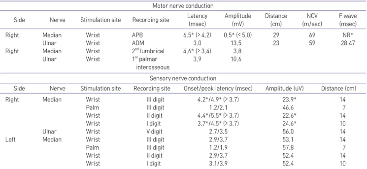
Electrodiagnostic examination was performed on the same day in the bilateral upper extremities nerves (Table 1). Right median motor NCS showed prolon- gation of distal latency and reduced amplitude (6.5 msec, 0.5 mV). The right “median-second-lumbrical and ulnar-interossei (M2L-UI) comparison study” re- vealed positive finding (M2L, 4.6 msec; UI: 3.9 msec, M2L-UI difference > 0.6 msec), but the amplitude of second lumbrical muscle recorded compound motor unit action potential (CMAP) was normal (right, 3.8 mV; left, 3.6 mV) (Table 1). Sensory nerve response of right median nerve with middle finger recording re- vealed prolonged latency and reduced amplitude with wrist stimulation (wrist: 4.2 msec, 23.9 μV; palm: 1.2 msec, 46.6 μV). The median sensory responses with thumb and index fingers recordings were also obtained to figure out the selectivity in fascicular involvement but showed no difference among three fingers. The F- wave was unobtainable with the right median nerve
Table 1. Motor and Sensory Nerve Conduction Study Data
Motor nerve conduction Side Nerve Stimulation site Recording site Latency
(msec)
Amplitude (mV)
Distance (cm)
NCV (m/sec)
F wave (msec)
Right Median Wrist APB 6.5* (> 4.2) 0.5* (< 5.0) 29 69 NR*
Ulnar Wrist ADM 3.0 13.5 23 59 28.47
Right Median Wrist 2nd lumbrical 4.6* (> 3.4) 3.8
Ulnar Wrist 1st palmar
interosseous
3.9 10.6
Sensory nerve conduction
Side Nerve Stimulation site Recording site Onset/peak latency (msec) Amplitude (uV) Distance (cm)
Right Median Wrist III digit 4.2*/4.9* (> 3.7) 23.9* 14
Palm III digit 1.2/2.1 46.6 7
Wrist II digit 4.4*/5.5* (> 3.7) 22.6* 14
Wrist I digit 3.7*/4.5* (> 3.7) 24.6* 10
Ulnar Wrist V digit 2.7/3.5 56.0 14
Left Median Wrist III digit 2.9/3.7 53.1 14
Palm III digit 1.2/1.9 57.8 7
Wrist II digit 2.9/3.7 52.4 14
Wrist I digit 3.1/3.9 52.4 10
Abnormal cutoff value based on median-to-2nd lumbrical compared with ulnar-to-palmar interosseous > 0.6 msec. Reference values are rep- resented in parentheses and abnormal values are represented with asterisk
NCV: nerve conduction velocity, APB: abductor pollicis brevis, ADM: abductor digiti minimi, NR: no response
stimulations. The latency difference between the me- dian and ulnar nerves in M2L-UI study and the sensory conduction study revealed mild CTS of demyelination and some axonal degeneration, but the findings of motor conduction study were compatible with severe neuropathy with severe axonal degeneration and some demyelination.
Needle electromyogram (EMG) was done in the right upper extremity muscles. Abnormal spontaneous activ- ities and polyphasic, large amplitude and long duration motor unit potentials with discrete recruitment pat- terns were noted in the right APB muscle. Normal mo- tor unit potentials were noted in all the other muscles examined (Table 2). Sonography was performed on the same day. Mild swelling of the right median nerve was found at the distal wrist crease level (cross-sectional area: 0.137 cm2) which were also compatible with mild CTS. There were no other abnormal findings at the palm. In conclusion, the patient was diagnosed with a mild degree right carpal tunnel syndrome and concom- itant incomplete but severe neuropathy of right RMB of median nerve.
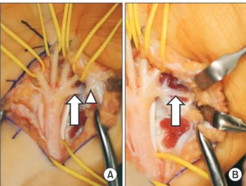
The patient underwent a surgery a month after the examination. On operation, the RMB was found as the transligamentous type with a whitish degenerative change, and severely compressed by thickened liga- ment and fascia of thenar muscles. Open carpal tunnel release and decompression and neurolysis of RMB of median nerve were done (Fig. 1).
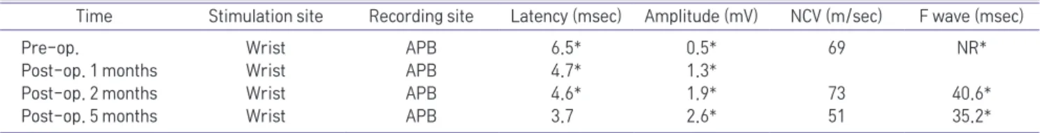
Follow-up NCS was done at one, two and five months after the surgery. On the first follow-up visit, tingling sense was reduced to half and the latency and
amplitude of median CMAP revealed improvement (4.7 msec, 1.3 mV). On the second follow-up visit, the sensory symptom disappeared and the right thumb abduction improved to 4/5 in MRC grade. Median CMAP latency and amplitude were 4.6 msec and 1.9 mV, respectively. The latency of right median sensory response also showed improvement with the middle finger recording (wrist: 3.5 msec, 24.0 μV; palm: 1.1 msec, 46.6 μV) (Table 3). On the third follow-up visit, strength of right thumb abduction was restored to al- most normal grade. Previously existed tenderness on right thenar muscle was disappeared. Latency and am- plitude of median CMAP (3.7 msec, 2.6 mV) were im- proved as well those of median sensory response (wrist:
3.6 msec, 34.0 μV; palm: 1.1 msec, 51.0 μV).
Table 2. Needle Electromyography Study
Muscle Insertional
activity
Spontaneous activity
Motor unit action potentials
N Polyphasia Amplitude Duration IP
Right Brachioradialis N - N F
Pronator teres N - N F
EDC N - N F
FDI N - N F
APB F&P (+) Polys Large Long DIS (3~4 MUAPs)
IP: interference pattern, N: normal, F: full, F&P: fibrillation potentials & positive sharp waves, R: reduced, DIS: discrete, MUAP: motor unit action potential, EDC: extensor digitorum communis, FDI: first dorsal interosseous, APB: abductor pollicis brevis, -: not applicable
A B
Fig. 1. The right recurrent motor branch of median nerve (arrow, A) was found perforating the ligament (arrowhead, A) and showed whitish degenerative change (arrow, B).
Discussion
Separate injury to the RMB of the median nerve is relatively rare, but several cases have been reported:
after repetitive compression damage due to cycling or weightlifting, compression due to mass-like lesions such as schwannoma and palmar ganglion, or iatrogen- ic injuries from carpal tunnel release.7,8 In most of these cases, abnormal median motor response with normal median sensory conduction study suggested neuropa- thy of RMB of the median nerve, but only few of them proved the neuropathy through surgery.
Typically, RMB of median nerve branches out distal to the flexor retinaculum and innervates APB, op- ponens pollis (OP), and superficial head of the flexor pollicis brevis (FPB) muscles. We can think of two dis- tinct mechanisms considering the compressive etiol- ogy of a RMB of the median nerve neuropathy. First is a compression of the nerve at the palm level which is closely associated with the anatomical variation of the nerve. Poisel classified variations into three subtypes, and named them extraligamentous, subligamentous, and transligamentous types. Among the types, trans- ligamentous type is at the highest risk for injury. It is known to be more vulnerable because not only it can easily be compressed by hypertrophy of muscle and ligament but also it can easily be damaged during carpal tunnel surgery if not properly identified. The incidence of transligamentous type is quite confusing9 as Ahn has provided relatively low incidence (1.1%) of transligamentous type in Koreans in his prospective ca- daveric study.10
The second is related to the intraneural topography
of motor fibers before an emergence of the recur- rent motor thenar branch and sensory fibers to thumb which are located at the most volar-radial quadrant.
This may explain the reason why the RMB of median nerve and sensory fibers to thumb are more susceptible in certain cases of CTS: selective fascicular involvement within the median nerve at the level of carpal tunnel.3-5
In this case, the patient had a history of frequent use of knife and pressing sushi rice. These activities may have caused compression of RMB of median nerve oth- er than CTS itself. The patient also had weakness and atrophy of thenar muscle rather than numbness of fin- gers. Tenderness on thenar eminence could be caused by chronic muscle pain considering his job which is not directly related to injury of RMB of median nerve. On electrodiagnostic examination, APB-recorded CMAP revealed very low amplitude and delayed latency while the sensory responses were mildly involved. Although M2L-UI study revealed positive finding, 2L-recorded CMAP was normal. We also evaluated the sensory re- sponses of thumb and index fingers to rule out the fascicular involvement of volar-radial side, but revealed no difference among thumb, index and middle fingers.
Ultrasonography also revealed the only mild swelling of median nerve at the wrist and no abnormal finding at palm. These findings lead us to make the diagnosis of separate neuropathy of RMB of median nerve, and this was proved by surgery. The RMB of median nerve was found as the transligamentous type and the reason of compressive neuropathy of the patient thought to be the anatomical variation of the nerve.
Identifying a neuropathy of RMB of median nerve could be difficult, as the case is rare and relatively
Table 3. Pre- and Post-operative Median Motor Conduction Study
Time Stimulation site Recording site Latency (msec) Amplitude (mV) NCV (m/sec) F wave (msec)
Pre-op. Wrist APB 6.5* 0.5* 69 NR*
Post-op. 1 months Wrist APB 4.7* 1.3*
Post-op. 2 months Wrist APB 4.6* 1.9* 73 40.6*
Post-op. 5 months Wrist APB 3.7 2.6* 51 35.2*
Abnormal values are represented with asterisk
NCV: nerve conduction velocity, op.: operation, APB: abductor pollicis brevis, NR: no response
unexpected. These patients typically complain more motor symptoms than sensory symptoms. Electrodi- agnostic examination is useful when utilized properly.
Findings are typically normal for median sensory nerve conduction and M2L-UI studies, but APB recorded CMAP amplitudes are significantly reduced.
In conclusion, we report a case of CTS patient who has concomitant neuropathy of RMB of median nerve.
If the patient only underwent an endoscopic surgery, a release of separately involved RMB could have been omitted. Through precise analysis of electrodiagnos- tic findings, correct diagnosis was achieved and could guide proper surgery.
References
1. Jablecki CK, Andary MT, So YT, Wilkins DE, Williams FH:
Literature review of the usefulness of nerve conduction studies and electromyography for the evaluation of patients with carpal tunnel syndrome. AAEM Quality Assurance Committee. Muscle Nerve 1993: 16: 1392-1414
2. Rizzello G, Longo UG, Franceschi F, Martinelli N, Meloni MC, Quadrini R, et al: Compression neuropathy of the motor
fibers of the median nerve at wrist level. Journal of the Chi- nese Medical Association 2009: 72: 268-270
3. Mackinnon SE, Dellon AL: Anatomic investigations of nerves at the wrist: I. Orientation of the motor fascicle of the me- dian nerve in the carpal tunnel. Ann Plast Surg 1988: 21: 32- 35
4. Mondelli M, Aretini A, Ginanneschi F, Padua L: Thenar mo- tor neuropathy electrophysiological study of 28 cases. J Clin Neurophysiol 2010: 27: 344-349
5. Stewart JD: Peripheral nerve fascicles: anatomy and clinical relevance. Muscle Nerve 2003: 28: 525-541
6. Bennett JB, Crouch CC: Compression syndrome of the re- current motor branch of the median nerve. J Hand Surg Am 1982: 7: 407-409
7. Shams OE, Al-Ghamdi SM: Isolated neuropathy of the recur- rent motor branch of the median nerve. Neurosciences (Ri- yadh) 2006: 11: 326-328
8. Worobel MA, Alexander B, Hirsch M, Nguyen V: Recurrent median nerve injury in a weight lifter. Am J Phys Med Reha- bil 2017: 96: e20-e23
9. Kozin SH: The anatomy of the recurrent branch of the me- dian nerve. J Hand Surg Am 1998: 23: 852-858
10. Ahn DS, Yoon ES, Koo SH, Park SH: A prospective study of the anatomic variations of the median nerve in the carpal tunnel in Asians. Ann Plast Surg 2000: 44: 282-287