Copyright ⓒ 2017 by Korean Society for Surgery of the Hand, Korean Society for Microsurgery, and Korean Society for Surgery of the Peripheral Nerve. All Rights reserved.
This is an Open Access article distributed under the terms of the Creative Commons Attribution Non-Commercial License (http://creativecommons.org/licenses/by-nc/4.0/)
Carpal tunnel syndrome is the most common com- pressive neuropathy of the upper extremity1. Moreover, several causative factors increase the carpal tunnel pres- sure, which include space-occupying mass, tenosynovitis, aberrant musculature, or congenital abnormal median nerve. A persistent median artery is a numerous reported anatomical variant, which is supposed to degenerate over time2,3. In addition, studies have shown its co-existence with bifid median nerve. Most cases of persistent median artery are known to be asymptomatic4, although some re-
ports demonstrated that co-existence of persistent median artery with bifid median nerve can lead to carpal tunnel syndrome5. However, a case of thrombosed persistent median artery with bifid median nerve is extremely rare.
Hence, we report an unusual case and surgical outcome of thrombosed persistent median artery with bifid median nerve causing acute carpal tunnel syndrome. Informed consent was obtained from the patient for purpose of the case report.
Microsurgery
이분 정중신경을 동반한 잔류 정중동맥 폐색에 의한 급성 수근관 증후군:
증례 보고
김동민
1ㆍ박종웅
2ㆍ최인철
31국군수도병원 정형외과, 2고려대학교 안암병원 정형외과, 3서울바른세상병원 정형외과
Thrombosed Persistent Median Artery with Bifid Median Nerve Causing Acute Carpal Tunnel Syndrome: A Case Report
Dongmin Kim
1, Jong Woong Park
2, In Cheul Choi
31Department of the Orthopedic Surgery, Armed Forces Capital Hospital, Seongnam, Korea
2Department of the Orthopedic Surgery, Korea University Anam Hospital, Seoul, Korea
3Department of the Orthopedic Surgery, Seoul Barunsesang Hospital, Seoul, Korea
Among the numerous causes of carpal tunnel syndrome, thrombosed persistent median artery with bifid median nerve is extremely rare. Our presentation shows that an unusual case and surgical outcome of thrombosed persistent median artery with bifid median nerve causing acute carpal tunnel syndrome. Care has to be taken that identification of abnormal anatomy in the carpal tunnel is essential, especially in abruptly occurred carpal tunnel syndrome. We suggest that Doppler ultrasound examination is very useful in an outpatient clinic-based for identification of abnormal structure, which makes more quickly and accurate diagnosis than magnetic resonance image.
Key Words: Carpal tunnel syndrome, Persistent median artery, Bifid median nerve, Occlusion
Received August 28, 2017, Revised October 3, 2017, Accepted October 19, 2017 Corresponding author: In Cheul Choi
Hand and Microsurgery Center, Seoul Barunsesang Hospital, 421 Siheung-daero, Geumcheon-gu, Seoul 08523, Korea TEL: +82-2-2111-9700, FAX: +82-2-2111-9999, E-mail: indolldr@hanmail.net
Case Report
sponding author’s hospital for pain at the volar aspect of the left wrist and paresthesia with sensory loss at the me- dian nerve territory from the thumb to the middle finger.
The patient had no prior history of taking anti-diabetic, anti-hypertensive, and any thrombogenic-related medica- tions. The chief complaint was pain that occurred acutely, and the symptoms persisted for five weeks. The patient’s numbness was not relieved by oral medication. On physi- cal examination, both Tinnel sign and Phalen test were
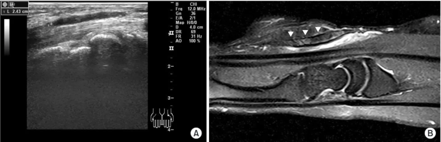
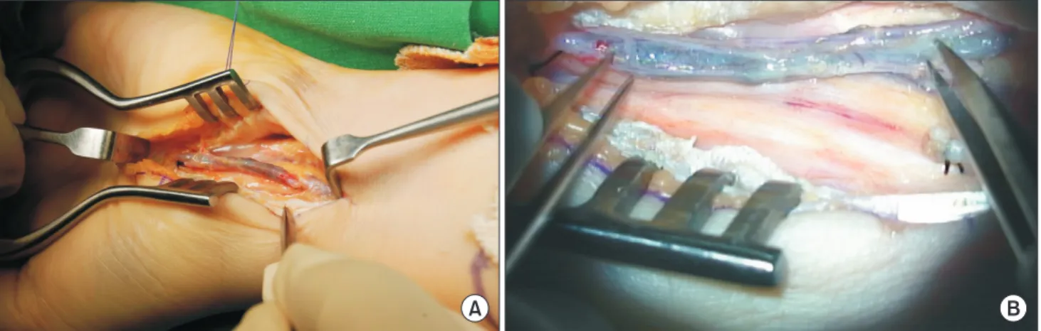
able to identify a persistent median artery between the di- vided median nerves on Doppler ultrasound examination (Fig. 1). The persistent median artery was completely obstructed in the carpal tunnel area, but blood flow was identified to be proximal to the carpal tunnel. In addition, we identified a thrombus — that was approximately 4 cm in length — of the persistent median artery at the wrist on both the magnetic resonance image and Doppler ultra- sound (Fig. 2). The surgery was performed with an oper- ating microscope. First, we performed open carpal tunnel release. The skin was incised using a curved longitudinal incision, passing the wrist crease. Subsequently, we could identify the split median nerve from the inlet of the carpal tunnel, which is proximally 2 cm. In addition, the radial- side branch of the median nerve was more dominant than the other side, and the persistent median artery was found between the split median nerve. The persistent median artery was completely obliterated in 5 mm diameter, and a 2-cm length of the artery was obliterated proximally and distally from the carpal tunnel inlet. Under the oper- ating microscope, the obstructed artery was resected and ligated proximally (Fig. 3). The patient had previous par- esthesia of the affected hand, which was resolved imme- diately postoperatively, and preoperative chief complaint was completely resolved at 9 months postoperatively.
A B
Fig. 2. (A) The thrombosis in about 4 cm length of the persistent median at the wrist on the ultrasound examination. (B) The thrombosis was indicated with arrowhead (arrowheads) on magnetic resonance image.
Fig. 1. The Doppler ultrasound. (A) Normal echogenicity and contour of the median nerve (dotted circle) in the healthy wrist. (B) Persistent median artery (lined circle) between the divided median nerves (dotted circles) in the affected wrist.
DISCUSSION
Carpal tunnel syndrome has typical symptoms, includ- ing paresthesia or numbness in the region of median nerve distribution1. However, patients typically do not complain about symptoms on the volar aspect of the wrist. If the patient has these symptoms, we should rule out other uncommon causes of compressive neuropathy of the wrist. Acute onset of median nerve entrapment symptom tends to result from trauma, swelling, infec- tions, inflammation, anomalous anatomy, coagulopathy, or tumorous conditions6. In this present case, atypical symptoms were noted, which were painful sensation at the volar aspect of the wrist and abrupt symptom onset.
These are the reasons why we performed further evalua- tion for the possibility of abnormal anatomy or mass-like lesion.
The incidence of bifid median nerve varies from 0.8%
to 21.0% in patients with carpal tunnel syndrome7. The various differences of the incidence maybe because of various diagnostic tools or ethnic groups. In addition, several reports have suggested that the bifid median nerve can be a causative factor for carpal tunnel syndrome8. One of the hypotheses is that the bifid median nerve may facilitate compression of the nerve in the carpal tunnel because of larger summated cross-sectional area com- pared with the cross-sectional area of a non-bifid median nerve8. In contrast, some authors recently reported that the bifid median nerve is not an independent risk factor
for development of carpal tunnel syndrome7,9.
Meanwhile, co-existence of the bifid median nerve and persistent median artery was suggested by several au- thors2,9. Embryologically, the upper limb develops at the end of four weeks to eight weeks gestation. Angiogenesis of the upper limb is followed with development of the upper limb. Briefly, the median artery is the main branch of the brachial artery at the forearm area at first, followed by the ulnar and radial arteries that play a major role for providing the blood supply below the forearm level while the median artery degenerates2. At this time, the persistent median artery can cause splitting of the median nerve, which is thus called bifid median nerve. There- fore, when we encounter either the bifid median nerve or persistent median artery, we should evaluate for the co- occurrence of both anatomical abnormalies. Generally, two imaging modalities can identify an abnormal finding in the carpal tunnel, the Doppler ultrasound and magnetic resonance imaging. Most of the cited reports have used a Doppler ultrasound for diagnosis of persistent median artery and bifid median nerve in the carpal tunnel. Based on our present case, we also recommend that the Dop- pler ultrasound examination is useful for identification of this abnormal anatomy in the carpal tunnel area in the outpatient clinic. In addition, a clear understanding of the abnormal anatomy is very important when performing surgery for carpal tunnel syndrome.
The management of thrombosed persistent median ar- tery with bifid median nerve causing acute carpal tunnel
A B
Fig. 3. (A) The persistent median artery was completely obliterated in each 2 cm length proximally and distally from the carpal tunnel inlet. (B) Under the operating microscope, the obstructed artery was resected and ligated proximally.
sidered treatment with anticoagulants as a conservative management. Although he did not mention the accurate period of symptom duration, the report suggested early detection of the thrombosed median artery can be of great help for preventing the need for any surgery or thrombo- lytics. Our present case also demonstrated an acute onset of compressive median nerve neuropathy, but the symp- toms of obliterated median artery had persisted for five weeks. That is why we considered surgery for the man- agement of the obstructed median artery and compressive median nerve neuropathy. Based on our experiences, we think that a problematic vessel in the past can also tend to be problematic in the future, and the carpal tunnel space and proximal area are sites vulnerable to compression of the contents in the carpal tunnel space anatomically. The persistent median artery can also be easily compressed around the carpal tunnel. Hence, we recommend surgical management for this abnormal finding rather than a con- servative treatment in the same case, although immediate medical therapy can help avoid surgery in the early stage.
In conclusion, careful identification of an abnormal anatomy in the carpal tunnel is essential, particularly in cases wherein carpal tunnel syndrome occurred abruptly.
Among the diagnostic tools, Doppler ultrasound exami- nation is very useful in an outpatient clinic, because it provides a quicker and more accurate diagnosis than magnetic resonance imaging. In addition, we suggest that surgical treatment for a thrombosed persistent median ar- tery in carpal tunnel syndrome provides a good result.
CONFLICTS OF INTEREST
The authors have nothing to disclose.
1. Rempel D, Evanoff B, Amadio PC, et al. Consensus cri- teria for the classification of carpal tunnel syndrome in epidemiologic studies. Am J Public Health. 1998;88:1447- 51.
2. Gassner EM, Schocke M, Peer S, Schwabegger A, Jaschke W, Bodner G. Persistent median artery in the carpal tun- nel: color Doppler ultrasonographic findings. J Ultrasound Med. 2002;21:455-61.
3. Kopuz C, Baris S, Gulman B. A further morphological study of the persistent median artery in neonatal cadavers.
Surg Radiol Anat. 1997;19:403-6.
4. Granata G, Caliandro P, Pazzaglia C, et al. Prevalence of bifid median nerve at wrist assessed through ultrasound.
Neurol Sci. 2011;32:615-8.
5. Kele H, Verheggen R, Reimers CD. Carpal tunnel syn- drome caused by thrombosis of the median artery: the im- portance of high-resolution ultrasonography for diagnosis.
Case report. J Neurosurg. 2002;97:471-3.
6. Tosti R, Ilyas AM. Acute carpal tunnel syndrome. Orthop Clin North Am. 2012;43:459-65.
7. Kasius KM, Claes F, Meulstee J, Verhagen WI. Bifid median nerve in carpal tunnel syndrome: do we need to know? Muscle Nerve. 2014;50:835-43.
8. Bayrak IK, Bayrak AO, Kale M, Turker H, Diren B. Bifid median nerve in patients with carpal tunnel syndrome. J Ultrasound Med. 2008;27:1129-36.
9. Walker FO, Cartwright MS, Blocker JN, et al. Prevalence of bifid median nerves and persistent median arteries and their association with carpal tunnel syndrome in a sample of Latino poultry processors and other manual workers.
Muscle Nerve. 2013;48:539-44.
10. Salter M, Sinha NR, Szmigielski W. Thrombosed persis- tent median artery causing carpal tunnel syndrome asso- ciated with bifurcated median nerve: a case report. Pol J Radiol. 2011;76:46-8.
이분 정중 신경을 동반한 잔류 정중동맥 폐색에 의한 급성 수근관 증후군: 증례 보고
김동민
1ㆍ박종웅
2ㆍ최인철
31국군수도병원 정형외과, 2고려대학교 안암병원 정형외과, 3서울바른세상병원 정형외과
수근관 증후군(carpal tunnel syndrome) 원인 중 드물게 잔류 정중동맥(persistent median artery)에 의한 수근 관내 정중 신경의 압박이 보고되어 있다. 잔류 정중동맥은 비교적 많이 보고된 해부학적 변이이지만, 이분 정중 신 경을 동반하며 혈전으로 인한 잔류 정중동맥의 폐색이 급성 수근관 증후군을 초래한 증례는 굉장히 드문 것으로 보 고된다. 본 증례는 이분 정중 신경을 동반한 잔류정중동맥의 폐색에 의한 급성 수근관 증후군 사례로 수근관 증후 군을 수술하는 외과의라면 손목통증을 동반한 신경증상이 있는 경우 술 전 초음파 및 도플러검사의 중요성을 염두 에 두어야 할 것이다.
색인단어: 수근관 증후군, 잔류 정중동맥, 이분 정중 신경, 폐색
접수일 2017년 8월 28일 수정일 2017년 10월 3일 게재확정일 2017년 10월 19일
교신저자 최인철
08523, 서울시 금천구 시흥대로 421, 서울바른세상병원 정형외과
TEL 02-2111-9700 FAX 02-2111-9999 E-mail indolldr@hanmail.net