CLINICAL ARTICLE
Korean J Neurotrauma 2013;9:1-5 ISSN 2234-8999
Introduction
Carpal tunnel syndrome (CTS) is the most common en- trapment neuropathy. In most cases of CTS, characteristic clinical findings alone may be sufficient for diagnosis.17,22) Although the electrodiagnostic tests (EDT) have been report- ed to be high sensitivity and specificity, other studies noted a substantial false-positive and false-negative rate of 10- 20%.6,7,19)
Since 1992, Buchberger et al.2) has been the first to quan- tify changes in CTS using ultrasonography (USG), many studies demonstrated a consistent and significantly increased median nerve cross-sectional area (CSA) in patients with
CTS. The most frequently used criterion for USG study is at the level of carpal tunnel inlet, where the median nerve is identified most easily.14,22) However, the normal range and pathologic threshold of median nerve CSA vary widely be- tween laboratories. The pathologic thresholds are ranging from 9 to 14 mm2.9,22)
The object of our study is to establish the clinical effica- cy of USG in patients with CTS through reestablish the nor- mal range and pathologic threshold of median nerve CSA at the level of carpal tunnel inlet in prospectively recruited patients with and without CTS; and also to correlate ultra- sonographic measurements with electrophysiological mea- surements of CTS severity.
Materials and Methods
Study population
Patients were enrolled prospectively at our institute from September 2008 to April 2012. The USG was carried out on 60 wrists of 48 CTS patients who confirmed by EDT ac-
The Diagnostic Value of Ultrasonography in Korean Carpal Tunnel Syndrome Patients
Jae-Hyun Shim, MD, Jae-won Doh, MD, PhD, Kyeong-Seok Lee, MD, PhD, Jai-Joon Shim, MD, Seok-Mann Yoon, MD, PhD and Hack-Gun Bae, MD, PhD
Department of Neurosurgery, Soonchunhyang University College of Medicine, Cheonan Hospital, Cheonan, Korea
Objective: The purpose of this study was to assess the diagnostic utility of the wrist ultrasonography (USG) in patients with and without carpal tunnel syndrome (CTS).
Methods: Individuals with electrodiagnostically proven CTS patients and healthy control subjects were enrolled prospec- tively. USG was done 60 wrists of 48 patients with CTS and 36 wrists of 18 controls. The USG analysis included median nerve cross sectional area (CSA) at the level of carpal tunnel inlet. We also evaluated the relationship between median nerve CSA at the level of carpal tunnel inlet and severity grade of nerve conduction test in CTS patients.
Results: The median nerve CSA at the level of carpal tunnel inlet was significantly larger in CTS patients (13.6 mm2 versus 7.7 mm2, p<0.0001). And there was an association between median nerve CSA and severity grade of nerve conduction stud- ies (p=0.036). Receiver operating characteristics (ROC) analysis yielded sensitivity of 86.7% and specificity of 88.9% using a cut-off value of 9 mm2. But the specificity was increased to 97.2%, although sensitivity was decreased to 78.3%, when us- ing cut-off value at 10.1 mm2.
Conclusion: Ultrasonographic measurement of the median nerve CSA at carpal tunnel inlet was useful in diagnosis of CTS.
According to ROC analysis, USG is used as a complementary test for electrodiagnostic test.
(Korean J Neurotrauma 2013;9:1-5) KEY WORDS: Carpal tunnel syndrome ㆍUltrasonography ㆍElectrodiagnosis.
Received: September 12, 2012 / Revised: October 28, 2012 Accepted: November 3, 2012
Address for correspondence: Jae-won Doh, MD, PhD Department of Neurosurgery, Soonchunhyang University College of Medicine, Cheonan Hospital, 31 Suncheonhyang 6-gil, Dongnam- gu, Cheonan 330-721, Korea
Tel: +82-41-570-3651, Fax: +82-41-572-9297 E-mail: [email protected]
cording to the American Association of Neuromuscular and Electrodiagnostic Medicine (AANEM) criteria. As control group, we performed USG on 36 wrists of 18 healthy adults who did not have signs and symptoms of CTS.
Clinically severe CTS was defined to be present in patients who had two or more of the following criteria: 1) nocturnal paresthesias and pain in the median nerve distribution in the hand, which cause awakes; 2) reproduction or aggrava- tion of paresthesias or pain by Tinel or Phalen signs; 3) ag- gravation of paresthesias by activities such as driving a car, riding a bike, holding a book, or holding a telephone; and 4) relief of symptoms by shaking the hand. These clinical cri- teria have been used previously in other studies.11,27) Atrophy of abductor pollicis brevis muscle (APBM) was confirmed by visual inspection and palpation of the muscle. Patients with a history of peripheral neuropathy, diabetes and who underwent previous CTS surgery were excluded in this study.
This study was approved by the institutional review board of our Hospital. All participants provided signed informed consent (SCH-2012-069). No compensation was provided.
Electrodiagnostic test
Nerve conduction studies (NCS) across the affected wrists were performed on all patients. The studies were performed according to the protocol of AANEM,23) while maintain- ing the skin temperature at 32℃. The median motor nerve conduction study was performed by supramaximal elec- trical stimulation of the median nerve at the wrist and re- cording from an active electrode placed over the motor point of the APBM 8 cm from the stimulation point with the ref- erence electrode over the metacarpophalangeal joint of the thumb. Distal motor latency and baseline-negative peak amplitude of compound muscle action potentials (CMAP) of median nerve were measured. Sensory nerve conduction study was performed by positioning the recording electrode on the index finger and stimulating the proximal median nerve at points 7 cm and 14 cm from the recording electrode.
Through these tests, distal sensory latency, baseline-negative peak amplitude of sensory nerve action potentials (SNAPs), and conduction velocity at the wrist segment were measured.
The severity of electrophysiological CTS impairment was
assessed according to the classification reported by Ste- vens23) and divided into 3 groups as Table 1.
Ultrasonography
The affected wrist was assessed with USG in patients group immediately after the EDT. In control subjects, both wrists were assessed with USG. USG was performed using a 7-12 MHz linear-array transducer. A radiologist conduct- ed the measurement without any information of the EDT results. Subjects were examined lay down on a hard, flat sur- face with the arm supinated, the wrist in neutral position, and fingers semi-extended. Median nerve measurements were performed at carpal tunnel inlet, which defined as the proximal margin of the flexor retinaculum between the scaphoid tubercle and the pisiform bone. The distal wrist crease served as an external landmark for initiation of scan- ning. The CSA measurements were performed by tracing the margin of the inner border of the perineural hyperecho- genic rim surrounding the hypoechogenic median nerve with electronic calipers.
Statistical analysis
Statistical analyses were performed using the SPSS 17.0 software package (SPSS Inc., Chicago, IL, USA), and sta- tistical significance was set at p<0.05. Student’s t-test was used to test for differences in the patients and healthy con- trols. For statistical significance between 3 grade groups of NCS, one-way analysis of variance test was employed.
Receiver operating characteristics (ROC) curves were configured to establish the cut-off points of median nerve CSA with optimal sensitivity and specificity for establish- ing a diagnosis of CTS.
Results
Study population
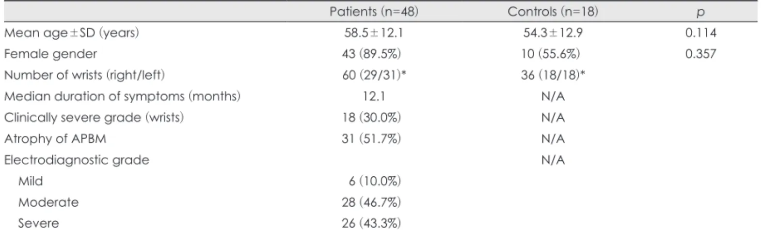
Forty-eight (60 wrists) patients and 18 (36 wrists) healthy controls were included in the study. The age and sex distri- bution of patient and control group are shown in Table 2. The median symptom duration of CTS patients was 12.1 months.
Of the 60 wrists, 18 cases were clinically severe CTS and TABLE 1. Electrophysiological carpal tunnel syndrome grading scheme according to the classification reported by Stevens23)
DSL (ms) Samp (µV) DML (ms) Mamp (mV)
Mild >3.5 <20 - -
Moderate Abnormal median sensory latencies as above >4.2 <5
Severe Abnormal median motor/sensory latencies as above, with absent SNAP or thenar CMAP abnormal spontaneous activity on needle examination
DSL: distal sensory latency, Samp: sensory amplitude, DML: distal motor latency, Mamp: motor amplitude, SNAP: sensory nerve action potential, CMAP: compound motor action potential
31 cases showing atrophy of APBM.
Electrodiagnostic test
The severity of CTS according to classification reported by Stevens was mild in 6 wrists, moderate in 28 wrists and severe in 26 wrists. The mean values of NCS were follow- ings: sensory latency, 3.05 ms; sensory amplitude, 11.76 µV;
sensory velocity, 29.78 m/s; motor latency, 5.45 ms; and motor amplitude, 9.18 mV. CMAPs were absent in 2 cases and median SNAPs were completely absent in 13 cases.
Ultrasonography and statistical analysis
The mean value of CSA was 13.6±4.8 mm2 (range 6.2- 31.2 mm2) in patient group, and 7.7±1.2 mm2 (range 5.4- 10.5 mm2) in control group. The mean CSA at carpal tun- nel inlet was significantly larger in patients than in controls (p<0.0001). The ROC analysis demonstrated the area under
the curve at 0.935 for median nerve CSA (95% confidence interval, 0.86-0.98; p<0.0001)(Figure 1). The cut-off value which defined to the best sensitivity over specificity ratio was at 9 mm2 (sensitivity of 86.7% and specificity of 88.9%).
The 2 standard deviation (SD) above the mean control val- ue was 10.1 mm2. With the cut-off value of 10.1 mm2 the specificity was increased to 97.2%, whereas the sensitivity was slightly decreased to 78.3%.
Mean median nerve CSA of the patient group who classi- fied with mild, moderate and severe NCS grade was 9.4 mm2, 13.3 mm2, and 14.9 mm2, respectively (Table 3). There was an association between median nerve CSA and sever- ity grade of NCS (p=0.036)(Figure 2). The severe grade of NCS showed significantly larger median nerve CSA.
TABLE 2. Summary of carpal tunnel syndrome patients and healthy control subjects
Patients (n=48) Controls (n=18) p
Mean age±SD (years) 58.5±12.1 54.3±12.9 0.114
Female gender 43 (89.5%)* 10 (55.6%)* 0.357
Number of wrists (right/left) 60 (29/31)* 36 (18/18)*
Median duration of symptoms (months) 12.1 N/A
Clinically severe grade (wrists) 18 (30.0%)* N/A
Atrophy of APBM 31 (51.7%)* N/A
Electrodiagnostic grade N/A
Mild 06 (10.0%)*
Moderate 28 (46.7%)*
Severe 26 (43.3%)*
*the numbers in parenthesis denote the counts of right/left wrists. APBM: abductor pollicis brevis muscle
TABLE 3. Median nerve CSA according to NCS grade NCS grade Median nerve CSA (mm2) p
Mild 09.4±2.4
0.036*
Moderate 13.3±4.2
Severe 14.9±5.4
*statistically significant was obtained through one-way anal- ysis of variance test (p<0.05). NCS: nerve conduction test, CSA:
cross sectional area 1.0
0.8
0.6
0.4
0.2
0.0
0.0 0.2 0.4 0.6 0.8 1.0 1-specificity
Sensitivity
FIGURE 1. Receiver operating characteristics curve for ultraso- nographic assessments of median nerve cross-sectional area at carpal tunnel inlet. The area under the curve is 0.935 (95% con- fidence interval, 0.86-0.98; p<0.0001).
25.0
20.0
15.0
10.0
5.0
Mild Moderate Severe Controls
Median nerve CSA
(mm
2)
FIGURE 2. Box plot showing median nerve cross-sectional area (CSA) according to severity of nerve conduction study, and con- trol subjects.
Discussion
This study has demonstrated that median nerve CSA at the carpal tunnel inlet is significantly increased in electro- diagnostically proven CTS patients compared with healthy controls.
The advantage of USG is less invasive than EDT, which can make one discomfort from the electrical stimulations.
And, USG may also be advantage in advanced CTS patients with severe APBM atrophy where NCS shows no more re- sponse.14) Anatomical variations of median nerve and other morphological changes, such as cysts, neuromas, and aber- rations of muscles and nerves, at the wrist are relatively com- mon.1,4,8,13,16) Therefore USG can provide an anatomical pro- file which is important when minimal invasive surgery, such as endoscopic median nerve release, is performed.
Mean normal value of median nerve CSA at the carpal tunnel inlet have varied among reports, ranging from 6.1 to 10.4 mm2.3,21,24,28) In our healthy control group, mean val- ue of median nerve CSA at the carpal tunnel inlet was 7.7 mm2, which fall within the range of previous reports. Some studies considered that differing demographic and biomet- ric features, such as older age, male gender, body mass in- dex, and handedness, may contribute to the range of normal value.3,5) However, the debates are still remains, other stud- ies found no significant association between biometric char- acteristics of subjects and median nerve CSA at wrist and forearm in their well-matched case control study.6,9) Thus, it can be said that the range of normal values for median nerve CSA in the literatures more likely reflects variations in study design and USG technique.
The patients who proven electrodiagnostically CTS had significantly increased median nerve CSA than control sub- jects. There is no clear explanation why median nerve is found to be thickened or enlarged on USG in CTS. One possible pathophysiology can be assumed based on the re- sult of experimental study for entrapment neuropathy. The cascade of the biological response to compression in periph- eral nerves includes endoneurial edema, demyelination, in- flammation, distal axonal degeneration, fibrosis, growth of new axons, remyelination, and thickening of the perineuri- um and endothelium. The degree of axonal degeneration is associated with the amount of endoneurial edema.18,20) There- fore, a more severe grade of median neuropathy as defined by EDT can produce increased endoneurial edema. And this increased swelling will appear as higher median nerve CSA in USG. This was confirmed by our result of significant cor- relation between median nerve CSA and the NCS severity scale.
ROC analysis in previous studies yields sensitivities rang- ing from 62% to 97.9% and specificities ranging from 83%
to 100% using a cut-off value which makes best sensitivity over specificity ratio.1,9,16,26,28) Our ROC analysis yielded a cut-off value of 9 mm2 with 86.7% sensitivity and 88.9%
specificity. Those results are similar to previous reports and comparable with sensitivities and specificities of the EDT results. Therefore, if EDT is not available or not tolerable, ultrasonographic assessment of median nerve CSA can be used as a first-line test for diagnosis of CTS. In this setting, a cut-off value with maximal sensitivity and specificity should be used.
The sensitivity and specificity of EDT for diagnosis of CTS has been estimated to be 80-90% and 82-85%, re- spectively.10,13,15,25) Eventually, approximately 10 to 15% of subjects are the “milder” cases of CTS, those with clinical symptom of CTS but normal NCS results. Koyuncuoglu et al.13) found that such “milder” cases of CTS were signifi- cantly higher median nerve CSA than healthy control sub- jects. And they confirmed the diagnosis of CTS in 30% of subjects using cut-off value of 10.5 mm2, which predicted specificity as 94.7%.29) Some authors reported very high specificity for diagnosis of CTS using 2SD above the mean control value of median nerve CSA.9,14) Our ROC analysis also yielded better specificity (97.2%) using cut-off value of 10.1 mm2 (but decreased sensitivity to 78.3%). Thus, USG has a better specificity than NCS when using cut-off value of median nerve CSA as 2SD above the mean control value. The USG assessments of median nerve CSA using cut-off value with high specificity can be used as a comple- mentary with EDT. If EDT is not available or not tolerable, USG can be used as a stand-alone test for diagnosis of CTS.
In this setting, a cut-off value with maximal sensitivity and specificity based on ROC analysis should be used. If there is high clinical suspicion for CTS, but the EDT results are not sufficient for diagnosis of CTS, the USG could be used to confirm the diagnosis. In this case, a cut-off value of me- dian nerve CSA with high specificity (e.g., 2SD above the mean control value) should be used.
Our study has several limitations. First, we included both wrists from each control subjects. And there were relative- ly small number of control subjects. Second, we measured median nerve CSA only at the level of carpal tunnel inlet.
The value of USG for CTS diagnosis could be more in- creased by measuring median nerve CSA in multiple loca- tion at the wrist12,14) or considering other ultrasonographic features (such as flattening ratio of the median nerve, pal- mar bowing of the flexor retinaculum) not included in this study.
Conclusion
In this study, we showed that ultrasonographic assess- ments of the median nerve CSA at carpal tunnel inlet were useful in diagnosis of CTS. And when using a cut-off value of 10.1 mm2 may help the diagnosis of CTS in patients with negative EDT results.
■ The authors have no financial conflicts of interest.
REFERENCES
1) Bayrak IK, Bayrak AO, Kale M, Turker H, Diren B. Bifid median nerve in patients with carpal tunnel syndrome. J Ultrasound Med 27:1129-1136, 2008
2) Buchberger W, Judmaier W, Birbamer G, Lener M, Schmidauer C. Carpal tunnel syndrome: diagnosis with high-resolution sonog- raphy. AJR Am J Roentgenol 159:793-798, 1992
3) Cartwright MS, Shin HW, Passmore LV, Walker FO. Ultrasono- graphic reference values for assessing the normal median nerve in adults. J Neuroimaging 19:47-51, 2009
4) Chloros GD, Cartwright MS, Walker FO, Wiesler ER. Sonography and electrodiagnosis in carpal tunnel syndrome diagnosis, an anal- ysis of the literature. Eur J Radiol 71:141-143, 2009
5) Duncan I, Sullivan P, Lomas F. Sonography in the diagnosis of car- pal tunnel syndrome. AJR Am J Roentgenol 173:681-684, 1999 6) El Miedany YM, Aty SA, Ashour S. Ultrasonography versus nerve
conduction study in patients with carpal tunnel syndrome: substan- tive or complementary tests? Rheumatology (Oxford) 43:887- 895, 2004
7) Grundberg AB. Carpal tunnel decompression in spite of normal electromyography. J Hand Surg Am 8:348-349, 1983
8) Hammer HB, Haavardsholm EA, Kvien TK. Ultrasonographic measurement of the median nerve in patients with rheumatoid ar- thritis without symptoms or signs of carpal tunnel syndrome. Ann Rheum Dis 66:825-827, 2007
9) Hunderfund AN, Boon AJ, Mandrekar JN, Sorenson EJ. Sonogra- phy in carpal tunnel syndrome. Muscle Nerve 44:485-491, 2011 10) Iyer VG. Understanding nerve conduction and electromyograph-
ic studies. Hand Clin 9:273-287, 1993
11) Kasius KM, Claes F, Verhagen WI, Meulstee J. Ultrasonography in severe carpal tunnel syndrome. Muscle Nerve 45:334-337, 2012 12) Klauser AS, Halpern EJ, De Zordo T, Feuchtner GM, Arora R,
Gruber J, et al. Carpal tunnel syndrome assessment with US: val- ue of additional cross-sectional area measurements of the median nerve in patients versus healthy volunteers. Radiology 250:171-177, 13) Koyuncuoglu HR, Kutluhan S, Yesildag A, Oyar O, Guler K, Ozden 2009 A. The value of ultrasonographic measurement in carpal tunnel
syndrome in patients with negative electrodiagnostic tests. Eur J Radiol 56:365-369, 2005
14) Nakamichi K, Tachibana S. Ultrasonographic measurement of median nerve cross-sectional area in idiopathic carpal tunnel syn- drome: Diagnostic accuracy. Muscle Nerve 26:798-803, 2002 15) Nathan PA, Keniston RC, Meadows KD, Lockwood RS. Predic-
tive value of nerve conduction measurements at the carpal tunnel.
Muscle Nerve 16:1377-1382, 1993
16) Pastare D, Therimadasamy AK, Lee E, Wilder-Smith EP. Sonog- raphy versus nerve conduction studies in patients referred with a clinical diagnosis of carpal tunnel syndrome. J Clin Ultrasound 37:389-393, 2009
17) Phalen GS. The carpal-tunnel syndrome. Clinical evaluation of 598 hands. Clin Orthop Relat Res 83:29-40, 1972
18) Rempel D, Dahlin L, Lundborg G. Pathophysiology of nerve com- pression syndromes: response of peripheral nerves to loading. J Bone Joint Surg Am 81:1600-1610, 1999
19) Rempel D, Evanoff B, Amadio PC, de Krom M, Franklin G, Fran- zblau A, et al. Consensus criteria for the classification of carpal tunnel syndrome in epidemiologic studies. Am J Public Health 88:
1447-1451, 1998
20) Rempel DM, Diao E. Entrapment neuropathies: pathophysiology and pathogenesis. J Electromyogr Kinesiol 14:71-75, 2004 21) Sarría L, Cabada T, Cozcolluela R, Martínez-Berganza T, García S.
Carpal tunnel syndrome: usefulness of sonography. Eur Radiol 10:
1920-1925, 2000
22) Seror P. Sonography and electrodiagnosis in carpal tunnel syn- drome diagnosis, an analysis of the literature. Eur J Radiol 67:146- 152, 2008
23) Stevens JC. AAEM minimonograph #26: the electrodiagnosis of carpal tunnel syndrome. American Association of Electrodiagnos- tic Medicine. Muscle Nerve 20:1477-1486, 1997
24) Swen WA, Jacobs JW, Bussemaker FE, de Waard JW, Bijlsma JW.
Carpal tunnel sonography by the rheumatologist versus nerve con- duction study by the neurologist. J Rheumatol 28:62-69, 2001 25) Werner RA, Andary M. Electrodiagnostic evaluation of carpal
tunnel syndrome. Muscle Nerve 44:597-607, 2011
26) Wiesler ER, Chloros GD, Cartwright MS, Smith BP, Rushing J, Walker FO. The use of diagnostic ultrasound in carpal tunnel syn- drome. J Hand Surg Am 31:726-732, 2006
27) Witt JC, Hentz JG, Stevens JC. Carpal tunnel syndrome with nor- mal nerve conduction studies. Muscle Nerve 29:515-522, 2004 28) Wong SM, Griffith JF, Hui AC, Tang A, Wong KS. Discriminatory
sonographic criteria for the diagnosis of carpal tunnel syndrome.
Arthritis Rheum 46:1914-1921, 2002
29) Yesildag A, Kutluhan S, Sengul N, Koyuncuoglu HR, Oyar O, Gul- er K, et al. The role of ultrasonographic measurements of the me- dian nerve in the diagnosis of carpal tunnel syndrome. Clin Ra- diol 59:910-915, 2004