ABSTRACT
Background: Over the past few decades, demographics information has changed
significantly in patients with surgically resected lung cancer. Herein, we evaluated the recent trends in demographics, surgery, and prognosis of lung cancer surgery in Korea.
Methods: Patients with surgically resected primary lung cancer from 2002 to 2016 were retrospectively analyzed. Multivariable Cox regression analysis was conducted to identify prognostic factors for overall survival. The annual percent change (APC) and statistical significance were calculated using the Joinpoint software.
Results: A total of 7,495 patients were enrolled. Over the study period, the number of lung cancer surgeries continued to increase (P < 0.05). The proportion of women to total subjects has also increased (P < 0.05). The proportion of elderly patients (≥ 70 years) as well as those with tumors measuring 1–2 cm and 2–3 cm significantly increased in both genders (all P < 0.05). The proportion of patients with adenocarcinoma, video-assisted thoracic surgery, sublobar resection, and pathological stage I significantly increased (P < 0.05). The 5-year overall survival rate of lung cancer surgery increased from 61.1% in 2002–2006 to 72.1% in 2012–2016 (P < 0.001). The operative period was a significant prognostic factor in multivariable Cox analysis (P < 0.001).
Conclusion: The mean age of patients with lung cancer surgery increased gradually, whereas tumor size reduced. Prognosis of lung cancer surgery improved with recent increases in the frequency of adenocarcinoma, video-assisted thoracic surgery, sublobar resection, and pathological stage I. The operation period itself was also an independent prognostic factor for overall survival.
Keywords: Time Trend; Demography; Surgical Resection; Surgical Outcome;
Non-Small Cell Lung Cancer; Republic of Korea
INTRODUCTION
Despite a continuous decline in smoking rates for decades, lung cancer remains the most commonly diagnosed cancer and the leading cause of cancer-related deaths worldwide.1 In 2018, lung cancer was newly diagnosed in 2.1 million people (11.6% of the total number of
Original Article
Received: Jul 16, 2019 Accepted: Oct 6, 2019 Address for Correspondence:
Sehoon Choi, MD, PhD
Department of Thoracic and Cardiovascular Surgery, Asan Medical Center, University of Ulsan College of Medicine, 88 Olympic-ro 43-gil, Songpa-gu, Seoul 05505, Korea.
E-mail: [email protected]
© 2019 The Korean Academy of Medical Sciences.
This is an Open Access article distributed under the terms of the Creative Commons Attribution Non-Commercial License (https://
creativecommons.org/licenses/by-nc/4.0/) which permits unrestricted non-commercial use, distribution, and reproduction in any medium, provided the original work is properly cited.
ORCID iDs Jae Kwang Yun
https://orcid.org/0000-0001-5364-5548 Han Pil Lee
https://orcid.org/0000-0002-4234-1220 Geun Dong Lee
https://orcid.org/0000-0003-1890-7455 Hyeong Ryul Kim
https://orcid.org/0000-0002-6691-7693 Yong-Hee Kim
https://orcid.org/0000-0003-2177-4876 Dong Kwan Kim
https://orcid.org/0000-0003-1984-0352 Seung-Il Park
https://orcid.org/0000-0002-8729-0498 Sehoon Choi
https://orcid.org/0000-0002-9961-9289
Jae Kwang Yun ,1 Han Pil Lee ,2 Geun Dong Lee ,1 Hyeong Ryul Kim ,1 Yong-Hee Kim ,1 Dong Kwan Kim ,1 Seung-Il Park ,1 and Sehoon Choi 1
1 Department of Thoracic and Cardiovascular Surgery, Asan Medical Center, Ulsan University College of Medicine, Seoul, Korea
2Department of Thoracic and Cardiovascular Surgery, Kangwon National University Hospital, Gangneung, Korea
Recent Trends in Demographics, Surgery, and Prognosis of Patients with Surgically Resected Lung Cancer in a Single Institution from Korea
Oncology & Hematology
Disclosure
The authors have no potential conflicts of interest to disclose.
Author Contributions
Conceptualization: Choi S. Data curation: Yun JK, Lee GD, Kim HR, Kim YH, Kim DK, Park SI.
Formal analysis: Yun JK. Investigation: Yun JK, Lee HP. Supervision: Choi S. Validation: Gwag HB, Park SJ. Visualization: Yun JK, Lee HP.
Writing - original draft: Yun JK.
cancer cases), and 1.8 million people (18.4% of the total number of cancer cases) died of lung cancer worldwide.1 In Korea, lung cancer has been the most common cause of cancer death since 1999 and accounted for 22.6% (17,399/76,855) of all cancer deaths in 2015.2 Surgical resection is the only curative treatment that improves survival for early-stage lung cancer,3 and in Korea, 22% of newly diagnosed lung cancer cases are candidates for surgery.4 Over the past few decades, clinical information changed significantly in patients with surgically resected lung cancer. Patient screening was performed via low-dose computed tomography (LDCT) and an increased proportion of factors such as women gender, stage I, and adenocarcinoma have contributed to improved survival outcome of lung cancer surgery.5,6 As such, it is crucial to analyze the demographics, tumor-related background, and prognosis that can affect the incidence of disease and treatment modalities. In line with other countries,7-9 there have been two studies analyzing the trend on lung cancer surgery in Korea4,10 However, one study based on Korean national data does not have information on primary diagnosis, histologic type, and survival outcome.4 In addition, the other study included a relatively small number of patients and is out of date.10 Therefore, we sought to investigate recent trends and surgical outcomes of lung cancer surgery conducted in Korea on a large cohort collected by a single institute between 2002 and 2016.
METHODS
Patients
We reviewed the records of patients who underwent pulmonary resection for primary lung cancer between January 2002 and December 2016 from a prospectively collected database at Asan Medical Center, Seoul, Korea. Patients who underwent surgery for diagnostic intent were excluded from the study. Each cancer case of patients with more than one primary lung cancer was included. The patient population was pathologically staged according to the recommendations by the 8th edition American Joint Committee on Cancer (AJCC) in a retrospective manner.11 Tumor histology was categorized according to the World Health Organization classification.12 Lobectomy with systematic lymph node dissection was adopted as a standard procedure for primary lung cancer, but some patients with old age, borderline lung function, and early stage adenocarcinoma received sublobar resection. Patients with clinical stage I-IIIA, including N2 node metastasis, were indicated for surgical resection. All patients were followed either until death or the last follow-up date (March 1, 2019).
Follow-up information on all patients was obtained via notes taken during clinical follow- up, which was conducted every 3–6 months during the first 2 years after surgery and every 6–12 months thereafter. Chest CT scans were performed in sync with clinical visits or at any time when disease recurrence was suspected. Treatment modalities and chemotherapeutic regimens in relapsed cases were determined at the discretion of the attending physician.
Outcome measurements
Considering that the number of comorbid diseases per patient is superior to individual comorbidities (hypertension, diabetes mellitus, smoking, chronic bronchitis, coronary artery disease, cardiac arrhythmia, congestive heart failure, liver cirrhosis, cerebral vascular event, renal insufficiency, history of malignant disease, and prior thoracic surgery) in a predictive model for postoperative mortality,13 we considered it to be a categorical variable.
Major complications were evaluated according to the joint definitions provided by the
Society of Thoracic Surgeons and the European Society of Thoracic Surgeons.14 Postoperative mortality was defined as any deaths occurring within the first 30 days after operation and those occurring during the same hospitalization. Overall survival (OS) was defined as the time interval between the date of operation and the date of death, which was obtained by reviewing the records from the Korean National Security Death Index Database.
Statistical analysis
Continuous variables were presented as means and standard deviations, and the categorical variables were presented as a percentage. To effectively perform a trend analysis, the operation years were divided into three categories over 5-year intervals (2002–2006 vs.
2007–2011 vs. 2012–2016). For comparing among the three groups, we used parametric test (ANOVA) for continuous variables and χ2 test for categorical variables. Survival analysis was conducted using the Kaplan–Meier method and the results were assessed using the log-rank test. Cox proportional hazards model was used for univariable and multivariable analyses to identify prognostic factors for OS. The final multivariable model was selected by adopting the stepwise model selection approach (P ≤ 0.1 for entering the model and P ≤ 0.05 for staying in the model). Cox proportional hazard analysis was also utilized to adjust for baseline covariates and to find significant differences between adjacent operation year categories.
Annual percent changes (APC) in incidences, proportions, and their statistical significance were calculated using the Joinpoint software ver. 4.6.0.0 from the Surveillance Research Program of the United States National Cancer Institute.15 The other statistical calculations were performed using R version 3.4.2 (The R Foundation for Statistical Computing, Vanderbilt University, Nashville, TN, USA) by employing the Survival, ggplot2, GGally, survminer, and rms packages. P values of < 0.05 were considered to be statistically significant.
Ethics statement
This study was approved by the Institutional Review Board of Asan Medical Center (2019- 0923). The requirement for informed consent was waived.
RESULTS
Patient characteristics
From January 2002 to December 2016, a total of 7,485 patients with primary lung cancer were included in this study. Characteristics of the enrolled patients are summarized in Table 1.
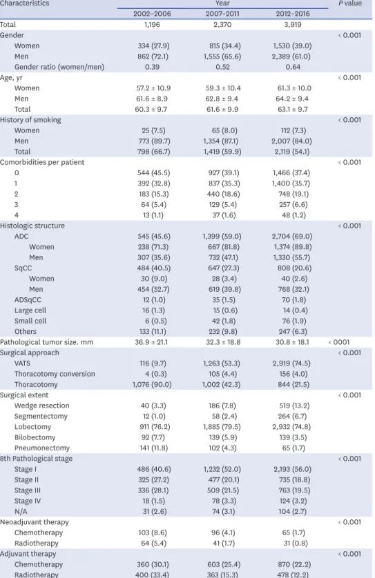
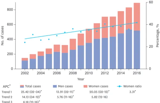
Over the study period, the number of lung cancer surgeries in both genders has continued to increase (Table 1 and Fig. 1). Especially, the proportion of women to the total number of surgeries has increased significantly from 24.0% to 41.9% (APC, 3.31; P < 0.05) (Fig. 1). The mean age of patients with lung cancer surgery in both genders increased gradually, and the number of comorbidities per patient also correspondingly increased (Table 1). The overall smoking rate among patients declined from 66.7% to 54.1% owing to the increased proportion of women with low smoking rates (Table 1). The proportion of patients with adenocarcinoma, sublobar resection (wedge resection or segmentectomy), and pathological stage I has significantly increased from 45.6% to 69.0% (P < 0.001), 4.3% to 19.9% (P < 0.001), and 40.6% to 56.0% (P < 0.001), respectively (Table 1 and Fig. 2). The proportion of patients with video-assisted thoracic surgery (VATS) has also significantly increased up to 84.4% in 2016;
especially in 2008, a dramatic increase has been observed (APC 45.17; P < 0.05) (Fig. 2). As the
Table 1. Demographic and clinical characteristics of enrolled patients (n = 7,485) according to the operation period
Characteristics Year P value
2002–2006 2007–2011 2012–2016
Total 1,196 2,370 3,919
Gender < 0.001
Women 334 (27.9) 815 (34.4) 1,530 (39.0)
Men 862 (72.1) 1,555 (65.6) 2,389 (61.0)
Gender ratio (women/men) 0.39 0.52 0.64
Age, yr < 0.001
Women 57.2 ± 10.9 59.3 ± 10.4 61.3 ± 10.0
Men 61.6 ± 8.9 62.8 ± 9.4 64.2 ± 9.4
Total 60.3 ± 9.7 61.6 ± 9.9 63.1 ± 9.7
History of smoking < 0.001
Women 25 (7.5) 65 (8.0) 112 (7.3)
Men 773 (89.7) 1,354 (87.1) 2,007 (84.0)
Total 798 (66.7) 1,419 (59.9) 2,119 (54.1)
Comorbidities per patient < 0.001
0 544 (45.5) 927 (39.1) 1,466 (37.4)
1 392 (32.8) 837 (35.3) 1,400 (35.7)
2 183 (15.3) 440 (18.6) 748 (19.1)
3 64 (5.4) 129 (5.4) 257 (6.6)
4 13 (1.1) 37 (1.6) 48 (1.2)
Histologic structure < 0.001
ADC 545 (45.6) 1,399 (59.0) 2,704 (69.0)
Women 238 (71.3) 667 (81.8) 1,374 (89.8)
Men 307 (35.6) 732 (47.1) 1,330 (55.7)
SqCC 484 (40.5) 647 (27.3) 808 (20.6)
Women 30 (9.0) 28 (3.4) 40 (2.6)
Men 454 (52.7) 619 (39.8) 768 (32.1)
ADSqCC 12 (1.0) 35 (1.5) 70 (1.8)
Large cell 16 (1.3) 15 (0.6) 14 (0.4)
Small cell 6 (0.5) 42 (1.8) 76 (1.9)
Others 133 (11.1) 232 (9.8) 247 (6.3)
Pathological tumor size. mm 36.9 ± 21.1 32.3 ± 18.8 30.8 ± 18.1 < 0001
Surgical approach < 0.001
VATS 116 (9.7) 1,263 (53.3) 2,919 (74.5)
Thoracotomy conversion 4 (0.3) 105 (4.4) 156 (4.0)
Thoracotomy 1,076 (90.0) 1,002 (42.3) 844 (21.5)
Surgical extent < 0.001
Wedge resection 40 (3.3) 186 (7.8) 519 (13.2)
Segmentectomy 12 (1.0) 58 (2.4) 264 (6.7)
Lobectomy 911 (76.2) 1,885 (79.5) 2,932 (74.8)
Bilobectomy 92 (7.7) 139 (5.9) 139 (3.5)
Pneumonectomy 141 (11.8) 102 (4.3) 65 (1.7)
8th Pathological stage < 0.001
Stage I 486 (40.6) 1,232 (52.0) 2,193 (56.0)
Stage II 325 (27.2) 477 (20.1) 735 (18.8)
Stage III 336 (28.1) 509 (21.5) 763 (19.5)
Stage IV 18 (1.5) 78 (3.3) 124 (3.2)
N/A 31 (2.6) 74 (3.1) 104 (2.7)
Neoadjuvant therapy < 0.001
Chemotherapy 103 (8.6) 96 (4.1) 65 (1.7)
Radiotherapy 64 (5.4) 41 (1.7) 31 (0.8)
Adjuvant therapy < 0.001
Chemotherapy 360 (30.1) 603 (25.4) 870 (22.2)
Radiotherapy 400 (33.4) 363 (15.3) 478 (12.2)
Continuous variables were presented as means ± standard deviations, and categorical variables as number (%).
ADC = adenocarcinoma, SqCC = squamous cell carcinoma, ADSqCC = adenosquamous cell carcinoma, VATS = video-assisted thoracic surgery.
proportion of patients with stage I patients increased, the rates of neoadjuvant and adjuvant therapy decreased (Table 1). Postoperative mortality decreased gradually from 2.8% to 0.3%.
The incidences of detailed postoperative complications over time are described in Table 2.
0 2002 2004 2006 2008 2010 2012 2014 2016
600
400
200
No. of cases Percentage, %
Year 800
0 60
20 40
Trend 1 Trend 2 Trend 3 APC*
25.42 (02–04)* 14.13 (04–12)*
6.18 (12–16)* Total cases
13.91 (02–11)* 5.76 (11–16)*
Men cases
20.05 (02–12)* 5.82 (12–16) Women cases
3.31* Women ratio
Fig. 1. Changes in the number of lung cancer surgeries for total, men, and women patients and the proportion of women to total patients according to the year. The numbers at the bottom of each variable in the legend indicate APC during the years shown in parentheses.
APC = annual percent change.
*Significantly different from zero at (P < 0.05).
0 2002 2004 2006 2008 2010 2012 2014 2016
60
40
20
Percentage, %
Year 80
Trend 1 Trend 2 Trend 3 APC
19.36* Sublobar resection 45.17 (02–08)*
7.33 (08–16) VATS operation
−1.41 (02–05) 10.57 (05–08)* 1.06 (08–16)* Pathological stage I 5.62 (02–10)*
2.47 (10–16)* Adenocarcinoma
Fig. 2. Changes in the proportion of adenocarcinoma, pathological stage I, VATS operation, sublobar resection according to the year. The numbers at the bottom of each variable in the legend indicate APC during the years shown in parentheses.
APC = annual percent change, VATS = video-assisted thoracic surgery.
*Significantly different from zero at (P < 0.05).
Time trend of age
The age distribution of lung cancer surgery showed a different trend depending on gender and time (Fig. 3). There was a significant increase in the proportion of patients who were aged ≥ 70 years in both genders (APC for men and women: 7.56 and 6.41, respectively; all P < 0.05). The proportion of men in other age groups declined significantly (APC for < 50 years, 50–59 years, and 60–69 years −3.70, −1.73, and −1.49; all P < 0.05), whereas only the proportion of women aged < 50 years showed a significant decrease (APC, −11.39; P < 0.05).
Patients aged 60–69 years accounted for the largest percentage of lung cancer surgeries, regardless of gender, and women were younger than men (Table 1).
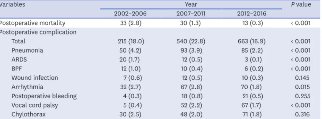
Table 2. Postoperative outcomes according to operation period
Variables Year P value
2002–2006 2007–2011 2012–2016
Postoperative mortality 33 (2.8) 30 (1.3) 13 (0.3) < 0.001
Postoperative complication
Total 215 (18.0) 540 (22.8) 663 (16.9) < 0.001
Pneumonia 50 (4.2) 93 (3.9) 85 (2.2) < 0.001
ARDS 20 (1.7) 12 (0.5) 3 (0.1) < 0.001
BPF 12 (1.0) 10 (0.4) 6 (0.2) < 0.001
Wound infection 7 (0.6) 12 (0.5) 10 (0.3) 0.145
Arrhythmia 32 (2.7) 67 (2.8) 70 (1.8) 0.015
Postoperative bleeding 4 (0.3) 18 (0.8) 21 (0.5) 0.255
Vocal cord palsy 5 (0.4) 52 (2.2) 67 (1.7) < 0.001
Chylothorax 30 (2.5) 48 (2.0) 71 (1.8) 0.316
Values are presented as number (%).
ARDS = acute respiratory distress syndrome, BPF = bronchopleural fistula.
0 2002 2004 2006 2008 2010 2012 2014 2016 40
30
20
10
Percentage, %
Year Year
50
2008
2004 2006 2012
2002 2010 2014 2016
Trend 1 Trend 2 Trend 3 APC
7.56*
≥ 70 yr 9.53 (02–06)*
−0.37 (06–16) 60–69 yr
−6.62 (02–06) 8.30 (06–10)
−2.76 (10–16) 50–59 yr
−11.39 (02–12)* 1.89 (10–16)
< 50 yr
6.41*
≥ 70 yr
−1.49* 60–69 yr
−1.73* 50–59 yr
−3.70*
< 50 yr
Women Men
Fig. 3. Changes in age-specific distribution according to the year. The numbers at the bottom of each variable in the legend indicate APC during the years shown in parentheses.
APC = annual percent change.
*Significantly different from zero at (P < 0.05).
Time trend of tumor size
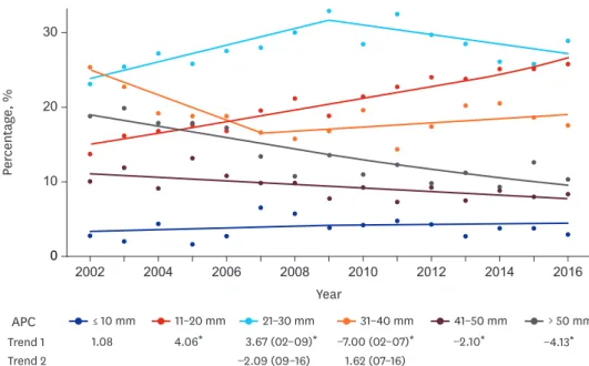
Tumor size was divided into six categories at 1-cm size intervals, and the distribution over time was plotted in Fig. 4. A significant increase was observed in tumors measuring 1–2 cm and 2–3 cm (APC for 1–2 cm and 2–3-cm sized tumor, 4.06 and 3.67; all P < 0.05). In particular, only the proportion of 1–2-cm sized tumors have significantly increased since 2009. On the contrary, the proportions of tumors measuring 3–4 cm, 4–5 cm, and > 5 cm in size have shown a significant decrease, with the largest reduction in the proportion of tumors measuring > 5 cm in size from 20% to 11% (APC, −4.13; P < 0.05). The proportion of tumors measuring < 1 cm in size was relatively consistent.
Time trend in OS
The median follow-up time was 50 months. At the end of follow-up, 2,495 of the 7,485 patients (33.3%) died, and the remaining 4,990 patients (66.7%) were alive or censored. The median survival time (MST) and 5-year survival rate for OS were 107 months (95% confidence interval [CI], 102–113) and 68.9%.
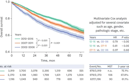
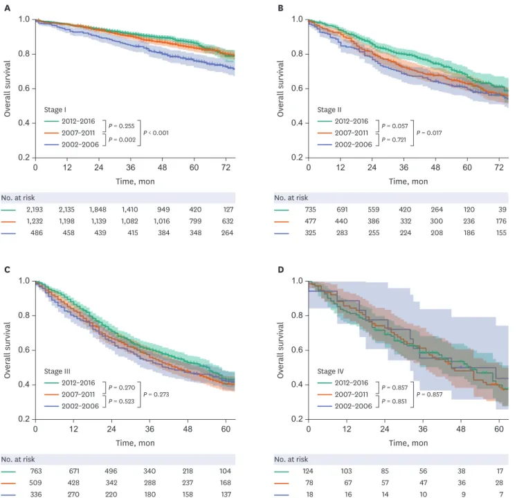
As shown in Fig. 5, a significant escalation in the survival curve was observed with each passing operation year period (all P < 0.001). The 5-year OS rate was 61.1%, 67.8%, and 72.1% in 2002–2006, 2007–2011, and 2012–2016, respectively. Multivariable Cox proportional hazard analyses were performed to identify independent prognostic factors for OS such as age, gender, operation year, smoking history, number of comorbidities, histologic type, surgical approach, surgical extent, pathological stage, neoadjuvant radiotherapy, adjuvant chemotherapy, and adjuvant radiotherapy (Table 3). After adjusting them as covariates, the survival outcome in the recent period (2012–2016) was superior to that in the previous periods (P < 0.001) (Fig. 5). We also performed a subgroup analysis of OS according to pathological stage (Fig. 6). Significant differences were found in patients with pathological stages I and II (P < 0.001 in stage I and 0.017 in stage II) (Fig. 6A and B). In contrast, there
0 2002 2004 2006 2008 2010 2012 2014 2016
20
centagPere, % 10
Year 30
Trend 1 Trend 2
APC 21–30 mm
3.67 (02–09)*
−2.09 (09–16)
≤ 10 mm 1.08
11–20 mm 4.06*
41–50 mm
−2.10*
> 50 mm
−4.13* 31–40 mm
−7.00 (02–07)* 1.62 (07–16)
Fig. 4. Changes in tumor size distribution according to the year. The numbers at the bottom of each variable in the legend indicate APC during the years shown in parentheses.
APC = annual percent change.
*Significantly different from zero at (P < 0.05).
were no significant differences in patients with pathological stages III and IV (P < 0.273 in stage III and 0.857 in stage IV) (Fig. 6C and D).
DISCUSSION
In this study, we evaluated recent trend analysis of demographics, surgery, and prognosis of lung cancer surgery for a large cohort from a single Korean institute over a 15-year period. Overall, despite the decrease in smoking rates, the number of lung cancer surgeries continued to increase. The mean age of patients with lung cancer surgery increased gradually, whereas tumor size declined. The proportions of patients with adenocarcinoma, VATS operation, sublobar resection, and pathological stage I was also increased significantly. These changes led to the improved 5-year OS rate from 61.1% to 72.1% in patients who received lung cancer surgery.
The main cause of the continued increase of lung cancer surgeries can be explained by the aging population in Korea. From 1999 to 2015, the crude incidence rate per 100,000 individuals (total number of cases divided by the mid-year population for the particular year) of lung cancer was increased from 42.1 to 66.8 in men and from 15.1 to 28.4 in women.
During the period, the age-standardized lung cancer incidence rate (a weighted average of the rates by age, where the weights are the percentage of persons in the corresponding age group of the standard population)15 changed from 50.9 to 42.3 in men and from 12.4 to 14.3 in women, which continued to decline since 2002 for men and 1998 for women.2,16 This means that the Korean population is increasingly concentrated in the age group where lung cancer is common, that is, the elderly individuals. This phenomenon is directly related to the increasing age of patients who received lung cancer surgery in our study. While the proportions of other age groups have been similar or decreased, the proportion of elderly patients (≥ 70 years) in both genders rapidly increased (Fig. 2). Considering the improved
0.40
48
24 36
12 60 72
1.0
0.8
erOvall survival 0.6
Time, mon
Multivariate Cox analysis adjusted for several covariates,
such as age, gender, pathologic stage, etc.
2012–2016 2007–2011 2002–2006
No. at risk 3,919 2,370 1,196
Event/No.
879/3,919 979/2,370 637/1,196
MST n.a.
106 90
5-year rate 72.1%
67.8%
61.1%
3,700 2,198 1,049
3,078 1,983 949
2,295 1,799 850
1,519 1,631 778
696 1,272 695
225 1,004 551
Years 12–16 vs. 02–06 12–16 vs. 07–11 07–11 vs. 02–06
HR 0.79 0.81 0.97
P value
< 0.001
< 0.001 0.604 Years
P < 0.001 P < 0.001 P < 0.001
Fig. 5. Kaplan-Meier estimates of overall survival according to the operation period. The P values in the survival graph were calculated using the log-rank test, and the P values in the table were assessed by multivariate Cox analysis.
HR = hazard ratio, MST = median survival time, n.a. = not available.
perioperative care for elderly patients and the report from United Nations,17 the number of elderly individuals and the frequency of lung cancer surgery for elderly patients have been expected to increase over the years.
Contrary to squamous cell carcinoma, the proportion of adenocarcinoma continued to increase and become the most common histologic type in both men and women (Table 1).
This trend is similar to that previously reported in the study conducted based on the data from the Korea Central Cancer Registry18 and those conducted in other countries.9,19,20 The US Surgeon General noted that since the 1950s, the predominance of adenocarcinoma in histologic type can be attributed to changes in the design and consumption of cigarettes, such as lower tar, lower nicotine, and filtered cigarettes.21 However, this argument is challenged by the contradictory evidences that the trend was observed long before the changes in cigarettes and that changes in cigarette design has no relation with the increase in the incidence of adenocarcinoma in epidemiological studies.22-24 Moreover, adenocarcinoma is more common in never-smokers, especially among Asian women.25 These findings suggest Table 3. Multivariable Cox proportional hazard analyses for overall survival
Variables Overall survival
Hazard ratio 95% Confidence Interval P value Age
< 70 year 1 Reference
≥ 70 year 1.87 1.70–2.04 < 0.001
Gender
Women 1 Reference
Men 1.18 1.03–1.35 0.020
Years
2002–2006 1 Reference
2007–2011 0.97 0.87–1.08 0.604
2012–2016 0.79 0.70–0.90 < 0.001
History of smoking 1.29 1.12–1.47 < 0.001
Comorbidities per patient, No.
0 1 Reference
1 1.21 1.10–1.34 < 0.001
2 1.46 1.31–1.64 < 0.001
3 1.70 1.45–1.99 < 0.001
4 2.36 1.81–3.10 < 0.001
Histologic structure
Non-small cell lung cancer 1 Reference
Small cell lung cancer 2.02 1.58–2.61 < 0.001
Surgical approach
VATS 1 Reference
Thoracotomy conversion 1.50 1.21–1.84 < 0.001
Thoracotomy 1.31 1.18–1.45 < 0.001
Surgical extent
Sublobar resection 1 Reference
Anatomic resection 0.78 0.68–0.89 < 0.001
8th Pathological stage
Stage I 1 Reference
Stage II 2.09 1.86–2.35 < 0.001
Stage III 4.01 3.54–4.54 < 0.001
Stage IV 4.75 3.91–5.77 < 0.001
Neoadjuvant therapy
Radiotherapy 1.37 1.01–1.86 0.042
Adjuvant therapy
Chemotherapy 0.85 0.77–0.93 0.002
Radiotherapy 1.14 1.03–1.27 0.012
VATS = video-assisted thoracic surgery.
that other unidentified etiologic factors might be related to the increase in the incidence of adenocarcinoma.
Recently, lung cancer in never-smokers (LCINS) has gained attention owing to the increasing number of related cases and its distinctive nature. LCINS is regarded as a different disease entity from lung cancer among smokers not only because most affected patients are women having adenocarcinoma but also because of distinct underlying molecular mechanisms.26 Global statistics estimate that LCINS accounts for 25% of the total lung cancer cases worldwide.27 Its prevalence is especially higher in Eastern Asia, constituting 33% and 38% of
0.20 1.0
0.8
0.6
erOvall survival 0.4
0.20
48
24 36
12 60 72
1.0
0.8
0.6
erOvall survival 0.4
Time, mon Stage I
P = 0.255 P = 0.002 P < 0.001
48
24 36
12 60 72
Time, mon
A B
No. at risk 2,193 1,232 486
2,135 1,198 458
1,848 1,139 439
1,410 1,082 415
949 1,016 384
420 799 348
127 632 264
No. at risk 735 477 325
691 440 283
559 386 255
420 332 224
264 300 208
120 236 186
39 176 155 2012–2016
2007–2011 2002–2006
2012–2016 2007–2011 2002–2006
0.20 1.0
0.8
0.6
erOvall survival 0.4
0.20
48
24 36
12 60
1.0
0.8
0.6
erOvall survival 0.4
Time, mon
48
24 36
12 60
Time, mon
C D
No. at risk 763 509 336
671 428 270
496 342 220
340 288 180
218 237 158
104 168 137
No. at risk 124
78 18
103 67 16
85 57 14
56 47 10
38 36 9
17 28 7 2012–2016
2007–2011 2002–2006
2012–2016 2007–2011 2002–2006 Stage II
P = 0.057 P = 0.721 P = 0.017
Stage III
P = 0.270 P = 0.523 P = 0.273
Stage IV
P = 0.857
P = 0.851 P = 0.857
Fig. 6. Stage-specific Kaplan-Meier estimates of overall survival according to the operation period. Kaplan-Meier survival curve for (A) pathological stages I, (B) pathological stage II, (C) pathological stage III, and (D) pathological stage IV. The P values in the survival graph were calculated using the log-rank.
lung cancer cases in Japan28 and Korea,29 respectively. During the study period, the number and the proportion of LCINS surgeries have increased owing to the increasing number of women patients with low smoking rates (Table 1). The overall LCINS incidence based on the Korea Central Cancer Registry also showed an increasing trend similar to that observed in our results.18
The widespread use of LDCT screening system for lung cancer has enabled the detection of early-stage lung cancer.5 Early detection of lung cancer patients has resulted in smaller tumor size (Fig. 4) and increased proportions of VATS operation, sublobar resection, and pathological stage I (Fig. 2). The increasing trend of pathological stage with a low malignant potential has also reduced the rate of bilobectomy, pneumonectomy, neoadjuvant therapy, and adjuvant therapy (Table 1). Along with the increase in the proportion of adenocarcinoma, the abovementioned factors have contributed to an improved survival rate of lung cancer surgery. The 5-year OS rate significantly improved from 61.1% to 72.1% (P < 0.001).
Nonetheless, this trend was not just caused by demographics changes. Operation period alone remained a significant prognostic factor in multivariable Cox analysis (P < 0.001).
Based on our data, relatively greater increase in survival rate was observed in patients with pathological stage I or II than that in the patients with advanced stage (Fig. 6). The reasons for these findings can be explained by several factors, such as improved surgical technique, standardized postoperative care, and development of adjuvant therapy. Especially in adjuvant therapy, platinum-based chemotherapy30 and target therapy (tyrosine kinase inhibitors in patients with activating mutations in epidermal growth factor receptor)31 might have contributed to improving the prognosis of recurrent lung cancer as well as primary lung cancer. However, because this study was targeted at patients who underwent lung cancer surgery, these findings could not be generalized to the entire population of lung cancer patients. Considering medical advances within the last decades, such as development of angiogenesis inhibitors, molecular therapy, and immunotherapy, survival rate of patients with advanced stage has improved and will continue to improve in the future.
Our current study has a few limitations stemming from its retrospective nature, as well as using observational data from a single institution. First, we acknowledge that our results cannot represent the trend for all the lung cancer surgeries performed in Korea, given that our research is not based on national data. However, being one of the largest tertiary referral hospitals having patients from all over of the country, our hospital encountered approximately 800 cases of primary lung cancer surgery in 2014, compared with 6,000 cases (including recurred cases) in Korea.4 Thus, we believe that our data, with relatively consistent quality and standardized protocols, fully reflect the trend for lung cancer surgery in Korea.
In addition, because our results are based on patients undergoing surgical treatment, the survival curves might be different from those of general patients with lung cancer, especially those with stage III and IV.
In conclusion, this study demonstrated important temporal changes in the demographics, surgery, and prognosis of lung cancer surgery at Asan Medical Center from 2002 to 2016.
The number of lung cancer surgeries and the proportion of patients who underwent VATS operation or sublobar resection, or those who were women or non-smokers or had adenocarcinoma or pathological stage I, increased significantly. These changes led to an improvement in the 5-year OS rate from 61.1% to 72.1% in patients who underwent lung cancer surgery.
ACKNOWLEDGMENTS
Sehoon Choi has full access to all of the data in the study and takes responsibility for the integrity of the data and accuracy of data analysis.
REFERENCES
1. Bray F, Ferlay J, Soerjomataram I, Siegel RL, Torre LA, Jemal A. Global cancer statistics 2018: GLOBOCAN estimates of incidence and mortality worldwide for 36 cancers in 185 countries. CA Cancer J Clin
2018;68(6):394-424.
PUBMED | CROSSREF
2. Jung KW, Won YJ, Kong HJ, Lee ES; Community of Population-Based Regional Cancer Registries. Cancer statistics in Korea: incidence, mortality, survival, and prevalence in 2015. Cancer Res Treat 2018;50(2):303-16.
PUBMED | CROSSREF
3. Howington JA, Blum MG, Chang AC, Balekian AA, Murthy SC. Treatment of stage I and II non-small cell lung cancer: diagnosis and management of lung cancer, 3rd ed: American College of Chest Physicians evidence-based clinical practice guidelines. Chest 2013;143(5 Suppl):e278S-e313S.
PUBMED | CROSSREF
4. Park S, Park IK, Kim ER, Hwang Y, Lee HJ, Kang CH, et al. Current trends of lung cancer surgery and demographic and social factors related to changes in the trends of lung cancer surgery: an analysis of the national database from 2010 to 2014. Cancer Res Treat 2017;49(2):330-7.
PUBMED | CROSSREF
5. Aberle DR, Adams AM, Berg CD, Black WC, Clapp JD, Fagerstrom RM, et al. Reduced lung-cancer mortality with low-dose computed tomographic screening. N Engl J Med 2011;365(5):395-409.
PUBMED | CROSSREF
6. Sawabata N, Miyaoka E, Asamura H, Nakanishi Y, Eguchi K, Mori M, et al. Japanese lung cancer registry study of 11,663 surgical cases in 2004: demographic and prognosis changes over decade. J Thorac Oncol 2011;6(7):1229-35.
PUBMED | CROSSREF
7. Morgant MC, Pagès PB, Orsini B, Falcoz PE, Thomas PA, Barthes FP, et al. Time trends in surgery for lung cancer in France from 2005 to 2012: a nationwide study. Eur Respir J 2015;46(4):1131-9.
PUBMED | CROSSREF
8. Strand TE, Bartnes K, Rostad H. National trends in lung cancer surgery. Eur J Cardiothorac Surg 2012;42(2):355-8.
PUBMED | CROSSREF
9. Meza R, Meernik C, Jeon J, Cote ML. Lung cancer incidence trends by gender, race and histology in the United States, 1973–2010. PLoS One 2015;10(3):e0121323.
PUBMED | CROSSREF
10. Lee JG, Lee CY, Bae MK, Byun CS, Kim DJ, Chung KY. Changes in the demographics and prognoses of patients with resected non-small cell lung cancer: a 20-year experience at a single institution in Korea. J Korean Med Sci 2012;27(12):1486-90.
PUBMED | CROSSREF
11. Detterbeck FC, Boffa DJ, Kim AW, Tanoue LT. The eighth edition lung cancer stage classification. Chest 2017;151(1):193-203.
PUBMED | CROSSREF
12. Travis WD, Brambilla E, Nicholson AG, Yatabe Y, Austin JH, Beasley MB, et al. The 2015 World Health Organization classification of lung tumors: impact of genetic, clinical and radiologic advances since the 2004 classification. J Thorac Oncol 2015;10(9):1243-60.
PUBMED | CROSSREF
13. Bernard A, Rivera C, Pages PB, Falcoz PE, Vicaut E, Dahan M. Risk model of in-hospital mortality after pulmonary resection for cancer: a national database of the French Society of Thoracic and Cardiovascular Surgery (Epithor). J Thorac Cardiovasc Surg 2011;141(2):449-58.
PUBMED | CROSSREF
14. Fernandez FG, Falcoz PE, Kozower BD, Salati M, Wright CD, Brunelli A. The Society of Thoracic Surgeons and the European Society of Thoracic Surgeons general thoracic surgery databases: joint standardization of variable definitions and terminology. Ann Thorac Surg 2015;99(1):368-76.
PUBMED | CROSSREF
15. Noone A, Howlader N, Krapcho M, Miller D, Brest A, Yu M, et al. SEER cancer statistics review, 1975–
2015. https://seer.cancer.gov/csr/1975_2015/. Updated 2018. Accessed April 3, 2019.
16. Shin HR, Won YJ, Jung KW, Kong HJ, Yim SH, Lee JK, et al. Nationwide cancer incidence in Korea, 1999–2001; first result using the national cancer incidence database. Cancer Res Treat 2005;37(6):325-31.
PUBMED | CROSSREF
17. World population ageing 1950–2050. http://www.un.org/. Updated 2015. Accessed April 15, 2019.
18. Park JY, Jang SH. Epidemiology of lung cancer in Korea: recent trends. Tuberc Respir Dis (Seoul) 2016;79(2):58-69.
PUBMED | CROSSREF
19. Zhou C. Lung cancer molecular epidemiology in China: recent trends. Transl Lung Cancer Res 2014;3(5):270-9.
PUBMED
20. Lortet-Tieulent J, Soerjomataram I, Ferlay J, Rutherford M, Weiderpass E, Bray F. International trends in lung cancer incidence by histological subtype: adenocarcinoma stabilizing in men but still increasing in women. Lung Cancer 2014;84(1):13-22.
PUBMED | CROSSREF
21. National Center for Chronic Disease Prevention and Health Promotion (US) Office on Smoking and Health. The Health Consequences of Smoking-50 Years of Progress: A Report of the Surgeon General. Atlanta, GA:
Centers for Disease Control and Prevention (US); 2014.
22. Chen F, Bina WF, Cole P. Declining incidence rate of lung adenocarcinoma in the United States. Chest 2007;131(4):1000-5.
PUBMED | CROSSREF
23. Brooks DR, Austin JH, Heelan RT, Ginsberg MS, Shin V, Olson SH, et al. Influence of type of cigarette on peripheral versus central lung cancer. Cancer Epidemiol Biomarkers Prev 2005;14(3):576-81.
PUBMED | CROSSREF
24. Marugame T, Sobue T, Nakayama T, Suzuki T, Kuniyoshi H, Sunagawa K, et al. Filter cigarette smoking and lung cancer risk; a hospital-based case--control study in Japan. Br J Cancer 2004;90(3):646-51.
PUBMED | CROSSREF
25. Dela Cruz CS, Tanoue LT, Matthay RA. Lung cancer: epidemiology, etiology, and prevention. Clin Chest Med 2011;32(4):605-44.
PUBMED | CROSSREF
26. Yano T, Haro A, Shikada Y, Maruyama R, Maehara Y. Non-small cell lung cancer in never smokers as a representative ‘non-smoking-associated lung cancer’: epidemiology and clinical features. Int J Clin Oncol 2011;16(4):287-93.
PUBMED | CROSSREF
27. Couraud S, Zalcman G, Milleron B, Morin F, Souquet PJ. Lung cancer in never smokers--a review. Eur J Cancer 2012;48(9):1299-311.
PUBMED | CROSSREF
28. Yano T, Miura N, Takenaka T, Haro A, Okazaki H, Ohba T, et al. Never-smoking nonsmall cell lung cancer as a separate entity: clinicopathologic features and survival. Cancer 2008;113(5):1012-8.
PUBMED | CROSSREF
29. Cho J, Choi SM, Lee J, Lee CH, Lee SM, Kim DW, et al. Proportion and clinical features of never-smokers with non-small cell lung cancer. Chin J Cancer 2017;36(1):20.
PUBMED | CROSSREF
30. Arriagada R, Bergman B, Dunant A, Le Chevalier T, Pignon JP, Vansteenkiste J, et al. Cisplatin-based adjuvant chemotherapy in patients with completely resected non-small-cell lung cancer. N Engl J Med 2004;350(4):351-60.
PUBMED | CROSSREF
31. Lynch TJ, Bell DW, Sordella R, Gurubhagavatula S, Okimoto RA, Brannigan BW, et al. Activating mutations in the epidermal growth factor receptor underlying responsiveness of non-small-cell lung cancer to gefitinib. N Engl J Med 2004;350(21):2129-39.
PUBMED | CROSSREF