High Prevalence of Sarcopenia in Korean Patients after Hip Fracture: A Case-Control Study
Sarcopenia-related falls and fractures are increasing worldwide due to the aging population. The purpose of this study was to 1) evaluate anthropometric characteristics related to hip fracture in Korean patients, 2) investigate sarcopenia prevalence in hip fracture (HF) and non-hip fracture (NF) groups, and 3) investigate the correlation between sarcopenia and osteoporosis. This case-control study examined 359 HF and 1,614 NF normal populations using Korea National Health and Nutrition Examination Survey data.
We performed whole-body dual energy X-ray absorptiometry to analyze body composition using the skeletal muscle mass index (SMI: lean mass/height2) and bone mineral density (BMD). In the HF group, using the AWGS definition, the prevalence of sarcopenia in women and men was 44.3% and 68.2%, respectively; in the NF group, it was 7.1% and 16.1%, respectively. Lower appendicular SMI (P < 0.001), leg muscle mass (P < 0.001), and higher prevalence of sarcopenia (P < 0.001) were observed in the HF group after adjustment for age and gender. In multivariate analysis, sarcopenia (OR = 6.52; 95%
CI = 4.67-9.09), age (OR = 1.15; 95% CI = 1.13-1.17), and osteoporosis (OR = 1.87;
95% CI = 1.35-2.58) were associated with the occurrence of a hip fracture. This study showed a higher prevalence of sarcopenia in patients with hip fractures compared with a normal population, and higher prevalence of sarcopenia in men.
Keywords: Dual Energy X-ray Absorptiometry; Hip Fracture; Sarcopenia; Skeletal Muscle Mass
Jun-Il Yoo,1 Yong-Chan Ha,2 Hyeok-Bin Kwon,2 Young-Kyun Lee,1 Kyung-Hoi Koo,1 and Moon-Jib Yoo3
1Department of Orthopaedic Surgery, Seoul National University Bundang Hospital, Seongnam, Korea;
2Department of Orthopaedic Surgery, Chung-Ang University College of Medicine, Seoul, Korea;
3Department of Orthopaedic Surgery, Dankook University College of Medicine, Cheonan, Korea Received: 8 April 2016
Accepted: 3 June 2016 Address for Correspondence:
Yong-Chan Ha, MD
Department of Orthopaedic Surgery, Chung-Ang University College of Medicine, 102 Heukseok-ro, Dongjak-gu, Seoul 06973, Korea
E-mail: [email protected]
http://dx.doi.org/10.3346/jkms.2016.31.9.1479 • J Korean Med Sci 2016; 31: 1479-1484
INTRODUCTION
Several studies in developed countries have reported a decreas- ing trend in hip fracture incidence (1-4). However, studies in Korea showed a 2-fold increase in the total number of hip frac- tures, and the incidence rate of hip fracture in women increas- ed steeply during a 10-year study period (5). Hip fractures in el- derly patients are very serious because of high mortality, loss of independence, lower quality of life, and high socioeconomic burden (6,7). Although most risk factors such as aging, gender, and medical comorbidities are inevitable, aging is the most im- portant risk factor for hip fractures; thus, identifying modifiable risk factors is extremely important.
To prevent falls and related fragility fractures, the role of mus- cles has been emphasized for maintaining functional perfor- mance. The mass and strength of skeletal muscles naturally de- crease with age, and this loss accelerates after age 65, with a risk of adverse outcomes such as physical disability, poor quality of life, and death (1). This condition is called sarcopenia, which is characterized by decreased muscle mass and impaired muscle function (2-4).
Sarcopenia and osteoporosis have become increasingly sig- nificant as the population of older people increases, and are
clinically very important and common pathological states. In skeletal tissues, muscle and bone interact mechanically and functionally (4). Therefore, aging and various pathological states influence muscle and bone simultaneously. Sarcopenia is con- sidered an indicator of the development of frailty (8) and loss of independence in the elderly. Furthermore, this condition is as- sociated with increased physical disability, resulting in a risk of falls (6). However, the impact of sarcopenia on osteoporotic frac- tures has rarely been reported.
The purpose of this study was to 1) evaluate anthropometric characteristics related to hip fracture in Korean patients, 2) in- vestigate sarcopenia prevalence in hip fracture (HF) and non- hip fracture (NF) groups, and 3) investigate the correlation be- tween sarcopenia and osteoporosis.
MATERIALS AND METHODS Patients
Between November 2011 and December 2014, fresh hip frac- ture patients (≥ 50 years) were eligible and assigned to the HF group. Exclusion criteria were inability to perform whole-body dual energy X-ray absorptiometry (DXA). Body composition and whole bone mineral density (BMD) were assessed using Musculoskeletal Disorders
DXA (DPX-NT; GE Medical Systems Lunar, Madison, WI, USA).
During the study period, 424 hip fracture patients aged 50 years and older were admitted to the study institution. Of these, 34 (8.1%) were excluded because there was no time to perform DXA preoperatively due to the need for urgent surgical repair, and 31 (7.3%) were excluded because of lack of informed con- sent.
The final study population of 359 participants in the HF group (272 females, 78.3 ± 9.8 years; 87 males, 75.3 ± 8.6 years) did not differ significantly from nonparticipants with regard to age, sex, height, or body weight (Table 1).
As a control, we used data from KNHANES IV-2008. Of a total 10,589 who participated in KNHANES IV-2008, 2,816 popula- tion were selected aged over 50 years and measured BMD by DXA (QDR 4500A, Hologic Inc., Waltham, MA, USA). Of these, 1,202 were excluded from the study because they received only lumbar or hip DXA and lacked data for whole-body DXA. Final- ly, 1,614 patients (950 females, 64.1 ± 9.1 years; 664 males, 63.3 ± 8.8 years) underwent whole-body DXA and were assigned to the NF group (Table 1).
Anthropometric measurements
Body composition was measured by whole-body DXA (DPX- NT; GE Medical Systems Lunar, Madison, WI, USA). Bone min- eral content, fat mass, and lean soft tissue mass were measured separately for each part of the body, including the arms and legs.
The lean soft tissue masses of the arms and legs were nearly equal to the skeletal muscle mass. As absolute muscle mass correlates with height, the skeletal muscle mass index (SMI) was calculat- ed by the following formula: lean mass/height2 (kg/m2), which is directly analogous to body mass index (BMI: weight/height2 [kg/m2]). Arm SMI was defined as (arm lean mass/height2 [kg/
m2]). Leg SMI was defined as (leg lean mass/height2 [kg/m2]).
Appendicular SMI was defined as the sum of the arm and leg SMI.
Sarcopenia was defined according to the criteria for the Asia Working Group for Sarcopenia (AWGS) (SMI below 5.4 kg/m2 in women and below 7.0 kg/m2 in men) (7) and European Work- ing Group on Sarcopenia in Older people (EWGSOP) (SMI be- low 5.5 kg/m2 in women and below 7.26 kg/m2 in men).
We simultaneously measured whole body BMD, including the lumbar spine, by DXA for all participants in the study. Os- teoporosis was defined as a BMD 2.5 standard deviations (SD) below the peak bone mass of a young, healthy, gender- and race- matched reference population according to the World Health Organization (WHO) diagnostic classification.
Biochemical analyses
In the HF group, detailed information on the 25-hydroxyvita- min D (25(OH)D) assay was provided previously (9). Serum 25(OH)D levels were determined with a radioimmunoassay kit (DiaSorin, Stillwater, MN, USA).
In the NF (control) group, serum 25(OH)D and parathyroid hormone (PTH) levels were measured using a gamma counter (1470 Wizard; Perkin Elmer, Turku, Finland) and LIAISON (Di- aSorin) with radioimmunoassay (25(OH)D 125I RIA Kit; DiaSo- rin) and chemiluminescence immunoassay (N-tact PTH Assay kit; DiaSorin), respectively.
Statistical analysis
We used Student’s t-test to compare characteristics in the study groups. To compare the prevalence of sarcopenia, we used the χ2 test. To find a significant relationship between appendicular SMI and BMD, Pearson’s correlation was carried out in each group. We evaluated the appendicular SMI value for continu- ous variables by using a general linear model for control, with covariates of age and sex. The general linear model is a general- ization of a multiple linear regression model in the case of more than one dependent variable.
Prevalence of sarcopenia was calculated in 4 age groups (ages Table 1. Characteristics of participants, body composition and skeletal muscle mass index in both females and males
Characteristics Females Males
HF group (n = 272) NF group (n = 950) P value HF group (n = 87) NF group (n = 664) P value
Age, yr 78.27 ± 9.79 64.1 ± 9.13 < 0.001 75.33 ± 8.65 63.3 ± 8.77 < 0.001
Height, cm 153.58 ± 6.06 153.06 ± 5.94 0.201 167.06 ± 6.35 165.92 ± 6.07 0.104
Weight, kg 53.52 ± 9.8 56.3 ± 8.41 < 0.001 60.61 ± 9.22 64.95 ± 10.07 < 0.001
BMI, kg/m2 22.54 ± 3.95 23.99 ± 3.09 < 0.001 21.72 ± 3.07 23.53 ± 3.03 < 0.001
Total femur BMD, g/cm2 0.6 ± 0.15 0.82 ± 0.15 < 0.001 0.7 ± 0.17 0.85 ± 0.14 < 0.001
Femur neck BMD, g/cm2 0.46 ± 0.1 0.82 ± 0.15 < 0.001 0.55 ± 0.14 0.61 ± 0.12 < 0.001
Total spine BMD, g/cm2 0.62 ± 0.12 0.66 ± 0.13 < 0.001 0.62 ± 0.14 0.68 ± 0.13 < 0.001
Whole body T-score -3.14 ± 1.12 -1.78 ± 1.26 < 0.001 -2.58 ± 1.13 -0.71 ± 1.26 < 0.001
Arm lean mass, kg 3.13 ± 1.21 3.41 ± 0.56 < 0.001 3.99 ± 1.94 5.64 ± 0.91 < 0.001
Leg lean mass, kg 9.03 ± 3.38 11.34 ± 1.6 < 0.001 10.94 ± 4.87 16.15 ± 2.48 < 0.001
Appendicular SMI, kg/m2 5.14 ± 1.9 6.32 ± 0.7 < 0.001 5.41 ± 2.29 7.89 ± 0.91 < 0.001
25(OH)D 15.07 ± 10.21 21.8 ± 7.72 < 0.001 14.55 ± 9.24 25.4 ± 7.45 < 0.001
PTH 70.62 ± 75.76 60.68 ± 26.02 0.001 75.4 ± 96.96 58.74 ± 24.5 0.001
BMI, body mass index; BMD, bone mineral density; SMI, skeletal muscle index; PTH, parathyroid hormone.
less than 70, 70 to 74, 75 to 80, and more than 80 years). The Mantel-Haenszel method was used for testing significance for age- and gender-adjusted prevalence of sarcopenia. To deter- mine the presence of sarcopenia as an independent variable in predicting the occurrence of hip fracture selected as a depen- dent variable, we used the stepwise multiple logistic regression model. The regression model also included the patient charac- teristics of age, sex, whole body BMD, weight, and height, which are known to be key predictors of skeletal muscle mass.
The strength of association of the chosen variables and the occurrence of hip fracture were reported as the odds ratio (OR) and 95% confidence interval (CI) compared to a reference group.
Statistical analyses were carried out using SPSS for Windows software (version 22.0; SPSS, Chicago, IL, USA). A P value of
< 0.05 was considered significant.
Ethics statement
The study design and protocol were approved by the institution- al review board of the Chung-Ang University Hospital, CAUH- IRB No. c2016048 (1785). Written informed consent was waived all patients involved in this study. The use of Korea National Health and Nutrition Examination Survey (KNHANES) data was reviewed and approved by the ethics committee of the Ko- rea Centers for Disease Control and Prevention (KCDC) (2011-02CON-21-C).
RESULTS
In the HF group, using the AWGS definition, the prevalence of sarcopenia in women and men was 44.3% and 68.2%, respec- tively; in the NF group, it was 7.1% and 16.1%, respectively. Sar- copenia prevalence was significantly higher in both men and women in the HF group than in the NF group (Table 2). When compared by age distribution, all age groups of women in the HF group had significantly higher prevalence of sarcopenia than the NF groups. The HF group of men aged less than 75 years had higher prevalence of sarcopenia than the NF group (P < 0.001).
However, men aged more than 75 years did not have significant- ly different prevalence of sarcopenia in either group.
In the HF group, using EWGSOP criteria, the prevalence of sarcopenia in women and men was 47.1% and 80.5%, respec- tively; in the NF group, it was 9.9% and 22.4%, respectively.
After adjusting for differences in age and the ratio of females in the HF and NF groups, general linear model analysis was used to compare the characteristics, body composition, and SMI of patients in both study groups (Table 3). No significant differences were observed for height, weight, and BMI after con- trolling for age and gender. Whole-body lean mass, BMD, and PTH were significantly lower in the HF group than in the NF group (P < 0.001). However, 25(OH)D was not significantly dif- ferent between the HF and NF groups (P = 0.238).
A stepwise logistic regression analysis was carried out to iden-
Table 2. Prevalence of sarcopenia in both women and men from each age group
Age, yr Prevalence in women Prevalence in men
HF group NF group P value HF group NF group P value
< 70 34.8% (16/46) 4.7% (32/680) < 0.001 52.6% (10/19) 10.4% (50/481) < 0.001
70-74 41.2% (14/34) 9.3% (12/129) < 0.001 87.5% (21/24) 22.7% (25/110) < 0.001
75-80 15.5% (13/84) 48.3% (29/60) < 0.001 64.7% (11/17) 44.7% (21/47) 0.157
80 < 46.6% (61/131) 17.5% (10/57) < 0.001 64.0% (16/25) 42.3% (11/26) 0.121
Total 44.3% (120/271) 7.1% (67/950) < 0.001 68.2% (58/85) 16.1% (107/664) < 0.001
HF, hip fracture; NF, non-hip fracture.
Table 3. Comparison of characteristics of patients between two groups after adjusting age and gender
Characteristics HF group NF group P value
Height, cm 158.09 ± 8.69 153.38 ± 8.32 0.756
Weight, kg 56.88 ± 9.93 53.58 ± 8.75 0.068
BMI, kg/m2 22.75 ± 3.56 22.73 ± 2.93 0.062
Total femur BMD, g/cm2 0.62 ± 0.16 0.8 ± 0.15 < 0.001
Femur neck BMD, g/cm2 0.48 ± 0.12 0.58 ± 0.12 < 0.001
Total spine BMD, g/cm2 0.62 ± 0.12 0.65 ± 0.14 < 0.001
Whole body T-score -3.04 ± 1.06 -2.07 ± 1.32 < 0.001
Arm lean mass, kg 3.5 ± 1.48 3.64 ± 0. 97 0.003
Leg lean mass, kg 9.83 ± 3.88 11.18 ± 2.29 < 0.001
Appendicular SMI, kg/m2 5.32 ± 2.02 6.24 ± 0.82 < 0.001
25(OH)D 15.54 ± 10.37 22.41 ± 8.82 0.238
PTH 68.13 ± 85.66 65.8 ± 29.33 < 0.001
BMI, body mass index; BMD, bone mineral density; SMI, skeletal muscle index; PTH, parathyroid hormone.
Table 4. Stepwise logistic regression analysis for hip fracture
Variables B OR 95% CI P value
Age 0.14 1.15 1.13-1.17 < 0.001
Presence of sarcopenia 1.87 6.52 4.67-9.09 < 0.001 Presence of osteoporosis 0.62 1.87 1.35-2.58 < 0.001 OR, odds ratio; CI, confidence interval.
tify predictive factors for the occurrence of a hip fracture. We found that older age, sarcopenia, and osteoporosis were signi fi- cant factors for the occurrence of a hip fracture (OR = 1.15, OR = 6.52, OR = 1.87; P < 0.001, P < 0.001 and P < 0.001, respectively) (Table 4). There was no significant correlation between appen-
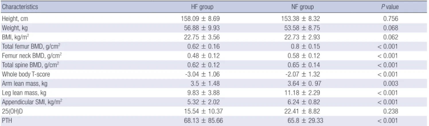
Fig. 1. Correlation of appendicular skeletal muscle mass index (SMI) and femur bone mineral density (BMD). (A) The hip fracture (HF) group. (B) The non-fracture (NF) group.
Correlation of appendicular SMI and femur neck BMD in (C) the HF group and (D) the NF group. Correlation of appendicular SMI and spine BMD in (E) the HF group and (F) the NF group.
Femur BMD (g/m2 )
Appendicular SMI (kg/m2)
0 2 4 6 8 10 12 1.5
1.0
0.5
0
R = 0.072, P = 0.194
Femur BMD (g/m2)
Appendicular SMI (kg/m2)
4 6 8 10 12
1.25
1.00
0.75
0.50
0.25 R = 0.131, P < 0.001
A B
Femur neck BMD (g/m2 )
Appendicular SMI (kg/m2)
0 2 4 6 8 10 12 1.2
0.9
0.6
0.3
0
R = 0.055, P = 0.323
Femur neck BMD (g/m2)
Appendicular SMI (kg/m2)
4 6 8 10 12
1.2
1.0
0.8
0.6
0.4
0.2
0 R = 0.12, P < 0.001
C D
Spine BMD (g/m2 )
Appendicular SMI (kg/m2)
0 2 4 6 8 10 12 1.2
1.0 0.8 0.6 0.4 0.2
0 R = 0.03, P = 0.585
Spine BMD (g/m2)
Appendicular SMI (kg/m2)
4 6 8 10 12 1.2
1.0
0.8
0.6
0.4
0.2 R = 0.123, P < 0.001
E F
dicular SMI and whole-body BMD in the HF (P = 0.194, P = 0.323, P = 0.585, respectively). However, there was a positive correlation between appendicular SMI and whole-body BMD in the NF group (R = 0.13, P < 0.001; R = 0.12, P < 0.001; R = 0.12, P < 0.001, respectively) (Fig. 1).
DISCUSSION
Sarcopenia and osteoporosis have similar pathophysiology and are important risk factors for fragility fractures (4,10). Hip frac- ture is especially significant in disability and frailty, and is known to be related to high mortality, decreased activities of daily liv- ing, and increased socioeconomic burden in elderly patients (11,12). The present study evaluated the prevalence of sarcope- nia in patients with hip fractures diagnosed by DXA after hip fracture occurrence, and was defined using AWGS criteria. This case-control study showed that the prevalence of sarcopenia and osteoporosis in patients with hip fracture was 6.5 and 1.8 times, respectively, more common than in controls. However, in the HF group, there was no significant correlation between appendicular SMI and whole-body BMD (P = 0.194, P = 0.323, P = 0.585, respectively).
This study showed that the prevalence of sarcopenia in wom- en and men in the HF group was 44.3% and 68.2%, respectively.
Hida et al. (13) reported that the prevalence of sarcopenia in hip fracture patients based on Japanese criteria (appendicular SMI < 5.46 kg/m2 in women, < 6.87 kg/m2 in men) was 44.7%
in women and 81.1% in men. Di Monaco et al. (14) reported that the prevalence of sarcopenia in patients with hip fracture based on the New Mexico Elder Health Survey (height adjusted appendicular SMI < 2 SD in a young reference group) was 64%
in women and 95% in men. These findings did not correspond with another study, in which Gonzalez et al. (15) evaluated the prevalence of sarcopenia in 479 patients with hip fracture. They reported that the prevalence of sarcopenia was 17.1% (12.4% in men, 18.3% in women) using the EWGSOP definition. This prev- alence of sarcopenia in the Gonzalez study is lower than in oth- er studies by 3 to 4 times. The reason for these differences in prevalence is mainly due to use of different criteria. Neverthe- less, despite these different definitions, the prevalence of sarco-
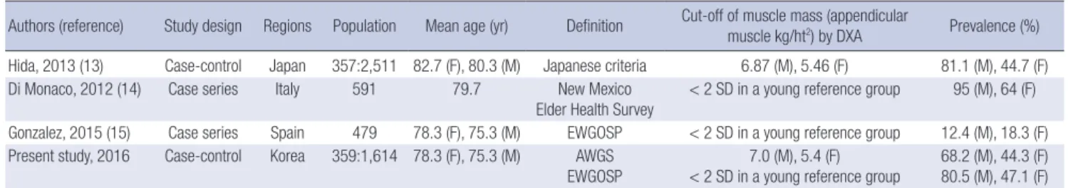
Table 5. Summaries of studies of sarcopenia in patients with hip fracture
Authors (reference) Study design Regions Population Mean age (yr) Definition Cut-off of muscle mass (appendicular
muscle kg/ht2) by DXA Prevalence (%) Hida, 2013 (13) Case-control Japan 357:2,511 82.7 (F), 80.3 (M) Japanese criteria 6.87 (M), 5.46 (F) 81.1 (M), 44.7 (F)
Di Monaco, 2012 (14) Case series Italy 591 79.7 New Mexico
Elder Health Survey < 2 SD in a young reference group 95 (M), 64 (F) Gonzalez, 2015 (15) Case series Spain 479 78.3 (F), 75.3 (M) EWGOSP < 2 SD in a young reference group 12.4 (M), 18.3 (F) Present study, 2016 Case-control Korea 359:1,614 78.3 (F), 75.3 (M) AWGS
EWGOSP 7.0 (M), 5.4 (F)
< 2 SD in a young reference group 68.2 (M), 44.3 (F) 80.5 (M), 47.1 (F) EWGSOP, European Working Group on Sarcopenia in Older people; AWGS, Asian Working Group for Sarcopenia; DXA, dual X-ray absorptiometry; F, female, M, male; SD, stan- dard deviation.
penia in men is higher than in women (Table 5).
Patients with hip fractures had lower serum 25(OH)D levels in this study. However, we could not identify a relationship be- tween sarcopenia and serum 25(OH)D levels. Gumieiro et al.
(16) evaluated the association between serum levels of 25(OH) D3 with mid-upper arm muscle circumference, handgrip stren- gth, and length of hospital stay in 102 patients with hip fracture.
They reported that serum levels of 25(OH)D3 were related to muscle strength rather than mass. In longitudinal observational studies, lower serum levels of vitamin D were associated with significantly decreased skeletal muscle mass during follow-up.
Liu et al. (17) performed a community-based study on vitamin D levels and skeletal muscle mass in 568 patients. They mea- sured 25(OH)D3 and appendicular SMI at baseline and 6 years later, and reported that appendicular SMI was significantly de- creased in the group with lower 25(OH)D3. However, we did not measure muscle strength and it was impossible to confirm a correlation with serum 25(OH)D3. Therefore, a prospective follow-up study is necessary to evaluate the effect of vitamin D on sarcopenia.
As a causal factor of the occurrence of hip fracture in multi- variate analysis in this study, the OR of sarcopenia was 3.5-fold higher than that of osteoporosis in patients with hip fracture.
This finding demonstrates that sarcopenia is a greater risk fac- tor in patients with hip fracture. This corresponds to another study, in which Hida et al. (13) reported that the presence of sarcopenia (P = 0.02), older age (P < 0.001), and lower whole- body BMD (P < 0.001) were significant risk factors for hip frac- ture in elderly patients. However, in evaluating the relationship between sarcopenia and osteoporosis in the HF group, there was no significant correlation between appendicular SMI and whole-body BMD. Hida et al. (13) reported the same results.
They found no significant correlation in the HF group (P = 0.257) between appendicular SMI and whole-body BMD.
There were several limitations. First, this study was cross-sec- tional and retrospective. Therefore, selection bias might be in- evitable. To overcome this, we designed a case-control study and found significant differences between the HF and NF groups.
Second, there could be errors in measured values because the whole-body DXA machines used for the patient group were dif-
ferent from those used for the control group. Hologic conver- sion formula is adjusting with Lunar data only for BMD data of spine and hip in two different devices (18). However, lean body mass and other site were not established conversion formula between two devices. Therefore, direct conversion is impossible and further studies are necessary to develop conversion formu- la for whole body DXA data between two devices. Finally, com- parison with other studies is very limited due to different defi- nitions of sarcopenia. However, the AWGS recently suggested guidelines for Asian populations. It is possible to compare re- sults with other studies in Asians. However, no standardization has been made of body composition for Korean patients.
In conclusion, the present study showed a higher prevalence of sarcopenia and osteoporosis in patients with hip fractures compared with normal populations, and that sarcopenia prev- alence is higher in men. Further study is needed to confirm the association with sarcopenia in patients with hip fracture.
ACKNOWLEDGMENT
Database of KNHANES was provided by the Korea Centers for Disease Control and Prevention. The authors would like to thank the Korea Centers for Disease Control and Prevention.
DISCLOSURE
The authors have no potential conflicts of interest to disclose.
AUTHOR CONTRIBUTION
Study design: Yoo JI, Ha YC, Kwon HB. Data acquisition: Yoo JI, Ha YC, Yoo MJ. Data analysis: Kwon HB, Lee YK. Writing manu- script: Yoo JI, Ha YC, Koo KH. Approval of final manuscript and ensuring research integrity: all authors.
ORCID
Jun-Il Yoo http://orcid.org/0000-0002-3575-4123 Yong-Chan Ha http://orcid.org/0000-0002-6249-0581 Hyeok-Bin Kwon http://orcid.org/0000-0001-5095-4521 Young-Kyun Lee http://orcid.org/0000-0001-6564-4294 Kyung-Hoi Koo http://orcid.org/0000-0001-5251-2911 Moon-Jib Yoo http://orcid.org/0000-0003-2101-1641 REFERENCES
1. Cassell E, Clapperton A. A decreasing trend in fall-related hip fracture in- cidence in Victoria, Australia. Osteoporos Int 2013; 24: 99-109.
2. Melton LJ 3rd, Khosla S, Crowson CS, O’Connor MK, O’Fallon WM, Riggs BL. Epidemiology of sarcopenia. J Am Geriatr Soc 2000; 48: 625-30.
3. Vanitallie TB. Frailty in the elderly: contributions of sarcopenia and vis-
ceral protein depletion. Metabolism 2003; 52: 22-6.
4. Kaji H. Interaction between muscle and bone. J Bone Metab 2014; 21: 29- 40.
5. Ha YC, Park YG, Nam KW, Kim SR. Trend in hip fracture incidence and mortality in Korea: a prospective cohort study from 2002 to 2011. J Kore
an Med Sci 2015; 30: 483-8.
6. Lloyd BD, Williamson DA, Singh NA, Hansen RD, Diamond TH, Finnegan TP, Allen BJ, Grady JN, Stavrinos TM, Smith EU, et al. Recurrent and inju- rious falls in the year following hip fracture: a prospective study of inci- dence and risk factors from the Sarcopenia and Hip Fracture study. J Ger
ontol A Biol Sci Med Sci 2009; 64: 599-609.
7. Chen LK, Liu LK, Woo J, Assantachai P, Auyeung TW, Bahyah KS, Chou MY, Chen LY, Hsu PS, Krairit O, et al. Sarcopenia in Asia: consensus re- port of the Asian Working Group for Sarcopenia. J Am Med Dir Assoc 2014;
15: 95-101.
8. Frisoli A Jr, Chaves PH, Ingham SJ, Fried LP. Severe osteopenia and osteo- porosis, sarcopenia, and frailty status in community-dwelling older wom- en: results from the Women’s Health and Aging Study (WHAS) II. Bone 2011; 48: 952-7.
9. Park Y, Kwon SJ, Ha YC. Association between urinary sodium excretion and bone health in male and female adults. Ann Nutr Metab 2016; 68:
189-96.
10. Reginster JY, Beaudart C, Buckinx F, Bruyère O. Osteoporosis and sarco- penia: two diseases or one? Curr Opin Clin Nutr Metab Care 2016; 19:
31-6.
11. Yoon HK, Park C, Jang S, Jang S, Lee YK, Ha YC. Incidence and mortality following hip fracture in Korea. J Korean Med Sci 2011; 26: 1087-92.
12. Lee SR, Ha YC, Kang H, Park YG, Nam KW, Kim SR. Morbidity and mor- tality in Jeju residents over 50-years of age with hip fracture with mean 6-year follow-up: a prospective cohort study. J Korean Med Sci 2013; 28:
1089-94.
13. Hida T, Ishiguro N, Shimokata H, Sakai Y, Matsui Y, Takemura M, Terabe Y, Harada A. High prevalence of sarcopenia and reduced leg muscle mass in Japanese patients immediately after a hip fracture. Geriatr Gerontol Int 2013; 13: 413-20.
14. Di Monaco M, Castiglioni C, Vallero F, Di Monaco R, Tappero R. Sarcope- nia is more prevalent in men than in women after hip fracture: a cross- sectional study of 591 inpatients. Arch Gerontol Geriatr 2012; 55: e48-52.
15. González-Montalvo JI, Alarcón T, Gotor P, Queipo R, Velasco R, Hoyos R, Pardo A, Otero A. Prevalence of sarcopenia in acute hip fracture patients and its influence on short-term clinical outcome. Geriatr Gerontol Int Forthcoming 2015.
16. Gumieiro DN, Murino Rafacho BP, Buzati Pereira BL, Cavallari KA, Tanni SE, Azevedo PS, Polegato BF, Mamede Zornoff LA, Dinhane DI, Innocenti Dinhane KG, et al. Vitamin D serum levels are associated with handgrip strength but not with muscle mass or length of hospital stay after hip frac- ture. Nutrition 2015; 31: 931-4.
17. Liu G, Lu L, Sun Q, Ye X, Sun L, Liu X, Zong G, Jin Q, Li H, Lin X. Poor vita- min D status is prospectively associated with greater muscle mass loss in middle-aged and elderly Chinese individuals. J Acad Nutr Diet 2014; 114:
1544-1551.e2.
18. Ganda K, Nguyen TV, Pocock N. Gender disparity in BMD conversion: a comparison between Lunar and Hologic densitometers. Arch Osteopo
ros 2014; 9: 180.